Case Report
Acute Intestinal Obstruction Revealing Metachronous Gastrointestinal Adenocarcinoma in a Small Bowel Neuroendocrine Tumor: A Case Report
Sara Cavallari, Tullio Piardi*, Ana Diaz Cives and Reza Kianmanesh
Department of General, Digestive and Endocrine Surgery, Hôpital Robert Debré, Centre Hospitalier Universitaire de Reims, France
*Corresponding author: JTullio Piardi, Service de Chirurgie Générale, Digestive et Endocrinienne. Centre Hospitalier Universitaire de Reims, avenu du Général Koenig, 51092 Reims Cedex, France
Published: 27 Jun, 2016
Cite this article as: Cavallari S, Piardi T, Cives AD,
Kianmanesh R. Acute Intestinal
Obstruction Revealing Metachronous
Gastrointestinal Adenocarcinoma in a
Small Bowel Neuroendocrine Tumor: A
Case Report. Clin Surg. 2016; 1: 1022.
Abstract
Neuroendocrine tumors are cancers that develop in the diffuse neuroendocrine system. The small intestine is one of the most common sites where gastrointestinal neuroendocrine cancers develop and the most common histological types of malignant tumors of the small intestine. In the literature the relationship between neuroendocrine tumors and the development of secondary primary malignancies, whether synchronous or metachronous, is well described. Usually, these involve the colorectal, the gastro urinary tract and the bronco pulmonary system, while the localization in the small intestine is uncommon. We describe here the case of a patient followed-up for an ileal resection, which occurred in emergency for intestinal obstruction; the latter was due to a jejunal adenocarcinoma linked to a neuroendocrine tumor. This report illustrates the rare association of small intestinal neuroendocrine tumor with secondary small bowel malignancies.
Introduction
Carcinoid tumor was coined by Oberndofer 1907 [1] to describe a small neoplasia arising from neuroendocrine cells and characterized by a propensity to produce peptides, neuroamines and
other vasoactive substances. The literature has expanded the concept of carcinoid, later replaced
by the term Neuro Endocrine Tumor (NET) [2]. NETs that originate from the cells of the diffuse
neuroendocrine system of the gastrointestinal tract (GI-NET) are considered rare [3]; they are the
second most common neoplasia of the small intestine (Si-NET) [4]. The localisation of the tumor
in jejunum/ileum tract is the third most common primary site, after lung and rectum [5]. The
incidence rates have increased in the more recent years: Si-NETs are 0.67-0.81/100,000/years [6].
The mean age at diagnosis is between 60 and 65 years [6], and male to female ratio is 1.4/1.0 [5].
NET association with secondary primary malignancies (SPM) is an increasing phenomenon
[7]. The occurrence of other malignancies is estimated to range up to 55% [8]; they can have a synchronous or metachronous presentation [9]. The majority of cases are localized in the colorectal tract and genitourinary tract [10,11].
We report here the case of a patient who first underwent an ileal resection for neuroendocrine
carcinoma, and who after 8 months had an emergency exploratory laparotomy for jejunum
occlusion.
Case Report
A 65-year-old man was referred with generalised abdominal pain, vomiting and obstipation of
6 months duration. His medical history was hyperuricemia, dyslipidemia, implant of right hip for
algodistrophy, laparoscopic sigmoidectomy for diverticula, appendectomy, resection of Meckel's
diverticulum and bilateral inguinal hernia. MRI enterography revealed a mass in the right iliac
fossa with dilatation of the upstream. The tumour markers were normal (CA 19.9 20; CEA 0.9;
Chromogranina A 55). A contrast-enhanced CT scan of the abdomen confirmed the presence of
an ileal mass without secondary localisation. The patient underwent an ileal resection in September
2015. The laparotomy showed a tumor restricted to the ileum terminal (27 cm) without hepatic
metastasis, but suspected dissemination in the pelvic peritoneum.
The histopathological examination concluded for Si-NET (mitotic index 1, Ki 67 was 2%;
Immunoistochemistry positivity of Chromogranina and Synaptophysin). 4 lymph nodes out of 12
were metastatic (mitotix index 5 and Ki 67 3%). The presence of 2 metastatic nodules of the pelvic
peritoneum was noted. The global tumor stage was pT3 (m) (2) N1
(4N+/12N) L1 V1 Pn1 M1 R0. In conclusion, it was a Si-NET stage IV
and grading G2 [12,13]. The patient had a good postoperative
recuperation, and the hospital discharge was on day7. A treatment
with analogues of somatostatina was required. After 4 months, an
abdominal hepatic MRI showed hypervascular lesion of the spleen,
suspecting relapse without liver metastasis or abdominal localisation.
Progressively the patient developed abdominal pain, associated with
obstipation and nausea.
Figure 1
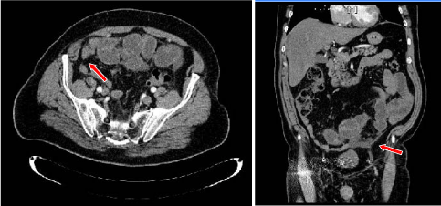
Figure 1
CT showing a disparity in caliber between distended proximal small
bowel loops and collapsed distal small bowel loops.
Figure 2
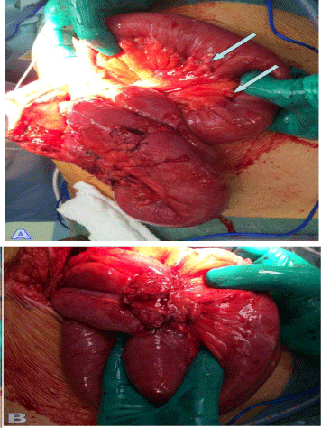
Figure 2
Perioperative photos - A: nodules of peritoneal carcinomatosis; B:
the tumor with retraction of the mesentery.
Due to the continuity of symptoms, a CT-scan was performed in
April 2016. A small bowel obstruction at the jejuno-ileum junction in
the area of the surgical intervention was detected, probably caused by
adhesions, but without signs of ischemic distress (Figure 1).
After 15 days the patient required urgent hospital admission with
worsening of panic symptomatology: severe bloating and abdominal
cramps, nausea, vomiting and constipation. The patient underwent
an emergency exploratory laparotomy that revealed a hard mass in
the mid-jejunum encasing the jejunal loops and mesentery associated
with a peritoneal carcinosis (Figure 2).
In a histopathological examination peritoneal carcinomatosis was
diagnosed with parietal, focal and diffuse infiltration of the ileal wall
with a well differentiated adenocarcinoma. The latter morphological
and immunophenotypic appearance (CK7+, CDX2+, CK20-)
supported a primary intestinal origin. There was no residue of the
neuro-endocrine tumour previously diagnosed.
Discussion
Several study have investigated and provided evidence of SPM
incidence in patients with NET. In 1944, Pearson and Fitzgerald
described the high incidence (23%) of SPM in patients with carcinoid
tumors at autopsy [8]. The association between NET and other
malignancies is an increasingly appreciated phenomenon. In a
retrospective review of 69 patients with GI-NET, Gerstle et al. showed
that 29 (42%) had synchronous tumors and 3 (4%) had metachronous
tumors. In their study, Kamp et al demonstrated that the occurrence
of synchronous secondary primary intestinal malignancies is greater
in GI-NET patients compared with the general population. In another
French study of 270 patients with GI-NET 21 (12.8%) of them also
had a synchronous tumor [13].
The incidence of SPM in patients with GI-NET ranges from 12%
to 46% with an average of 17% [11]. The most common site of SPM
is the gastrointestinal tract (32-62%), followed by the genitourinary
tract (9-27%), breast (14-17%) and the lung system (9-13%) [1]. In
about one third of the cases a small bowel carcinoid tumor may be
associated to SPM, whether synchronous (22%) or metachronous
(9%) [9].
The major series before 1975 in English literature are reported in
(Table 1). The percentage of involvement of the small intestine as SPM
ranges from 0% to 17%, whereas that of Godwin' series combines into
one group ileum and cecum. Then, this sustains that the jejunumileum
SPM is really uncommon [14].
In an epidemiological study about Si-NET and adenocarcinoma
Zar et al. [15] have found that SPM are generally diagnosed within
the first years after diagnosis of a first tumor and that metachronous
tumor is defined according to the lesion diagnosed > 6 months [16].
In our case, the diagnosis of jejunum adenocarcinoma was 8 months
after the first tumor; so, it is a metachronous tumor.
Amin et al. [17] have considered the risk of metachronous
cancers in patients with SI-NET. Between 1973 and 2007 the authors
identified 8331 patients with Si-NET thanks to the Surveillance,
Epidemiology, and END Results database (SEER). They observed
that 33% had developed a metachronous primary tumor. They also
estimated that only 3% of SPM were localized in the small bowel.
Besides, metachronous malignancy may be associated to a genetic
predisposition, behavioural risk factors or common environmental
exposures. Exogenous mitotic effects of secretary products from a
primary tumor can also generate neoplastic transformation, even
a combination of all these factors [18]. Several studies have tried
to establish the relationship between NET and the development of
SPM. Some consider the secretion of biologically active compounds
by the neuroendocrine cells. Zuncker et al. [19] proposed that many
of the secreted peptides have growth factor properties and that non
carcinoid tumor cells can over express receptors for these regulatory
peptides. However, other authors have considered the role played by
non-neuroendocrine peptides in carcinogenesis.
Table 1

Table 1
NET: NeuroEndocrine Tumors; GI-NET: Gastrointestinal Neuroendocrine Tumors; SPM: Secondary Primary Malignancies; S: Synchronous; M: Metachronous; GI: Gatrointestinal; - data not available; *value that considers two series together; the separate cases that return a value of 17% and 18%; also, small intestine and cecum are treated in the same group.
Regarding diagnosis, Zar et al. [15] stressed the importance of an accurate research of synchronous primary malignancies in presence
of Si-NET. In our case, the diagnosis of Si-NET was incidental and
histological. However, even if during the first surgery the abdominal
cavity was explored no tumor was identified.
Therefore, an intensive follow up of patients is warmly
recommended for the prevention of late-stage diagnosis to monitor
the possible development of metachronous tumor. Habal et al. [11]
asserted that the overall prognosis depends primarily on the more
aggressive SPM. The authors [15] evaluated the cause of death in
patients who had been diagnosed SI-NET; they observed that 32%
of those patients had died within 30 days from diagnosis of SPM. In
our experience the patient had peritoneal carcinomatosis at diagnosis
of SPM. He developed an advanced malignant tumor in 8 months,
although he was under intensive follow-up. During this period,
the instrumental investigations had not revealed anything that
could identify SPM. The only relevant fact was signs of obstruction
syndrome, which is presumably falsely interpreted as postoperative
adhesion. The importance of an intensive follow-up was confirmed
in the Consensus guidelines for the management of patients with
digestive NET (ENETS 2016). For patients with G2 NET a check-up
every 3-6 months recommends a life-long follow-up [5], considering
that after 25 years only approximately 20% of all patients are free of
disease [20-25]. In our case, which was discussed in a multidisciplinary
meeting with endocrinologists and oncologists, the follow-up was set
at 3 months, taking into consideration the peritoneal localisation.
Unfortunately, the diagnosis of an advanced cancer was made.
Acknowledgement
The authors thank Doctor Jocelyne Wuibout for the proofreading of this research paper.
References
- Oeberndorfer S. Uber die "Kleinen Dunndamcarcinome". Verhandl Deutsch Pathol. 1907; 11: 113-16.
- Papotti M, De Herder WW. Neuroendocrine tumors. A multidisciplinary Approach. Karger. 2015.
- Modlin IM, Oberg K, Chung DC, Jensen RT, DeHerder WW, Thakken RV, et al. Gastroeteropancreatic neuroendocrine tumors. Lancet Oncol. 2008; 9: 61-72.
- Micheletto G, Scainnamea I, Zanoni A, Panizzo V, Rubino B, Danelli P. Intestinal neuroendocrine tumor. Case report and review of the literature. Ann Ital Chir. 2009; 80: 319-324.
- Jonathan Strosberg: Neuroendocrine tumours of the small intestine. Best Pract Res Clin Gastroenterol. 2012; 26: 755-773.
- Niederle B, Pape UF, Costa F, Gross D, Kelestimur F, Knigge U, et al. ENETS Consensus guidelines update for Neuroendocrine Neoplasms of the Jejunum and Ileum. Neuroendocrinology. 2016; 103: 125-138.
- Ellis L, Shale MJ, Coleman MP. Carcinoid tumors of the gastrointestinal tract: trends in incidence in England since 1971. Am J Gastroenterol. 2010; 105: 2563-2569.
- Pearson CM, Fitzgerald PJ. Carcinoid tumors - a reemphasis of their malignant nature: review of 140 case. Cancer. 1944; 2: 1005.
- Shebani KO, Souba WW. Prognostic and survival in patients with gastrointestinal tract carcinoid tumors. Ann Surg. 1999; 229: 815-823.
- Gerstle JT, Kauffman GJ, Koltun WA. The incidence, management and outcome of patients with gastrointestinal carcinoids and second primary malignancies. J Am Coll Surg. 1995; 180: 427-432.
- Habal N, Sims C, Bilchik AJ. Gastrointestinal carcinoid tumors and secondary primary malignancies. J Surg Oncol. 2000; 75: 301-306.
- Rindi G, Kloppel G, Couvelard A, Komminoth P, Korner M, Lopes JM, et al. TNM staging for a midgut and hindgut (neuro) endocrine tumors:a consensus proposal including a grading system. Virchows Arch. 2007; 451: 757-762.
- Klimstra DS, Arnold R, Cappella C. WHO classification of tumours of the digestive system. Lyon IACR 2010.
- Berner M. Digestive carcinoids and synchronous malignant tumors. Helv Chim Acta. 1993; 59: 757-766.
- Zar N, Garmo H, Holmberg L, Hellman P. Risk of secondary primary malignancies and causes of death in patients with adenocarcinoma and carcinoid of the small intestine. Eur J Cancer. 2008; 44: 718-725.
- Kamp K, Damhuis RAM, Feelders RA, De Herder WW. Occurrence of second primary malignancies in patients with neuroendocrine tumors of the digestive tract and pancreas. Endocr Relat Cancer. 2012; 19: 95-99.
- Amin S, Warner RRP, Itzkowitz SH, Kang Kiim M. The risck of metachronous cancers in patients with small-intestinal carcinoid tumors: a US population-based study. Endocrine-Related Cancer. 2012; 19: 381-387.
- Modlin IM, Oberg K, Chung DC, Jenser RT, De Herder WW, Thakker RV,et al. Gastropancreatic neuroendocrine tumours. Lancet Oncology. 2008; 9: 61-72.
- Zuncker KA, Longo WE, Modlin IM, Bilchik AJ, Adrian TE. Malignant diathesis from jejuno-ileual carcinoids. Am J Gastroenterol. 1989; 84: 182-186.
- Moertel CG. Karnofky memorial lecture. An odyssey in the land of small tumors. J Clin Oncol. 1987; 5: 1502-1522.
- Godwin DJ. Carcinoid Tumors. An analysis of 2837 cases. Cancer. 1975; 36: 560-569.
- Promegger R, Ensinger C, Steiner P, Sauper T, Profanter C, Margreiter R. Neuroendocrine tumors and second primary malignancy. A relationship with clinical impact? Anticancer Research. 2004; 24: 1049-1052.
- Clift AK, Drymousis P, Al-Nahhas A, Wasan H, Martin J, Holm S, et al. Incidence of second primary malignancies in patients with neuroendocrine tumours. Neuroendocrinology. 2015; 102: 26-32.
- Kothari T, Mangla J. Malignant tumors associated with carcinoid tumors of the gastrointestinal tract. J Clin Gastroenterol. 1981; 3: 43-46.
- Reina JJ, Serrano R, Codes M, Jiménez E, Bolanos M, Gonzalez E, et al. Second primary malignancies in patients with neuroendocrine tumors. Clin Transl Oncol. 2014; 16: 921-926.