Review Article
Clinical Status of Tissue Engineering and Regenerative Medicine in Cardiovascular Disease
Miyachi H1, Shoji T1, Sugiura T1, Miyamoto S1, Breuer KC1 and Shinoka T1,2*
1The Tissue Engineering Program and Center for Cardiovascular and Pulmonary Research, Nationwide Children’s Hospital, USA
2Department of Cardiothoracic Surgery, The Heart Center, Nationwide Children’s Hospital, USA
*Corresponding author: Toshiharu Shinoka, The Tissue Engineering Program and Department of Cardiothoracic Surgery, The Heart Center, Nationwide Children’s Hospital, 700 Children’s Drive, T2294, Columbus, OH 43205, USA
Published: 20 Jun 2016
Cite this article as: Miyachi H, Shoji T, Sugiura T,
Miyamoto S, Breuer KC, Shinoka T.
Clinical Status of Tissue Engineering
and Regenerative Medicine in
Cardiovascular Disease. Clin Surg.
2016; 1: 1021.
Abstract
Cardiovascular diseases, such as coronary artery disease, aortic disease, peripheral vascular disease, and heart failure, contribute to be significant causes of worldwide mortality despite modern advances in medicine and surgery. Current vascular grafts and prosthetic heart valves used to treat CVD have limitations such as the lack of growth capacity and risks of thrombosis, stenosis, and calcification. Similarly, Left Ventricular (LV) reconstruction surgery can reduce the dilated LV volume after cardiac remodeling, but is incapable of regenerating myocardium. Theoretically, cardiovascular tissue engineering and cardiac regenerative medicine have the potential to address the limitations of current grafts and prosthetic heart valves. An ideal Tissue Engineered Vascular Graft (TEVG) and heart valve (TEHV) is thrombus free, easily handled, biocompatible, durable, and maintains mechanical integrity as the scaffold degrades and remodels into native tissue. However, small-diameter (<6 mm) TEVGs have not yet shown clinical effectiveness, and TEHVs still have limitations for clinical use. Cell injection therapies, which induce myocardial regeneration, are promising approaches for myocardial repair. However, the beneficial effects on current cell injection therapies are mainly associated with the secretion of paracrine factors rather than direct differentiation of cardiac cells. Here we will review various advanced devices, approaches, and strategies to address current drawbacks, focusing on current clinical studies and ongoing clinical trials for TEVG, TEHV, and myocardial regeneration based on cardiovascular medicine.
Introduction
Cardiovascular diseases (CVDs), such as coronary artery disease (CAD), aortic disease, peripheral artery diseases (PAD), heart valvular disease, and heart failure are leading causes of death worldwide [1]. The use of expanded polytetrafluoroethylene (ePTFE, Goretex) or polyethylene terephthalate (PET, Dacron) for vascular graft implantation is common to treat patients with PADs and aortic disease [2,3]. However, synthetic materials show limitations associated with thrombosis, lack of durability, and inability to regenerate native tissue [4]. To treat three vessel disease, patients typically undergo Coronary Artery Bypass Grafting (CABG), where autologous tissue are harvested from either the internal mammary artery or the saphenous vein. However, autologous tissues may be in short supply, thus making it difficult to perform multiple or repeat operations. Heart valve replacement procedures are commonly performed to treat patients with heart valvular disease. There are two categories of prosthetic heart valves; mechanical and biological. Unfortunately, mechanical hearts valves have a significant risk of thromboembolic complication, resulting in the need for anticoagulation therapy, whereas, and biological heart valves have their own drawbacks such as poor long-term durability and ectopic calcification. Ischemic heart disease mortalities have decreased due to recent advances in medical device therapies and surgeries. However, the prevalence of congestive heart failure secondary to ischemic heart diseases increasing. In efforts to restore cardiac function after ischemic damage, LV reconstruction surgery and cell injection therapy have been proposed as myocardial repair approaches. Though vascular, valvular, and myocardial devices are readily available and have demonstrated clinical efficacy, they are not without limitations. Several cellular therapeutic approaches for cardiac repair have been evaluated in small and large animal models. Additionally, TEVGs and TEHVs have been created to address cardiovascular challenges, and have demonstrated long-term safety and efficacy in humans. However, the current cardiovascular tissue engineered devices and regenerative medicine therapeutic approaches are far from providing ideal, off-the-shelf clinical treatment. In this article, we will discuss therapeutic applications of Tissue Engineered Vascular Grafts (TEVGs), Tissue Engineered Heart Valves (TEHVs), and regenerative cardiac repair approaches that appear to be particularly promising effects in clinical studies and ongoing clinical trials.
Table 1

Table 1
Clinical trials of TEVG.
Tissue Engineered Vascular Graft and Heart Valve
The concept of tissue engineering was first proposed during
the mid-1980s in an effort to overcome the shortage of suitable
donor organs for transplantation. Tissue engineering is defined as
the fabrication of alternative materials for the purpose of restoring
biological and physiologic function at the site injury or defect, and
eventually integrating with a patient’s native tissue [5,6]. The ideal
tissue-engineered construct will mimic host tissue in that it consists
of cells, the extracellular matrix (ECM), and a signaling system. The
general concept of tissue engineering has three main components; 1)
a scaffold materials, 2) cells, and 3) biochemical and physiochemical
signaling [6].
Tissue engineered vascular graft
To date, hundreds of synthetic and/or biological TEVGs have
been developed and evaluated in studies involving a multitude
of models (Table 1). In 2001, we started a human clinical trial
implanting venous TEVGs for extra cardiac total cavo-pulmonary
connections. The alternative material scaffold was composed of a
woven fabric made of a 50:50 mixture of L-lactide and ε-caprolactone
(PLCL) reinforced with polyglycolic acid (PGA), and seeded with
autologous bone marrow mononuclear cells (BM-MNCs). Long-term
results demonstrated that the TEVG was clinically viable, as there
was no graft-related mortality or evidence of graft rupture, aneurysm,
infection, or ectopic calcification (Figure 1). Approximately 24% of
patients had graft stenosis, but all underwent successful percutaneous
angioplasties [7]. In animal models, various combinations of scaffold
materials, cells and chemical substances have been suggested. Of
synthetic degradable polymers, PGA, Polylactic acid (PLA), and
poly (ε-caprolactone) (PCL) have been the most widely used [8-
10]. Several groups have combined/ blended multiple materials to
take advantage of the best characteristics of individual materials.
The most well known of these co-polymers are PLCL, PLGA; Poly
(L-lactic-co-glycolide), PHA; Polyhydroxyalkanoates, and MPEGPDHA;
polyethylene glycol and dihydrozyacetone polycarbonate
[11-14]. Biological scaffolds are another approach to constructing
TEVGs. The group of evaluated biological scaffolds includes collagen,
fibrin, hydrogels, xenogenicsmall intestinal submucosa (SIS), and
decellularized vessels from both allogenic and xenogenicsources
[15-17]. As for cell sources to seed TEVGs, endothelial cells (ECs),
smooth muscle cells (SMCs), BM-MNCs, mesenchymal stem cells,
embryonic stem (ES) cells, and induced pluripotent stem (iPS) cells
have been investigated [7,18-21].
Figure 1
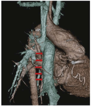
Figure 1
3-Dimensional CT imaging 1 year after TEVG implantation. The
TEVG was implanted into a patient with single ventricle physiology as extra
cardiac total cavo-pulmonary conduit. Red arrows show the TEVG.
There are greater challenges when designing and constructing an arterial TEVG versus a venous TEVG because it must be durable enough to withstand high arterial pressures. Thus, it is logical that many investigations have focused on creating arteriovenous (AV) shunt TEVGs as the next progression from venous scaffolds. L’Heureux et al. developed a Tissue Engineering by Self-Assembly (TESA) design approach, utilizing a new production method to form strong tissue without the use of synthetic biomaterials [22]. This patient-specific graft required autologous fibroblasts to be cultured for 6-9 months to produce sheets of tissue. The tissue sheets are then fused together around a stainless steel mandrel, dehydrated, and subsequently luminally seeded with autologous ECs [23]. Lifeline grafts (Cytograft Tissue Engineering. Novato, CA) were investigated clinically and of 3 clinical trial patients, 2 patients required interventions for stenosis (both eventually failed) within a year and 1 patient died due to infection [24]. These grafts are limited by their high production costs (>$15,000 per graft), long production times, and technically complicated constructions [25]. Therefore, major improvements with this approach are needed before this type of TEVG is translated clinically and commercially marketable. Artegraft is the commercial name for an AV shunt TEVG that took a biological design approach. The graft is composed of decellularized bovine carotid artery graft and favorably required fewer interventions than ePTFE grafts to maintain patency [26]. Omni flow II is a biosynthetic graft that is widely used in many European countries, but fewer South American and Asian nations. However, it is not approved for sale in the U.S., France, and Japan. This graft, composed of cross linked ovine collagen with a polyester mesh endoskeleton, has shown favorable long-term results for hemodialysis use in several studies [27,28]. The Humacyte graft, developed by Dahl et al. [29] is a promising TEVG. It is perhaps the closest to being clinically translated and serving as a readily available off-the-shelf conduit to be used in large and small diameter graft applications. The graft is constructed by culturing cadaveric human SMCs on tubular PGA scaffolds in a bioreactor that delivers cyclic radial strain. During the culture, SMCs secrete ECM proteins, and deposits collagen as the PGA degrades. The resulting tissue is then subsequently decellularized with detergents, which leaves behind a collage nous matrix. The Humacyte TEVG has shown good patency (7/8 in baboon AV shunt and 5/6 canine CABG models) [2]. Recently, two phase 2 clinical trials (n=60) investigating AV shunt TEVGs revealed a higher secondary patency of 89% at 1 year versus PTFE (55- 65% at 1 year)as reported in a multicenter study [30]. The Humacyte TEVG is also currently undergoing clinical trials for peripheral artery bypass applications. Despite several TEVGs displaying promising animal results [31-33], arterial TEVGs are not yet commercially available. The ideal arterial TEVG needs to be both technically and economically viable. Additionally, many cardiovascular diseases are time sensitive and considerations must be made in terms of how long it takes for a given TEVG to be produced.
Figure 2
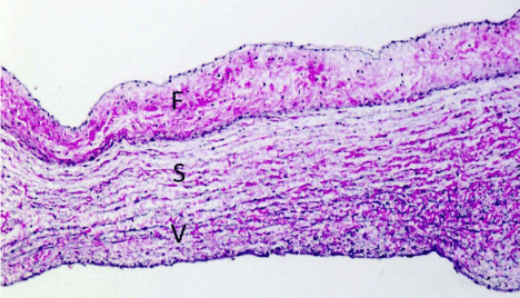
Figure 2
Tissue image of tri-layered structure of an aortic leaflet in sheep.
The three layers consist of fibrosa (F), spongiosa (S), and ventricularis (V).
Tissue engineered heart valve
The TEHV concept consists of an autologous cell-seeded
or unseeded three-dimensional (3D) biocompatible and/or
biodegradable scaffold. A scaffold provides the 3D template and
environment for cells to attach and proliferate into neo tissue. Ideally,
the neo tissue will eventually provide the TEHV with the proper
mechanical properties as the scaffold degrades. There have been many
groups that focused their TEHV research designs on mimicking
native heart valve structure. Human semi lunar valves (pulmonary
and aortic) consist of three semicircular leaflets (cusps) attached to a
fibrous annulus called roots [34]. Valve cusp thickness is generally less
than 1mm in depth, and is thicker at the base and tip. These flexible
valve leaflets (cusps) are composed of three distinct layers: the fibrosa,
spongiosa, and ventricular is (Figure 2) [34,35]. The fibrosa is located
closest to the aorta, composed of circumferentially oriented fibrillary
collagens (type I and III), and associated with a cusp’s mechanical
stiffness and strength [35]. The middle surface is the sponginosa,
and consists of proteoglycans interspersed with collagen fibers. The
layer works as a cushioned interface between the two outer layers to
facilitate valve movement and integrity. The ventricular is composed
of aligned elastic fibers interspersed with short collagen fibers. It
enables valve extension and recoiling under diastolic and systolic
pressures. In general, heart valves include a layer of ECs and valvular
interstitial cells (VICs) between the ECM.A recent studies revealed
that ECs play an important role in heart valve development by
undergoing endocardial-to-mesenchymal transformation [36]. The
VICs are differentiating into myofibroblasts [37], have features found
in both SMC and the fibroblast. Thus, VICs are also thought to play
a very important role in generating TEHVs neo tissue due to their
ECM production. We first introduced a TEHV concept in 1995 that
consisted of a polyglactin woven mesh, sandwiched between 2 nonwoven
PGA mesh sheets. The TEHV was seeded with myofibroblasts
and ECs and then used to reconstruct right posterior pulmonary
heart valve leaflets in a sheep model [38]. Subsequently, we also
investigated a TEHV made of a PGA and PLA co-polymer scaffold in
a lamb model [39]. Many synthetic based materials have been studied
in large animal models and include poly-hydrozyalkanoates (PHAs)
and poly-4-hydroxybutyrate (P4HB) [40,41], poly-hydroxyoctanoate
(PHO) [42], PGA and P4HB [43], PLCL and poly(D, L-lactide-coglycolide)
(PLGA) [44], and polygllycerolsebacate (PGS) [45] (Table 2). Scaffolds blending both biological and synthetic materials have
also been investigated. Porous chitosan-modified PCL scaffolds have
been designed and constructed to improve fibroblast cell attachment
[46]. A composite scaffold composed of PLCL, PLGA, and type 1
collagen has also been tested for TEHV efficacy. A variety of synthetic
material approaches have been studied and include applying P4HA to
mold PGA meshes, PGA/PLLA composite fibrous scaffolds, and PGSPCL
hybrid constructions [43,47-49]. To date however, synthetic
heart valves have not yet been clinically translated. Contrastingly,
decellularized biological-based TEHVs have shown promising
clinical results and some fixed varieties using this approach are
already commercially available (Table 2). TEHV scaffolds require
space for cell attachment, migration, and proliferation. However,
current commercially available non-decellularized xenogenic heart
valves have this ECM space occupied with fixed cells. Therefore we
excluded profiling unseeded, fixed xenogenic heart valve approaches
in this review. Dohmen et al. [50,51] investigated decellularized
allograft implantations seeded patient ECs for reconstruction of
the right ventricular outflow tract during the Ross operation and
displayed favorable long-term results [52]. Their commercially
available cryopreserved pulmonary allografts (CyrolifeInc,
Kennesaw, GA) are created using a proprietary process in the Auto
tissue Laboratories (Auto Tissue GmbH, Berlin, Germany). Cyrolife
subsequently developed a decellularized CyroValve SG pulmonary
human heart valve using Syner Graft technology and demonstrated
clinically acceptable outcomes when compared with conventional
pulmonary allografts [53]. Matrix P plus N is another decellularized
porcine pulmonary heart valve that is already clinically available in
Germany and developed by Auto Tissue GmbH. It has been tested for
right ventricular outflow tract (RVOT) reconstruction or pulmonary
valve replacement during Ross procedures. However, several recent
studies have presented controversial results. In a study involving 93
pediatric patients undergoing RVOT reconstruction using Matrix
P/P plus valves, 35.5% reported conduit failure and 29% conduit
dysfunction [54]. In contrast, a different study, where Matrix P valves
were implanted in 61 patients with congenital heart disease, showed
favorable mid-term performance [55].
Table 2
Table 2
Large animal and clinical trials of TEHV.
Ozaki et al. [56] recently investigated a novel aortic valve reconstruction method using autologous pericardium and had favorable mid-term results. They treated pericardium with a 0.6% glutaraldehyde solution for 10minutes, implanted the pericardium, and the tissue manually reformed into leaflets [56,57]. The advantages of allogenic materials, such as CyroValve SG pulmonary human heart valve, are their smaller immunogenic reactions and the absence of potential disease transmission when compared to xenografts. Advantageously, autologous materials, like the method demonstrated by Ozaki, may be able to overcome the general supply shortage that allogenic materials typically present, however repeated surgery is not feasible.
Cardiac Regenerative Therapy
Ischemic heart disease mortalities have decreased with advances in medical and device therapies, but congestive heart failure secondary to ischemic heart disease has increased world-wide. Acute myocardial infarction (MI) occurs when a coronary vessel occludes and, results in ischemic myocardium. Infracted myocardial are as elicits an inflammatory response which produces collage nous and/or non-contractile scar tissue. Subsequently, the ventricle will progressively dilate in a phenomenon known as ventricular remodeling, which contribute to decreased ventricular contractile function, cardiac failure, and congestive heart failure. Thus, finding a viable therapeutic strategy to treat post-infarction remodeling and progressive heart failure remains a challenge for clinicians. Several cardiac regenerative therapeutic strategies have been suggested such as; Cell-based therapeutic, biomaterial, and endogenous factor approaches (Figure 3). Several biomaterial-based strategies focused on viable cells retention and reinforcing damaged myocardial areas have been evaluated pre-clinically. Additionally, a cellular materialbased strategies and endogenous factor-stimulating bioactive molecules have not yet achieved clinical translation. Strategies and approaches that have not yet reached human trials will be disregarded for the purposes of this paper, instead this review will focus on the clinical status of cell-therapeutic and a cellular material-based approach investigations.
Figure 3
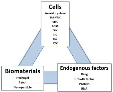
Figure 3
Three components of cardiac regenerative medicine. This
schema represents three component of cardiac regenerative medicine;
Cells, materials, and endogenous factors. Different combinations can be
considered.
Cell-based therapy
Cell-based therapies have emerged as promising treatments to
regenerate dead myocardium and improve LV function. Many studies
have evaluated the functional efficacy of isolated progenitor cells in
preclinical large animal models. Clinical trials have demonstrated
the safety and feasibility of using bone marrow-derived stem cells
[BM-MNCs or mesenchymal stem cells (MSCs)], adipose derived
stem cells (ADSCs), or heart-derived stem cells [cardiac stem cells
(CSCs) or cardio sphere-derived cells (CDCs)] in humans. However,
long-term clinical results regarding the use of aforementioned cell
populations to treat patients with MI, heart failure, and refractory
angina, are varied and display minimal improvement in LV function
[58-60]. Additionally, the benefits associated with adult stem cell use
are attributed to paracrine factor secretion as opposed to direct de
novo cardiac cell differentiation [61]. By promoting growth factor
and cytokine secretion, stem cell therapies reduce the scar volume
associated with MI and myocyte apoptosis, while simultaneously
activating endogenous cardiac stem cells to increase myocyte production and proliferation. Another factor to consider is that stem
cell injection approaches have had difficulties with cell retention
and achieving efficiently consistent outcomes [62]. Therefore, the
utilization of hydrogel and/or patches is being examined to increase
cell survivability and enable better localized cell delivery without
mechanical washout.
Table 3
Table 3
Clinical trials of cell therapy for myocardium repair
Skeletal myoblasts: In the late 1990s, several animal studies
revealed that skeletal myoblasts could differentiate into new
myocardium and improved post-infracted cardiac function [63-65].
However, the MAGIC trial failed to improve the cardiac function
and increased the risk for ventricular arrhythmia, thereby possibly
suggesting that human skeletal myoblasts are unable to differentiate
into cardiac myocyotes and synchronize native myocardial electrical
activity [66]. Therefore, these beneficial effects may be due to
paracrine secretion. Currently in the United States, the MARVEL
trial investigating catheter-delivered autologous skeletal myoblasts
(MyoCELL) injection therapy is ongoing [67]. Transplantation
of skeletal myoblast sheets has shown to improve cardiac function
in patients with heart failure due to ischemic heart disease [68].
However, the skeletal myoblast sheets are not available worldwide.
Bone marrow derived mononuclear cells (BM-MNCs): BMMNCs
have been widely investigated in preliminary clinical studies.
Though studies have demonstrated the feasibility of safely injecting
BM-MNCs at the site of ischemic injury, the clinical benefits of
this approach remains controversial. Several clinical trials such as
BOOST [59-69], FINCELL [70], REPAIR-AMI [71], and TOPCAREAMI [72,73], have shown that BM-MNC intracoronary injections
improved left ventricular ejection fraction (LVEF) when compared
to controls. However, the BOOST trial did not demonstrate any
tangible long-term benefit [59], and other clinical investigations,
such as the ASTAMI, BONAMI, Leuven-AMI, FOCUS-CCTRN,
and HEBE trials [74], resulted in negative outcomes. Additionally,
the TIME and Late TIME trials revealed that time of BM-MNC
injection had no correlation to inducing a clinical benefit when
treating acute MI [75,76]. Several ongoing clinical studies, such as the
REVITALIZE (NCT00874354), REGEN-AMI (NCT00765453), and
BARI (NCT01569178) trials, are still investigating the efficacy of BMMNC
injection approaches, however the overall negative outcomes
of these trials have shifted research focus toward the utilization of
other cell types.
Mesenchymal stem cells: Mesenchymal stem cells are derived
from bone marrow aspirates or adipose tissue. The former have
high engraftment and can induce endogenous cardiomyogenesis.
The TAC-HFT trial compared the use of MSCs and BM-MNCs in
patients with heart failure. The investigation revealed that both cell
types were safe to implant and favorably trended toward reverse
remodeling and regional contractility [77]. The POSEIDON trial
compared autologous and allogenic MSCs transplantation in patients
with ischemic cardiomyopathy. The investigation revealed that
allogenic cells did not induce an unwanted immune response, and
while each group did not increase LVEF, both displayed improved
functional capacity [78]. The cardiopoietic stem cell therapy in heart
failure (C-CURE) trial tested the ability of a cardiogenic cocktail
to enhance the therapeutic benefits of autologous MSCs. The trial
demonstrated both feasibility and safety, while producing a 7%
increase in LVEF and positive effects on exercise tolerance [79].
Clinically, two companies have focused on utilizing ADSCs as a
therapeutic approach. An ADSC known commercially as Adipocell
(U.S. stem cell, inc. Sunrise, FL) recently completed its ANGEL phase
1 trial, whereas Cytori cell (Cytori Therapeutics, inc. San Diego,
CA) is already available for sale in Europe. The APOLLO phase I/
II a trial that administrated Cytori cell to patients with ST-elevation
MI resulted in a positive trend towards improved cardiac function
and perfusion [80]. The PRECISE trial evaluated Cytori cell use in
patients with chronic myocardial ischemia and showed no ejection
fraction (EF) improvement or reductions in scar size, but resulted in
better patient symptoms and exercise tolerance [81]. The ATHENA
I and II trials, investigating Cytori cell use in patients with ischemic
heart failure demonstrated feasibility and safety, but resulted in no
differences in LVEF or volume when compared with controls [82].
Cardiac stem cells: CSCs are clonogenic, multipotent, selfrenewing,
and heart specific stem cells with the ability to differentiate
into cardiomyocytes, ECs, and vascular SMCs. CSCs are typically
isolated via antibody selection after homogenizing large pieces of
cardiac tissue. However, this method is applicable only to patients
that undergo cardiac surgery. A different and less-invasive isolation
approach is to culture CSCs acquired by single biopsy, but the
obtained volumes are significantly smaller. Alternatively, Smith
et al. [83] cultured tissue from percutaneous myocardial biopsy
specimens, formed cardio spheres, and subsequently expanded
them in a monolayer to isolate what is termed cardio sphere-derived
cells (CDCs). CDCs are a heterogeneous cell population that not
only contain adult CSCs, but also vascular cells and differentiated
progenitor cells [84]. The CADUCEUS phase 1 trial examined a
CDC therapeutic approach in 25 myocardial infracted patients [85].
CDCs extracted from right ventricular endomyocardial biopsies
and subsequently cultured for approximately 36 days, were injected
into the infracted coronary arteries6-12 weeks after acute MI. MRI
analysis of patients treated with CDCs showed reductions in scar
mass and increases in viable mass. However, LVEF and cardiac
volume did not improve by 1 year [86]. Similarly, the phase I/II
ALLSTAR trial (NCT01458405) is an ongoing study examining the
safety and efficacy of intracoronary CDCs injection in MI patients.
The ALCADIA (NCT00981006) trial is pilot study investigating the
safety and efficacy of utilizing autologous CDCs injection to treat
ischemic cardiomyopathy [87]. To compensate for poor CDCs
retention, a gelatin sheet containing bFGF was placed on epicardium
during CABG in conjunction with a CDC transendocardial injection.
Only 6 patients were enrolled, but cardiac MRI indicated a 12.1%
EF increase and 3.3% infarct size reduction. The stem cell infusion
in patients with ischemic cardiomyopathy (SCIPIO) trial is the firstin-human,
phase 1, randomized, open-label trial for autologous c-kit
(+) CSCs in patients with ischemic heart failure undergoing CABG
[88]. In the SCIPIO trial, c-kit (+) CSCs were isolated from a right
atrial appendage biopsy and administered via intracoronary infusion.
Investigators reported a significant increases in LVEF and decreases
in infracted size of >30%. However, the cardiogenic potential of these
cells is an area of significant controversy. A recent report has shown
that c-kit (+) cells can only generate cardiomyocytes at functionally
insignificant levels (<0.03%), and resulting new cardiomyocyte
formation is unlikely to be caused by CSC injection [89]. Additionally,
a separate study revealed that c-kit (+) cardiovascular precursor cells
are able to generate cardiomyocytes in neonatal mice hearts, but not
adult mice hearts [90].
The problem of cell-based therapies: Cell injection therapies
have shown only partially favorable results. Generally, isolated cells
are put in saline, and the suspensions are administered systemically
via intravenous infusion, directly myocardium injection (epicardially
or transendocardially), or perfusion into the coronary arteries
(intracoronary) or coronary sinus. These methods enabled delivery
of isolated cells to targeted areas of infracted myocardium. However,
a key challenge facing cellular therapeutic approaches is being able to
retain viable cells in targeted heart tissues and saline solutions have
not proven to be adequate in this regard. To address this issue, several
cell therapy techniques have been developed and investigated, such as
injectable hydrogels and biomaterial patches.
Biomaterial-based strategies
Injectable hydrogels are utilized to encapsulate, deliver and retain
cells to ischemic target areas for long term recovery. Hydrogels used
in cellular therapeutic approaches must have mechanical properties
to support the ventricular wall and also degrade without producing
toxic byproducts. Hydrogels are typically injected via three routes;
intracoronary, epicardially, or transendocardially. In vitro, some
hydrogels have demonstrated increased cell retention [91-93]. More
recently, in vitro and in vivo studies have focused on the application
of bioactive drug-releasing hydrogels. Bioactive molecules enclosed
in these hydrogels include prostaglandins [94], RNA [95], growth
factors [96], or bone morphogenetic protein-2 [97]. However, despite
promising data designing injectable cell-loaded hydrogels, there are
only two clinical studies that have investigated unseeded hydrogels as
a viable myocardial therapeutic approach. The first successful clinical
trial was performed with an intracoronary alginiate hydrogel [98].
The hydrogel was administered alone and preserved LV function. It
is hypothesized that this temporary scaffold replaces the damaged
extracellular matrix, and thereby reduces wall thinning and strain.
Another clinical trial consisted of 6 patients who were implanted
with Algisly-LVR epicardially through concomitant CABG or valve
surgery, and this strategy improved LV size and function. By applying
Laplace’s law to a failing dilated ventricle, this hydrogels injection
could theoretically increase wall thickness and reduce the chamber
diameter, resulting in decreased wall stress and improved LV function.
To date, however, there have been no clinical trials investigating a
combined cell therapy and hydrogel injection approach. Another
cardiac repair strategy is the use of a biomaterial patch that can be
applied epicardially onto a damaged heart. Biomaterial patchescan
supply therapeutic effects by releasing bioactive molecules, delivering
cells, providing mechanical support, and reducing dilatation. It is well
known the Dor procedure, which uses a Dacron patch to minimize
ventricle wall scarring, restores ventricular shape, increases LVEF,
and decreases LV volume. Biomaterial patches can also provide
the additional beneficial effects of cell therapy and degradable
materials to the Dor procedure. Biomaterial patches must be able to
provide stability, flexibility, and mechanical strength. In fact, several
electrospun biomaterials, such as PLGA, PCL, and PGS, have already
demonstrated beneficial results in vitro studies [99-101]. But there
have been no clinical trials utilizing biomaterial patches with cells
to date. To apply these cardiac regenerative therapies in a clinical
setting, more time and energy dedicated to studying in vitro and
in vivo research and surgical procedures is needed. One important
key is that administered cells promote cardiomyocyte proliferation,
improve cardiac function, and decreases long-term mortality.
Conclusion
This review focused on the clinical status of tissue engineering and regenerative medicine in cardiovascular disease. Currently, the development of vascular grafts and heart valves is trending towards biomaterial utilization instead of cell-based approaches. Contrastingly, investigations into cardiac repair therapies are primarily focused on cellular approaches. However, it is not essential that these trends continue, as new scientific discoveries and technological advances may warrant paradigm shifts in translational research. In order to reach clinical translation, therapeutic strategies must factor approaches that are less-invasive, cost effective, time-saving without breaching ethical concerns. Successful translation of complex multidisciplinary technologies clinically requires an active, multidisciplinary, and collaborative participation of clinicians, engineers, chemists, and biologists. Within this collaboration, promising future studies will further optimize tissue engineering technologies that will advance regenerative medicine and patient care.
References
- Mozaffarian D, Benjamin EJ, Go AS, Arnett DK, Blaha MJ, Cushman M,et al. Heart disease and stroke statistics--2015 update: a report from the American Heart Association. Circulation. 2015; 131: e29-322.
- Dahl SL, Kypson AP, Lawson JH, Blum JL, Strader JT, Li Y, et al. Readily available tissue-engineered vascular grafts. Science translational medicine. 2011; 3: 68ra9.
- Kurobe H, Maxfield MW, Breuer CK, Shinoka T. Concise review: tissueengineered vascular grafts for cardiac surgery: past, present, and future.Stem cells translational medicine. 2012; 1: 566-571.
- Tara S, Rocco KA, Hibino N, Sugiura T, Kurobe H, Breuer CK, et al.Vessel bioengineering. Circulation journal: official journal of the Japanese Circulation Society. 2014; 78: 12-19.
- Langer R, Vacanti JP. Tissue engineering. Science. 1993; 260: 920-926.
- Vacanti JP, Langer R. Tissue engineering: the design and fabrication of living replacement devices for surgical reconstruction and transplantation.Lancet. 1999; 354: SI32-134.
- Hibino N, McGillicuddy E, Matsumura G, Ichihara Y, Naito Y, Breuer C, et al. Late-term results of tissue-engineered vascular grafts in humans. The Journal of thoracic and cardiovascular surgery. 2010; 139: 431-436, 436. e1-e2.
- de Valence S, Tille JC, Mugnai D, Mrowczynski W, Gurny R, Moller M,et al. Long term performance of polycaprolactone vascular grafts in a rat abdominal aorta replacement model. Biomaterials. 2012; 33: 38-47.
- Hashi CK, Zhu Y, Yang GY, Young WL, Hsiao BS, Wang K, et al.Antithrombogenic property of bone marrow mesenchymal stem cells in nanofibrous vascular grafts. Proceedings of the National Academy of Sciences of the United States of America. 2007; 104: 11915-11920.
- Pektok E, Nottelet B, Tille JC, Gurny R, Kalangos A, Moeller M, et al.Degradation and healing characteristics of small-diameter poly(epsiloncaprolactone) vascular grafts in the rat systemic arterial circulation.Circulation. 2008; 118: 2563-2570.
- Roh JD, Nelson GN, Brennan MP, Mirensky TL, Yi T, Hazlett TF, et al.Small-diameter biodegradable scaffolds for functional vascular tissue engineering in the mouse model. Biomaterials. 2008; 29: 1454-1463.
- Thevenot PT, Nair AM, Shen J, Lotfi P, Ko CY, Tang L. The effect of incorporation of SDF-1alpha into PLGA scaffolds on stem cell recruitment and the inflammatory response. Biomaterials. 2010; 31: 3997-4008.
- Gogolewski S, Jovanovic M, Perren SM, Dillon JG, Hughes MK. Tissue response and in vivo degradation of selected polyhydroxyacids: polylactides(PLA), poly(3-hydroxybutyrate) (PHB), and poly(3-hydroxybutyrate-co-3-hydroxyvalerate) (PHB/VA). Journal of biomedical materials research. 1993; 27: 1135-1148.
- Zawaneh PN, Singh SP, Padera RF, Henderson PW, Spector JA, Putnam D. Design of an injectable synthetic and biodegradable surgical biomaterial. Proceedings of the National Academy of Sciences of the United States of America. 2010; 107: 11014-11019.
- Hiles MC, Badylak SF, Lantz GC, Kokini K, Geddes LA, Morff RJ. Mechanical properties of xenogeneic small-intestinal submucosa when used as an aortic graft in the dog. Journal of biomedical materials research. 1995; 29: 883-891.
- Weinberg CB, Bell E. A blood vessel model constructed from collagen and cultured vascular cells. Science. 1986; 231: 397-400.
- Swartz DD, Russell JA, Andreadis ST. Engineering of fibrin-based functional and implantable small-diameter blood vessels. American journal of physiology Heart and circulatory physiology. 2005; 288: H1451-460.
- Yue X, van der Lei B, Schakenraad JM, van Oene GH, Kuit JH, Feijen J,et al. Smooth muscle cell seeding in biodegradable grafts in rats: a new method to enhance the process of arterial wall regeneration. Surgery. 1988; 103: 206-212
- Sundaram S, Niklason LE. Smooth muscle and other cell sources for human blood vessel engineering. Cells, tissues, organs. 2012; 195: 15-25.
- Kurobe H, Tara S, Maxfield MW, Rocco KA, Bagi PS, Yi T, et al. Comparison of the biological equivalence of two methods for isolating bone marrow mononuclear cells for fabricating tissue-engineered vascular grafts. Tissue engineering Part C, Methods. 2015; 21: 597-604.
- Hibino N, Duncan DR, Nalbandian A, Yi T, Qyang Y, Shinoka T, et al. Evaluation of the use of an induced puripotent stem cell sheet for the construction of tissue-engineered vascular grafts. The Journal of thoracic and cardiovascular surgery. 2012; 143: 696-703.
- L'Heureux N, Paquet S, Labbe R, Germain L, Auger FA. A completely biological tissue-engineered human blood vessel. FASEB journal: official publication of the Federation of American Societies for Experimental Biology. 1998; 12: 47-56.
- McAllister TN, Maruszewski M, Garrido SA, Wystrychowski W,Dusserre N, Marini A, et al. Effectiveness of haemodialysis access with an autologous tissue-engineered vascular graft: a multicentre cohort study.Lancet. 2009; 373: 1440-1446.
- Wystrychowski W, McAllister TN, Zagalski K, Dusserre N, Cierpka L,L'Heureux N. First human use of an allogeneic tissue-engineered vascular graft for hemodialysis access. Journal of vascular surgery. 2014; 60: 1353-1357.
- McAllister TN, Dusserre N, Maruszewski M, L'Heureux N. Cell-based therapeutics from an economic perspective: primed for a commercial success or a research sinkhole? Regenerative medicine. 2008; 3: 925-937.
- NKennealey PT, Elias N, Hertl M, Ko DS, Saidi RF, Markmann JF, et al. A prospective, randomized comparison of bovine carotid artery and expanded polytetrafluoroethylene for permanent hemodialysis vascular access. J vascular surgery. 2011; 53: 1640-1648.
- Wang SS, Chu SH. Clinical use of omniflow vascular graft as arteriovenous bridging graft for hemodialysis. Artificial organs. 1996; 20: 1278-1281.
- Palumbo R, Niscola P, Calabria S, Fierimonte S, Bevilacqua M, Scaramucci L, et al. Long-term favorable results by arteriovenous graft with Omniflow II prosthesis for hemodialysis. Nephron Clinical practice. 2009; 113: c76-80.
- Dahl SL, Koh J, Prabhakar V, Niklason LE. Decellularized native and engineered arterial scaffolds for transplantation. Cell transplantation. 2003; 12: 659-666.
- Lawson JH, Glickman MH, Ilzecki M, Jakimowicz T, Jaroszynski A,Peden EK, et al. Bioengineered human acellular vessels for dialysis access in patients with end-stage renal disease: two phase 2 single-arm trials.Lancet. 2016; 387: 2026-2034.
- Mrowczynski W, Mugnai D, de Valence S, Tille JC, Khabiri E, Cikirikcioglu M, et al. Porcine carotid artery replacement with biodegradable electrospun poly-e-caprolactone vascular prosthesis. Journal of vascular surgery. 2014; 59: 210-219.
- Mahara A, Somekawa S, Kobayashi N, Hirano Y, Kimura Y, Fujisato T, et al. Tissue-engineered acellular small diameter long-bypass grafts with neointima-inducing activity. Biomaterials. 2015; 58: 54-62.
- Niklason LE, Gao J, Abbott WM, Hirschi KK, Houser S, Marini R, et al.Functional arteries grown in vitro. Science. 1999; 284: 489-493.
- Dohmen PM, Konertz W. Tissue-engineered heart valve scaffolds. Annals of thoracic and cardiovascular surgery: official journal of the Association of Thoracic and Cardiovascular Surgeons of Asia. 2009; 15: 362-367.
- Falk V, Walther T, Schwammenthal E, Strauch J, Aicher D, Wahlers T, et al. Transapical aortic valve implantation with a self-expanding anatomically oriented valve. European heart journal. 2011; 32: 878-887.
- Monaghan MG, Linneweh M, Liebscher S, Van Handel B, Layland SL,Schenke-Layland K. Endocardial-to-mesenchymal transformation and mesenchymal cell colonization at the onset of human cardiac valve development. Development. 2016; 143: 473-482.
- Schoen FJ. Evolving concepts of cardiac valve dynamics: the continuum of development, functional structure, pathobiology, and tissue engineering.Circulation. 2008; 118: 1864-1880.
- Shinoka T, Breuer CK, Tanel RE, Zund G, Miura T, Ma PX, et al. Tissue engineering heart valves: valve leaflet replacement study in a lamb model.The Annals of thoracic surgery. 1995; 60: S513-16.
- Shinoka T, Ma PX, Shum-Tim D, Breuer CK, Cusick RA, Zund G, et al. Tissue-engineered heart valves. Autologous valve leaflet replacement study in a lamb model. Circulation. 1996; 94: II164-168.
- Sodian R, Hoerstrup SP, Sperling JS, Daebritz S, Martin DP, Moran AM, et al. Early in vivo experience with tissue-engineered trileaflet heart valves. Circulation. 2000; 102: III22-29.
- Sodian R, Sperling JS, Martin DP, Egozy A, Stock U, Mayer JE, Jr, et al.Fabrication of a trileaflet heart valve scaffold from a polyhydroxyalkanoate biopolyester for use in tissue engineering. Tissue engineering. 2000; 6: 183-188.
- Stock UA, Nagashima M, Khalil PN, Nollert GD, Herden T, Sperling JS, et al. Tissue-engineered valved conduits in the pulmonary circulation. The Journal of thoracic and cardiovascular surgery. 2000; 119: 732-740.
- Hoerstrup SP, Sodian R, Daebritz S, Wang J, Bacha EA, Martin DP, et al.Functional living trileaflet heart valves grown in vitro. Circulation. 2000; 102: III44-49.
- Park H, Radisic M, Lim JO, Chang BH, Vunjak-Novakovic G. A novel composite scaffold for cardiac tissue engineering. In vitro cellular & developmental biology Animal. 2005; 41: 188-196.
- SMasoumi N, Jean A, Zugates JT, Johnson KL, Engelmayr GC, Jr. Laser microfabricated poly(glycerol sebacate) scaffolds for heart valve tissue engineering. Journal of biomedical materials research Part A. 2013; 101: 104-114.
- Mei N, Chen G, Zhou P, Chen X, Shao ZZ, Pan LF, et al. Biocompatibility of Poly (epsilon-caprolactone) scaffold modified by chitosan--the fibroblasts proliferation in vitro. Journal of biomaterials applications. 2005; 19: 323-339.
- Weber B, Scherman J, Emmert MY, Gruenenfelder J, Verbeek R, Bracher M, et al. Injectable living marrow stromal cell-based autologous tissue engineered heart valves: first experiences with a one-step intervention in primates. European heart journal. 2011; 32: 2830-2840.
- Eckert CE, Mikulis BT, Gottlieb D, Gerneke D, LeGrice I, Padera RF,et al. Three-dimensional quantitative micromorphology of pre- and post-implanted engineered heart valve tissues. Annals of biomedical engineering. 2011; 39: 205-222.
- Masoumi N, Annabi N, Assmann A, Larson BL, Hjortnaes J, Alemdar N,et al. Tri-layered elastomeric scaffolds for engineering heart valve leaflets. Biomaterials. 2014; 35: 7774-7785.
- Dohmen PM, Lembcke A, Hotz H, Kivelitz D, Konertz WF. Ross operation with a tissue-engineered heart valve. The Annals of thoracic surgery. 2002; 74: 1438-1442.
- Dohmen PM, Ozaki S, Verbeken E, Yperman J, Flameng W, Konertz WF.Tissue engineering of an auto-xenograft pulmonary heart valve. Asian cardiovascular & thoracic annals. 2002; 10: 25-30.
- Dohmen PM, Lembcke A, Holinski S, Pruss A, Konertz W. Ten years of clinical results with a tissue-engineered pulmonary valve. The Annals of thoracic surgery. 2011; 92: 1308-1314.
- Brown JW, Elkins RC, Clarke DR, Tweddell JS, Huddleston CB, Doty JR,et al. Performance of the CryoValve SG human decellularized pulmonary valve in 342 patients relative to the conventional CryoValve at a mean follow-up of four years. The Journal of thoracic and cardiovascular surgery. 2010; 139: 339-348.
- Perri G, Polito A, Esposito C, Albanese SB, Francalanci P, Pongiglione G, et al. Early and late failure of tissue-engineered pulmonary valve conduits used for right ventricular outflow tract reconstruction in patients with congenital heart disease. European journal of cardio-thoracic surgery: official journal of the European Association for Cardio-thoracic Surgery. 2012; 41: 1320-1325.
- Konertz W, Angeli E, Tarusinov G, Christ T, Kroll J, Dohmen PM, et al. Right ventricular outflow tract reconstruction with decellularized porcine xenografts in patients with congenital heart disease. The Journal of heart valve disease. 2011; 20: 341-347.
- Ozaki S, Kawase I, Yamashita H, Nozawa Y, Takatoh M, Hagiwara S, et al. Aortic valve reconstruction using autologous pericardium for patients aged less than 60 years. The Journal of thoracic and cardiovascular surgery. 2014; 148: 934-938.
- Ozaki S, Kawase I, Yamashita H, Uchida S, Takatoh M, Hagiwara S, et al.Aortic Valve Reconstruction Using Autologous Pericardium for Aortic Stenosis. Circulation journal: official journal of the Japanese Circulation Society. 2015; 79: 1504-1510.
- Schachinger V, Assmus B, Britten MB, Honold J, Lehmann R, Teupe C, et al. Transplantation of progenitor cells and regeneration enhancement in acute myocardial infarction: final one-year results of the TOPCARE-AMI Trial. Journal of the American College of Cardiology. 2004; 44: 1690-1699.
- Meyer GP, Wollert KC, Lotz J, Steffens J, Lippolt P, Fichtner S, et al. Intracoronary bone marrow cell transfer after myocardial infarction:eighteen months' follow-up data from the randomized, controlled BOOST(BOne marrOw transfer to enhance ST-elevation infarct regeneration)trial. Circulation. 2006; 113: 1287-1294.
- Assmus B, Fischer-Rasokat U, Honold J, Seeger FH, Fichtlscherer S,Tonn T, et al. Transcoronary transplantation of functionally competent BMCs is associated with a decrease in natriuretic peptide serum levels and improved survival of patients with chronic postinfarction heart failure:results of the TOPCARE-CHD Registry. Circulation research. 2007; 100: 1234-1241.
- Malliaras K, Zhang Y, Seinfeld J, Galang G, Tseliou E, Cheng K, et al.Cardiomyocyte proliferation and progenitor cell recruitment underlie therapeutic regeneration after myocardial infarction in the adult mouse heart. EMBO molecular medicine. 2013; 5: 191-209.
- Hastings CL, Roche ET, Ruiz-Hernandez E, Schenke-Layland K, Walsh CJ, Duffy GP. Drug and cell delivery for cardiac regeneration. Advanced drug delivery reviews. 2015; 84: 85-106.
- Taylor DA, Atkins BZ, Hungspreugs P, Jones TR, Reedy MC, Hutcheson KA, et al. Regenerating functional myocardium: improved performance after skeletal myoblast transplantation. Nature medicine. 1998; 4: 929-933.
- Murry CE, Wiseman RW, Schwartz SM, Hauschka SD. Skeletal myoblast transplantation for repair of myocardial necrosis. The Journal of clinical investigation. 1996; 98: 2512-2523.
- Ghostine S, Carrion C, Souza LC, Richard P, Bruneval P, Vilquin JT, et al. Long-term efficacy of myoblast transplantation on regional structure and function after myocardial infarction. Circulation. 2002; 106: I131-136.
- Menasche P, Alfieri O, Janssens S, McKenna W, Reichenspurner H,Trinquart L, et al. The Myoblast Autologous Grafting in Ischemic Cardiomyopathy (MAGIC) trial: first randomized placebo-controlled study of myoblast transplantation. Circulation. 2008; 117: 1189-2000.
- . Haider H, Lei Y, Ashraf M. MyoCell, a cell-based, autologous skeletal myoblast therapy for the treatment of cardiovascular diseases. Current opinion in molecular therapeutics. 2008; 10: 611-621.
- Sawa Y, Yoshikawa Y, Toda K, Fukushima S, Yamazaki K, Ono M, et al. Safety and Efficacy of Autologous Skeletal Myoblast Sheets (TCD-51073) for the Treatment of Severe Chronic Heart Failure Due to Ischemic Heart Disease. Circulation journal: official journal of the Japanese Circulation Society. 2015; 79: 991-999.
- Wollert KC, Meyer GP, Lotz J, Ringes-Lichtenberg S, Lippolt P,Breidenbach C, et al. Intracoronary autologous bone-marrow cell transfer after myocardial infarction: the BOOST randomised controlled clinical trial. Lancet. 2004; 364: 141-148.
- Huikuri HV, Kervinen K, Niemela M, Ylitalo K, Saily M, Koistinen P, et al. Effects of intracoronary injection of mononuclear bone marrow cells on left ventricular function, arrhythmia risk profile, and restenosis after thrombolytic therapy of acute myocardial infarction. European heart journal. 2008; 29: 2723-2732.
- Schachinger V, Erbs S, Elsasser A, Haberbosch W, Hambrecht R,Holschermann H, et al. Intracoronary bone marrow-derived progenitor cells in acute myocardial infarction. The New England journal of medicine. 2006; 355: 1210-1221.
- Assmus B, Schachinger V, Teupe C, Britten M, Lehmann R, Dobert N, et al. Transplantation of Progenitor Cells and Regeneration Enhancement in Acute Myocardial Infarction (TOPCARE-AMI). Circulation. 2002; 106: 3009-3017.
- Leistner DM, Fischer-Rasokat U, Honold J, Seeger FH, Schachinger V,Lehmann R, et al. Transplantation of progenitor cells and regeneration enhancement in acute myocardial infarction (TOPCARE-AMI): final 5-year results suggest long-term safety and efficacy. Clin Res Cardiol. 2011; 100: 925-934.
- Hirsch A, Nijveldt R, van der Vleuten PA, Tijssen JG, van der Giessen WJ, Tio RA, et al. Intracoronary infusion of mononuclear cells from bone marrow or peripheral blood compared with standard therapy in patients after acute myocardial infarction treated by primary percutaneous coronary intervention: results of the randomized controlled HEBE trial.European heart journal. 2011; 32: 1736-1747.
- Traverse JH, Henry TD, Ellis SG, Pepine CJ, Willerson JT, Zhao DX, et al. Effect of intracoronary delivery of autologous bone marrow mononuclear cells 2 to 3 weeks following acute myocardial infarction on left ventricular function: the LateTIME randomized trial. Jama. 2011; 306: 2110-2119.
- Traverse JH, Henry TD, Pepine CJ, Willerson JT, Zhao DX, Ellis SG, et al. Effect of the use and timing of bone marrow mononuclear cell delivery on left ventricular function after acute myocardial infarction: the TIME randomized trial. Jama. 2012; 308: 2380-2389.
- Valina C, Pinkernell K, Song YH, Bai X, Sadat S, Campeau RJ, et al.Intracoronary administration of autologous adipose tissue-derived stem cells improves left ventricular function, perfusion, and remodelling after acute myocardial infarction. European heart journal. 2007; 28: 2667-2677.
- Hare JM, Fishman JE, Gerstenblith G, DiFede Velazquez DL, Zambrano JP, Suncion VY, et al. Comparison of allogeneic vs autologous bone marrow-derived mesenchymal stem cells delivered by transendocardial injection in patients with ischemic cardiomyopathy: the POSEIDON randomized trial. Jama. 2012; 308: 2369-2379.
- Bartunek J, Behfar A, Dolatabadi D, Vanderheyden M, Ostojic M, Dens J, et al. Cardiopoietic stem cell therapy in heart failure: the C-CURE (Cardiopoietic stem Cell therapy in heart failure) multicenter randomized trial with lineage-specified biologics. Journal of the American College of Cardiology. 2013; 61: 2329-2338.
- Houtgraaf JH, den Dekker WK, van Dalen BM, Springeling T, de Jong R, van Geuns RJ, et al. First experience in humans using adipose tissuederived regenerative cells in the treatment of patients with ST-segment elevation myocardial infarction. Journal of the American College of Cardiology. 2012; 59: 539-540.
- Perin EC, Sanz-Ruiz R, Sanchez PL, Lasso J, Perez-Cano R, Alonso-Farto JC, et al. Adipose-derived regenerative cells in patients with ischemic cardiomyopathy: The PRECISE Trial. Am Heart J. 2014; 168: 88-95 e2.
- Henry TD, Pepine C, Lambert C, Traverse JH, Schatz R, Costa M, et al. The Athena Trials: Autologous Adipose-Derived Regenerative Cells (ADRCs) for Refractory Chronic Myocardial Ischemia with Left Ventricular Dysfunction. Catheter Cardiovasc Interv. 2016.
- Smith RR, Barile L, Cho HC, Leppo MK, Hare JM, Messina E, et al.Regenerative potential of cardiosphere-derived cells expanded from percutaneous endomyocardial biopsy specimens. Circulation. 2007; 115: 896-908.
- Lee ST, White AJ, Matsushita S, Malliaras K, Steenbergen C, Zhang Y, et al.Intramyocardial injection of autologous cardiospheres or cardiospherederived cells preserves function and minimizes adverse ventricular remodeling in pigs with heart failure post-myocardial infarction. Journal of the American College of Cardiology. 2011; 57: 455-465.
- Makkar RR, Smith RR, Cheng K, Malliaras K, Thomson LE, Berman D, et al. Intracoronary cardiosphere-derived cells for heart regeneration after myocardial infarction (CADUCEUS): a prospective, randomised phase 1 trial. Lancet. 2012; 379: 895-904.
- Malliaras K, Makkar RR, Smith RR, Cheng K, Wu E, Bonow RO, et al. Intracoronary cardiosphere-derived cells after myocardial infarction: evidence of therapeutic regeneration in the final 1-year results of the CADUCEUS trial (CArdiosphere-Derived autologous stem CElls to reverse ventricUlar dySfunction). Journal of the American College of Cardiology. 2014; 63: 110-122.
- Yacoub MH, Terrovitis J. CADUCEUS, SCIPIO, ALCADIA: Cell therapy trials using cardiac-derived cells for patients with post myocardial infarction LV dysfunction, still evolving. Glob Cardiol Sci Pract. 2013; 2013: 5-8.
- Bolli R, Chugh AR, D'Amario D, Loughran JH, Stoddard MF, Ikram S, et al. Cardiac stem cells in patients with ischaemic cardiomyopathy(SCIPIO): initial results of a randomised phase 1 trial. Lancet. 2011; 378: 1847-1857.
- van Berlo JH, Kanisicak O, Maillet M, Vagnozzi RJ, Karch J, Lin SC, et al. c-kit+ cells minimally contribute cardiomyocytes to the heart. Nature. 2014; 509: 337-341.
- Jesty SA, Steffey MA, Lee FK, Breitbach M, Hesse M, Reining S, et al.c-kit+ precursors support postinfarction myogenesis in the neonatal, but not adult, heart. Proceedings of the National Academy of Sciences of the United States of America. 2012; 109: 13380-13385.
- Liu Z, Wang H, Wang Y, Lin Q, Yao A, Cao F, et al. The influence of chitosan hydrogel on stem cell engraftment, survival and homing in the ischemic myocardial microenvironment. Biomaterials. 2012; 33: 3093-3106.
- Wang H, Shi J, Wang Y, Yin Y, Wang L, Liu J, et al. Promotion of cardiac differentiation of brown adipose derived stem cells by chitosan hydrogel for repair after myocardial infarction. Biomaterials. 2014; 35: 3986-3998.
- Wang T, Jiang XJ, Tang QZ, Li XY, Lin T, Wu DQ, et al. Bone marrow stem cells implantation with alpha-cyclodextrin/MPEG-PCL-MPEG hydrogel improves cardiac function after myocardial infarction. Acta biomaterialia. 2009; 5: 2939-2944.
- Wang T, Jiang XJ, Tang QZ, Li XY, Lin T, Wu DQ, et al. Bone marrow stem cells implantation with alpha-cyclodextrin/MPEG-PCL-MPEG hydrogel improves cardiac function after myocardial infarction. Acta biomaterialia. 2009; 5: 2939-2944.
- Hsueh YC, Wu JM, Yu CK, Wu KK, Hsieh PC. Prostaglandin E(2)promotes post-infarction cardiomyocyte replenishment by endogenous stem cells. EMBO molecular medicine. 2014; 6: 496-503.
- Monaghan M, Browne S, Schenke-Layland K, Pandit A. A collagen-based scaffold delivering exogenous microrna-29B to modulate extracellular matrix remodeling. Molecular therapy: the journal of the American Society of Gene Therapy. 2014; 22: 786-796.
- Cohen JE, Purcell BP, MacArthur JW, Jr., Mu A, Shudo Y, Patel JB, et al. A bioengineered hydrogel system enables targeted and sustained intramyocardial delivery of neuregulin, activating the cardiomyocyte cell cycle and enhancing ventricular function in a murine model of ischemic cardiomyopathy. Circulation Heart failure. 2014; 7: 619-626.
- Ebelt H, Hillebrand I, Arlt S, Zhang Y, Kostin S, Neuhaus H, et al.Treatment with bone morphogenetic protein 2 limits infarct size after myocardial infarction in mice. Shock. 2013; 39: 353-360.
- Frey N, Linke A, Suselbeck T, Muller-Ehmsen J, Vermeersch P, Schoors D, et al. Intracoronary delivery of injectable bioabsorbable scaffold (IK-5001) to treat left ventricular remodeling after ST-elevation myocardial infarction: a first-in-man study. Circulation Cardiovascular interventions. 2014; 7: 806-812.
- Prabhakaran MP, Kai D, Ghasemi-Mobarakeh L, Ramakrishna S. Electrospun biocomposite nanofibrous patch for cardiac tissue engineering. Biomedical materials. 2011; 6: 055001.
- Chen QZ, Bismarck A, Hansen U, Junaid S, Tran MQ, Harding SE, et al. Characterisation of a soft elastomer poly(glycerol sebacate) designed to match the mechanical properties of myocardial tissue. Biomaterials. 2008; 29: 47-57.
- Shin M, Ishii O, Sueda T, Vacanti JP. Contractile cardiac grafts using a novel nanofibrous mesh. Biomaterials. 2004; 25: 3717-3723.