Case Report
Bilioptysis with Hepatoiliobronchial Fistula: A Rare Hepatic Hydatidosis Complication
Juan Bellido Luque1*, Alvaro Ramirez Redondo1, Inmaculada Sanchez-Matamoros1, Fernando Oliva Mompeán2 and Angel Nogales Muñoz1
1Department of Hepatobiliopancreatic Surgery, Virgen de la Macarena, Spain
2Department of Gastrointestinal Surgery, Virgen de la Macarena, Spain
*Corresponding author: Juan Bellido Luque, Department of Hepatobiliopancreatic Surgery, Riotinto Hospital, Avda. la Esquila nº 6, Minas de Riotinto, 21660, Huelva, Spain
Published: 25 Sep, 2018
Cite this article as: Luque JB, Redondo AR, Sanchez-
Matamoros I, Mompeán FO, Muñoz
AN. Bilioptysis with Hepatoiliobronchial
Fistula: A Rare Hepatic Hydatidosis
Complication. Clin Surg. 2018; 3: 2126.
Abstract
We present a case of a patient with hepatic hydatidosis who, after an episode of cholangitis, presented
bilioptysis. A biliobronchial fistula and portal thrombosis were diagnosed, requiring surgical fistula
resection and bile duct prostheses placement as well as a vena cava filter. After the procedure, the
patient has not relapsed and continues with a vena cava filter due to his portal thrombosis.
Keywords: Biobronchial fistula; Hepatic hydatidosis; Bilioptysis; Right hepatectomy
Introduction
The Biliobronchial Fistula (BBF) is a rare communication between the biliary tract and the
bronchial tree [1]. The most frequent acquired causes are the rupture of hydatid cysts, hepatic
abscesses, trauma or iatrogenesis, being the congenital ones extremely rare [2]. The prevalence
estimated in a series of cases is 3.5% [3]. The most frequent symptomatology is bilioptysis, being
or not accompanied by cough, fever, jaundice, abdominal and thoracic pain, nausea and vomiting
[4,5].
Early diagnosis is important for further treatment due to difficult management. Magnetic
resonance cholangiography and CT scan are useful in this pathology although the demonstration of
bilirubin in sputum is a cost-effective measure as the first choice [1,6]. There are different treatments
for biliobronchial fistula, being invasive procedures the last option in this type of patient [6].
Case Presentation
A 68-year-old man with history of type II diabetes mellitus, moderate renal insufficiency,
hypertension and previous cholecystectomy, was admitted into Emergency Department due to
episode of cholangitis caused by streptococcus anginosus. During admission in the digestive unit,
complementary tests were performed, observing two 4.5 cm and 5.5 mm hydatid cysts in VIII and
VII liver segments respectively, with aerobilia and portal thrombosis (Figure 1). An Endoscopic
Cholangiopancreatography (ERCP) was performed and a communication with the biliary tract is
visualized, proceeding to perform sphincterotomy and biliary lavage with good results (Figure 2).
The cholangitis was solved and the patient was discharge after two weeks. The following year the
patient was admitted again due to vomiting and fever with initial diagnosis of basal pneumonia
due to right pulmonary mass, ruling out malignancy. In the CT scan performed during admission,
Pulmonary Thromboembolism (PTE) was observed with a large thrombus in the right pulmonary
artery. After anticoagulation, the PTE was solved but he kept presenting vomiting. A bilioptysis was
suspected and sputum biochemistry was performed, finding a bilirubin of 2 mg/dl. The diagnosis of
biliobronchial fistula was confirmed (Figure 3 and 4).
A scheduled surgery is proposed. The patient undergone to right hepatectomy with
diaphragmatic gap closure and percutaneous drain placement as well as an inferior vena cava filter.
During post-operatory course, a persistent bile leak is shown through the drain, and ERCP was
performed, identifying the leakage coming from the liver resection margin (Figure 5). The bile leak
is solved with a plastic stent placement that was removed at 6 months (Figure 6). After the prosthesis
removal, the patient presents episodes of deep venous thrombosis in both legs, so he continues with
an inferior vena cava filter. Currently the patient remains with good general condition and favorable
evolution after two years follow-up.
Figure 1
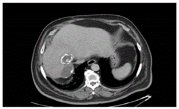
Figure 1
CT scan. Two partially calcified hydatid cysts in VII and VII
segments.
Figure 2
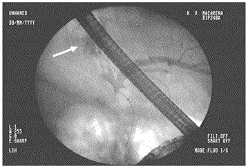
Figure 2
Preoperative ERCP. Cyst-biliar communication. Hydatid cyst is
filled with contrast (White arrow).
Figure 3
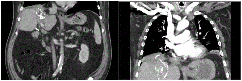
Figure 3
CT scan. Fistula hepato biliobronchial. Coronal view.
Figure 4
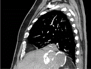
Figure 4
CT scan. Cyst-biliobronchial fistula. Sagital view.
Discussion
Biliobronchial fistula is an infrequent pathology that arises as a
complication, in our case, of a hydatid cyst that has been asymptomatic
for years but is complicated by an episode of cholangitis.
The early diagnosis of this pathology is a fundamental pillar.
The clinical diagnosis has vital importance, being the bilioptysis
the pathognomonic symptom of this pathology as in the case of our
patient. Sputum analysis helps to confirm the diagnosis to proceed to
more effective targeted treatment.
Among the most frequent diagnostic imaging are CT scan and
ERCP, both performed in our patient. The ERCP has high relevance
in this patient due to the therapeutic attitude with the previous
cholangitis and post-operatory bile leak.
There isn´t a gold standard in BBF treatment due to the few
studies carried out and low evidence that is currently available. The
treatment should be tailored according to each patient.
Among the non-surgical therapeutic options are the placement
of metallic or plastic biliary stent and fistulous tract embolization.
However, there is low experience with these methods and some
experts recommend only the placement of endoprostheses as
exclusive treatment in patients with low life expectancy.
Surgical invasive treatment should always be the last option but
must be taken into consideration whenever necessary. In our case,
right hepatectomy with diaphragmatic gap closure were performed
to solve the thoracic transit. Therapeutic ERCP with biliary stent
placement is useful in post-operatory bile leak after hepatectomy.
Figure 5
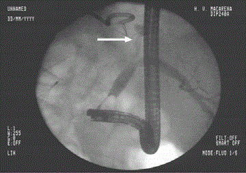
Figure 5
Postoperatory ERCP. Common and left hepatic ducts with contrast.
Bile leak identification coming from hepatic resection (White arrow).
Figure 6
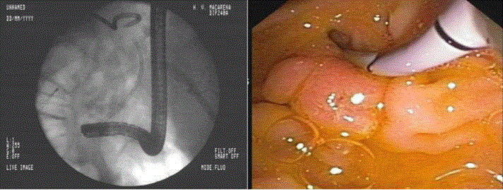
Figure 6
Postoperatory ERCP. Plastic stent is placed in common bile duct
to solve the bile leak.
References
- Nassar Y, Hida S, Richter S. A biliobronchial fistula in a patient with hepatocellcular carcinoma treated with chemoembolization diagnosed by hepatobiliary iminodiacetic acid scan and managed by endoscopic retrograde cholangiopancreatography. Gastroenterology Res. 2017;10(6):383-5.
- Chautems R, Buhler LH, Gold B, Giostra E, Poletti P, Chilcott M, et al. Surgical management and long-term outcome of complicated liver hydatid cyst caused by Echinococcus granulosus. Surgery. 2005;137(3):312-6.
- Uchikov AP, Safev GP, Stefanov CS, Markova DM. Surgical treatment of bronchobiliary fistulas due to complicated echinococcosis of the liver: Case report and literature review. Folia Med (Plovdiv). 2005;45(4):22-4.
- Sahin E, Enon S, Cangir AK, Kutlay H, Kavukcu S, Akay H, et al. Single-stage transthoracic approach for right lung and liver hydatid disease. J Thorac Cardiovasc Surg. 2003;126(3):769-73.
- Losada H, Vial M, Manterola C, Pienada V. Bronchobiliary fistula secondary to a hepatic hydatid cyst in transit to the thorax. Report of one case. Rev. Chilena de Cirugía. 2006;58:224-7.
- Liao GQ, Wang H, Zhu GY, Zhu KB, Lv FX, Tai S. Management of acquired bronchobiliary fistula: A systematic literature review of 68 cases published in 30 years. World J Gastroenterol. 2011;17(33):3842-9.