Case Report
Primary Ectopic Atypical Meningioma of the Middle Ear: A Case Report
Kaviraj Anand Rughoobur, Qiuyu Su, Xiaodan Zhu and Fanglei Ye*
Department of Otorhinolaryngology, Hospital of Zhengzhou University, Henan, China
*Corresponding author: Fanglei Ye, Department of Otorhinolaryngology, Hospital of Zhengzhou University, Henan, China
Published: 07 Sep, 2018
Cite this article as: Rughoobur KA, Su Q, Zhu X, Ye F.
Primary Ectopic Atypical Meningioma
of the Middle Ear: A Case Report. Clin
Surg. 2018; 3: 2096.
Abstract
Meningiomas are the second most common primary tumors of the Central Nervous System. Most cases occur in intracranial sites; but in very rare cases some do occur in extracranial regions. Ectopic meningiomas have no communication with intracranial meninges and misdiagnosis of this pathology may be encountered due to its anatomical complexity. The report below is one of the few uncommon documented cases of Primary Ectopic Atypical Meningioma of the middle ear which should increase the awareness of otolaryngologists over difficulties arising to diagnose this rare phenomenon.
Introduction
Meningiomas are tumors/neoplasms that arise from central nervous meninges- the arachnoidal
(meningothelial) cap cells [1]. This common type of tumor accounts for around 18% to 20% of
all intracranial lesions whereas less than 2% account for extracranial meningiomas [2,3]. These
extracranial meningiomas, based on their origins, can be classified into:
a) Primary/ ectopic – occurring from the ectopic site or
b) Secondary- usually extensions from intracranial masses [4,5].
Reported cases of ectopic meningiomas arise from skull bone, paranasal sinus, parotid gland,
middle ear, neck and skin.
Meningiomas are more common in females than males with overall incidence of (2:1) due
to estrogen or progesterone levels in women [5]. Most cases of meningiomas are usually benign
in nature. However, malignant transformation also occurs. According to the World Health
Organization Classification, meningiomas are categorized as follows:
• Grade 1- Benign. (80% )
• Grade 2- Atypical. (15% to 20%)
• Grade 3- Anaplastic. (1% to 3%)
Benign meningiomas grow slowly and have 5% recurrence in 5 years whereas atypical ones grow
quicker with 40% recurrence in 5 years. Anaplastic meningiomas are the worst type and have an
80% recurrence rate [3]. Benign lesions that are small (<2.5 cm), slow growing and asymptomatic
require close observation. Surgery may not be needed in such cases but in atypical cases, surgical
excision is the mainstay of management. Adjuvant therapy- radiation can also be considered in
some cases. Anaplastic meningiomas require both surgery and radiation therapy.
Case Presentation
A 46 year old female patient presented to the otology outpatient department of The First
Affiliated Hospital of Zhengzhou University with chief complaint of recurrent bilateral ear discharge
and hearing loss for past 30 years. A brief history revealed that she had tinnitus occasionally but
denied having otorrhea or any other neurological deficits. She attended local clinics for the above
symptoms whereby “ear drops” were given to her. While these improved her symptoms, she
unfortunately had repeated episodes over time which led to gradual increase in hearing loss and
dizziness. In our hospital, video otoscopy, Pure Tone Audiometry, Tympanometry and Temporal
Bone CT scan were ordered. Subsequently an initial diagnosis of Chronic Suppurative Otitis Media
in bilateral ears was made.
Video otoscopy
As illustrated in (Figure 1A, 1B), yellowish purulent effusion was
observed in right ear. Following cleaning of the ear canal, the tympanic
membrane could be clearly visualized. Tympanic membrane was
abnormal and handle of malleus could not be seen.
Pure tone audiometry
Audiometry in right ear revealed moderate to severe mixed
hearing loss whereas left ear was consistent with mild conductive
hearing loss (Figure 3A, 3B).
Tympanometry
Tybe B tympanogram was noted in both ears - suggesting
minimum or no movement of the tympanic membranes; possibly due
to presence of fluid or a mass in middle ear (Figure 4A, 4B).
Temporal bone CT scan
Assessing the axial view of Temporal bone CT scan, no deformity
was found in both auricles and the external auditory canals were
unobstructed. Mastoid air cells and the middle ear cavity in the right
ear had increased density. The ossicles were normal. In left ear, the
mastoid air cells were well pneumatized and left middle ear cavity
as well as ossicles was normal. Cochlea and Semicircular canals of
bilateral ears were normal. No dilatation of Internal Auditory Canal
on both sides (Figures 5A-5C). Consequently, Right Canal Wall down
Mastoidectomy + Ossicular Chain Reconstruction + Tympanoplasty
was done on the patient. During surgery, the following findings were
noted:
1. While drilling the mastoid bone, dark brown-like secretions
accumulated in the mastoid air cells were seen and granulations in
the middle ear cavity.
2. The granules had invaginated the pars-flaccida + the pars
tensa was perforated
3. Incus was destroyed as well as part of the malleus.
4. Stapes was intact.
5. Eustachian tube was unobstructed.
Figure 1
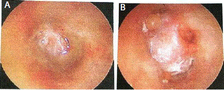
Figure 1
A) Yellowish purulent effusion was observed in right ear B) Following cleaning of the ear canal, the tympanic membrane could be clearly visualized.
Figure 2
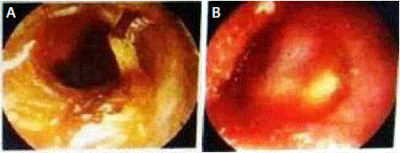
Figure 2
A) Illustrates Left ear filled with cerumen and purulent discharge B) After cleaning, an abnormal tympanic membrane and a swollen ear canal was observed.
Figure 3
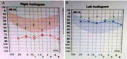
Figure 3
A) Audiometry in Right Ear revealed Moderate to Severe Mixed Hearing Loss B) Left Ear was consistent with Mild Conductive Hearing Loss.
Discussion
Up to now, very few cases of meningiomas extending to the
temporal bone and middle ear cavity have been reported. These
were mostly secondary extracranial conventional type meningiomas
extending from an intracranial lesion to the temporal bone or middle
ear [6]. Differential diagnosis of meningiomas in middle ear is:
schwannomas, paragangliomas, middle ear adenomas, metastatic
carcinomas, cholesteatomas, glomus tumors, retrotympanic
vascular tumors, and lipoma. Other than hearing loss (conductive,
sensorineural or mixed hearing loss), which is the most common
symptom [7], the following symptoms are also associated with
middle ear meningiomas: headaches, dizziness, vertigo, tinnitus,
otalgia, bleeding, “metallic taste,” and sometimes chronic cough. The
severity of symptoms usually depends on location of tumors. More
severe cases can present with epilepsy, speech disturbances, cognitive
impairment and visual disturbances. It has been reported that about
16% of Middle ear Meningiomas are usually accompanied by Chronic
Otitis Media. One theory behind this is that there is the possibility of
the tumor blocking the Eustachian Tube which prevents the drainage
of Middle Ear fluid [8]. Causes of meningiomas include exposure to
ionizing radiation, neurofibromatosis type 2- a genetic disorder and
some risk factors associated are trauma to head, use of cell phones
and also increased level of estrogen and/or progesterone, which can
explain the high incidence of the disease in women compared to
men [9]. Radiological Imaging like CT and MRI are very helpful in
differentiating middle ear meningiomas to other middle ear diseases.
They give an idea of the extent of tumor, whether there was absence
of ossicles or ossicular erosion and details of internal auditory canal
and associated structures [6]. As observed in this case, the patient was
initially diagnosed as bilateral Chronic Suppurative Otitis Media but
it is only after surgery, when histopathological report was obtained
that the definite diagnosis of Meningioma was confirmed. Since
conventional meningiomas have distinctive histological findings, it
is easy to diagnose them. But for rare types, as in the present case,
ectopic sites meningiomas can pose as a diagnostic challenge for
pathologists. Some may argue that biopsy of tympanic/middle ear
lesion during myringotomy may lead to earlier diagnosis but the risk
of damaging any vascular lesion or the facial nerve is high.
Treatment of choice for atypical meningiomas remains surgical
excision. Where complete removal of the meningioma is not possible,
radiotherapy is often warranted. It is also recommended that long
term follow up of patients be done together with a neurologist to
exclude recurrence. Patients and their family members also need to
be well educated about the prognosis of this disease and informed to
not ignore any associated symptoms.
Figure 4
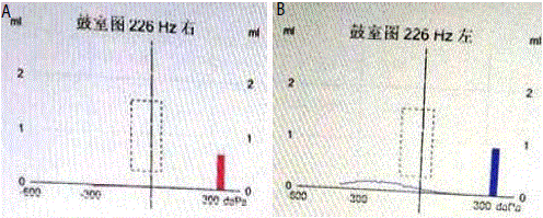
Figure 4
(A and B) Tybe B tympanogram was noted in both ears - suggesting minimum or no movement of the tympanic membranes; possibly due to presence of fluid or a mass in middle ear.
Figure 5
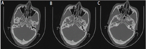
Figure 5
Temporal Bone CT SCAN.
Figure 6

Figure 6
Specimen was sent for histopathology examination which later confirmed the diagnosis of Primary Atypical Meningioma of Middle Ear.
Conclusion
Despite its rarity in otolaryngology, Primary Atypical Meningioma in middle ear is very likely to be misdiagnosed. Conventional tests such as Pure Tone Audiometry, Tympanometry and CT scans are helpful but not enough for diagnosis of this disease. So far, histopathological examination has been the only method to confirm the nature of the middle ear lesion. Once confirmed, it is imperative to follow up the patient closely and regularly with serial imaging for the rest of her life as the rate of recurrence is high. Since primary meningiomas are very rare, care must be taken to differentiate between intracranial or extracranial lesions as well as benign and malignant tumors.
References
- Sanei MH, Rabiei S, Eftekhari M, Jafari HR. Ectopic Meningioma (Hamartoma) of the Middle Ear: A Challenging Case in Frozen Section. Otol Neurotol. 2014;35(8): e231-e232.
- George M, Ikonomidis C, Pusztaszeri M, Monnier P. Primary meningioma of the middle ear: case report. J Laryngology & Otology. 2010;124(5):572–74.
- Karaaltın AB, Turgut FN, Yılmaz M, Batur S, Öztürk O. Isolated middle ear meningioma. Kulak Burun Bogaz Ihtis Derg. 2016;26(4):238-240.
- Chien WW, Saleh J, Kumar A, Primary Extracranial Meningioma of Middle Ear. Clinics in Surgery-General Surgery. 2016;1:1142.
- Sreenivas V, Hemanth V, Chaturvedi J. Meningioma Presenting as a Mass in the External Auditory Canal. Internet J Otorhinolaryngol. 2010;13(1):1-5.
- Manjaly JG, MTWatson G, Jones M. Primary meningioma of the middle ear: case report. JRSM Short Rep. 2011;2(11):92.
- Khadgi DR, Jinbai PDH. Primary Extracranial Meningioma in Middle Ear: A Rare Case Report. Int J Science Inventions Today. 2016;5(4);330-36.
- Singh DS, Bhatia DY, Dua DS, Sharma DJ, Sen DR. Extracranial Meningioma Presenting as an External Auditory Canal Polyp. Scholars J Med Case Rep. 2016;4(9):644-646.
- Ricciardiello F, Fattore L, Ester Liguori M, Oliva F, Luce A, Abate A, et al. Temporal bone meningioma involving the middle ear: A case report. Oncology Letters. 2015;10(4):2249-2252.