Research Article
Modified Watson Jones Technique for Lateral Ankle Stabilization Utilizing the Split Peroneus Longus
Elizabeth A Sanders*, Jurgen H Fernandez and Mark J Mendeszoon
Department of Foot and Ankle-Orthopedic Surgery, Precision Orthopaedic Specialties, USA
*Corresponding author: Elizabeth A Sanders, Department of Foot and Ankle-Orthopedic Surgery, Precision Orthopaedic Specialties, USA
Published: 06 Sep, 2018
Cite this article as: Sanders EA, Fernandez JH,
Mendeszoon MJ. Modified Watson
Jones Technique for Lateral Ankle
Stabilization Utilizing the Split Peroneus
Longus. Clin Surg. 2018; 3: 2093.
Abstract
Fourteen percent of patients require re-operation following a Brostrom anatomical augmentation.
Another study demonstrated 6% failure of a Brostrom augmentation secondary to athletic traumatic
injuries. Failure of primary anatomical augmentation has been noted to occur in patients with poor
quality tissue, generalized ligamentous laxity, or patients with longstanding instability greater than
ten years.
After failed anatomic lateral ankle stabilization procedures, reconstructive tenodesis procedures
have been described. The traditional Watson-Jones is described utilizing the entire peroneus
brevis tendon, leaving its distal insertion intact. Several modifications of the procedure have been
described. Harvesting peroneus longus tendon permits for greater length of the allograft and less
fraying of the fibers as compared to the peroneus brevis tendon, allowing for easier tubularization
and bone tunnel passage. We describe senior author’s (MJM) modified Watson-Jones lateral ankle
stabilization technique utilizing the split peroneus longus.
In the senior author’s experience, a modification of the Watson-Jones reconstruction procedure
utilizing the split peroneus longus is effective and reliable as a limb salvage procedure after a failed
anatomical lateral ankle reconstruction or following a failed anatomic lateral ankle stabilization
procedure.
Keywords: Lateral Ankle Instability; Lateral Ankle Stabilization; Watson-Jones; Brostrom; Revisional Lateral Ankle Reconstruction
Introduction
Approximately 20% to 25% of acute ankle sprains result in chronic ankle instability and results
from failure of functional rehabilitation [1]. When functional rehab after conservative treatments
fails, surgical repair should be considered.
When addressing an unstable lateral ankle, concomitant biomechanical deformities such as a
calcaneal varus, subtalar joint instability, limb length discrepancy, equinovarus deformity, adductus
foot, and a pronated forefoot must be evaluated and addressed for surgical success [2]. Associated
injuries such as osteochondral lesions, sinus tarsi syndrome, peroneal tendinopathy, or subtalar
joint instability should also be evaluated and addressed.
Historically, lateral ankle stabilization surgical procedures are performed to correct a single
ligamentous repair of the anterior talofibular ligament or double ligament repair of the anterior
talofibular and calcaneofibular ligaments. Other non-anatomic stabilization tenodesis procedures
have been described utilizing the peroneal tendons such as the split peroneus brevis procedure or
those depicted by Elmsli, Christman and Snook, Hambly, Whinfield, Lee, Nilsonne and Watson-
Jones [3]. In addition to peroneus brevis and longus tendon transfers, the Achilles and plantaris
tendons may be utilized the stabilize the ankle. Incorporating tendon allografts have also been
described [4].
A recent study demonstrated that 14% of patients require re-operation following a Brostrom
anatomical augmentation [5]. Another study demonstrated 6% failure of a Brostrom augmentation
secondary to athletic traumatic injuries [6]. Failure of primary anatomical augmentation has been
noted to occur in patients with poor quality tissue, generalized ligamentous laxity, or patients with
longstanding instability greater than ten years [7,8].
The traditional Watson-Jones is described utilizing the entire peroneus brevis tendon, leaving
its distal insertion intact. The tendon is passed through a tunnel in the fibula into the neck of the
talus, back through the fibula, and then attached to itself posteriorly
at the level of the fibula. Several modifications of the procedure have
been described due to the technical difficulty in obtaining adequate
tendon length for the transfer.
The peroneus brevis is inherently short, and the fibers tend
to fan out and separate easily, making tubularization of the fibers
and passage of the tendon through bone holes difficult. Harvesting
peroneus longus tendon permits for greater length of the allograft and
less fraying of the fibers allowing for easier tubularization and bone
tunnel passage.
In the experience of the senior author (MJM), following a failed
anatomic lateral ankle stabilization procedure, a modification of the
Watson-Jones reconstruction procedure utilizing the split peroneus
longus is effective and reliable.
Figure 1
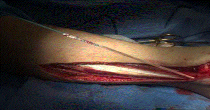
Figure 1
Harvesting the peroneus longus auto graft using the entire length
of the peroneus longus tendon and 25% width of the tendon. A modified whip
stitch is performed with fiber wire.
Figure 2
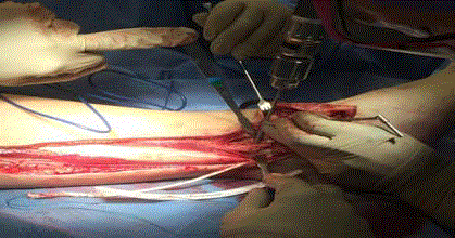
Figure 2
An anterior to posterior bone tunnel is made approximately 2.5-3.0
centimeters from the distal tip of the fibula. Another tunnel is made anterior
to posterior obliquely approximately 1.0-1.5 centimeters from the tip of the
fibula.
Figure 3
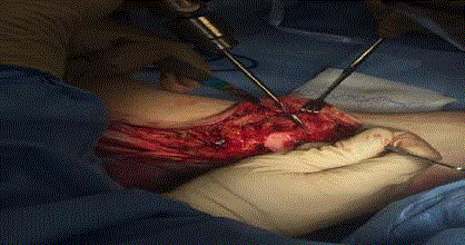
Figure 3
A slightly oblique dorsal to plantar bone tunnel is made in to the
neck of the talus midline and in to the inferior neck of the talus.
Figure 4
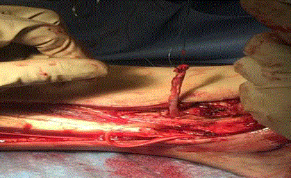
Figure 4
With a suture passer, the peroneus longus tendon is passed
from posterior to anterior in the proximal fibula drill hole. The tendon is then
passed underneath the extensor retinaculum of the lateral ankle toward the
talar neck drill hole.
Modified Watson-Jones Lateral Ankle Stabilization Using Split Perneus Longus Surgical Technique
A popliteal and adductor canal block is placed by anesthesia
prior in the pre-operative area. The patient is placed under general
anesthesia. A thigh tourniquet is applied. After sterile prep and
draping, an incision approximately 15-18 inches is marked from the
proximal posterior lateral leg along the peroneal tendons to behind
the fibula and curving anteriorly distally to the dorsal 4th metatarsal
base. To locate the sinus tarsi, an #18 gauge needle may be utilized.
Anatomic dissection is performed to visualize the peroneus longus.
Pertinent neurovascular structures are identified and retracted.
Ruptured anterior talofibular ligaments and calcaneofibular
ligaments or any residual hardware or anchors may be debrided at
this time while still preserving the extensor retinaculum. Dissection is
performed to visualize the neck of the talus. Attention is then drawn
to the peroneus longus. To harvest the auto graft tendon, a full length
of the peroneus longus is excised with approximately 25% width of
the tendon. A modified whip stitch is performed utilizing a fiber wire.
Bone tunnels are then created with sequential drilling using 2.5,
3.5, and 4.5 millimeter drill bits. An anterior to posterior tunnel is
made approximately 2.5 to 3 centimeters from the distal tip of the
fibula. A slightly oblique dorsal to plantar tunnel is made in the
talus neck midline and in to the inferior neck of the talus. The final
tunnel is made from anterior to posterior obliquely approximately 1.5
centimeters from the tip of the fibula.
Utilizing a suture passer, the peroneus longus auto graft tendon is
passed from posterior to anterior in the proximal fibula drill hole. The
tendon is then passed underneath the extensor retinaculum of the
lateral ankle, then passed from superiorly to inferiorly through the
talar neck. While placing the foot in dorsiflexion and slight eversion,
the tendon is then passed through the distal fibula hole anteriorly to
posteriorly, then sutured back onto itself utilizing non-absorbable
Ethibond #2 suture. Confirmation of stability of the ankle with the
procedure can be confirmed with a negative anterior drawer test. If
desired, a Brostrom augmentation utilizing a suture anchor may be
performed followed by retinaculum repair. Layered closure is then
performed.
Post-operatively, the patient is placed in a posterior splint in a
neutral position. One week post operatively, the splint is removed for
a wound check and the patient is placed into a non-weight bearing
below knee cast for an additional three weeks. After four weeks,
sutures are removed and the patient is partial weight bearing in a cast
or in a removable walking boot for an additional three weeks. Gradual
introduction to weight bearing into a shoe with an ankle brace is then
initiated. At ten weeks post-operatively, physical therapy is initiated.
Figure 5
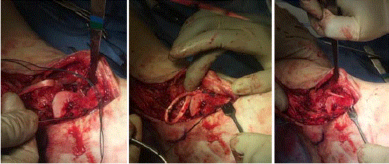
Figure 5
The tendon is passed from superiorly to inferiorly through the talar neck utilizing a suture passer.
Figure 6
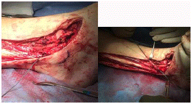
Figure 6
While placing the foot in dorsiflexion and slight eversion, the
tendon is then passed through the distal fibula hole anteriorly to posteriorly,
then sutured back onto itself utilizing a non-absorbable Ethibond #2 suture.
Discussion
In chronic ankle stability, direct primary ligament repair should
be the first choice of procedures. After failed anatomical lateral
ankle stabilization, tenodesis reconstructive procedures should be
considered.
Favorable results have been documented when utilizing the
modified Watson-Jones Lateral Instability Repair and range from
72% to 93% success with good to excellent results [7-12]. Studies
demonstrate good talar tilt and anterior-drawer stability radio
graphically post-operatively demonstrating rotational and lateral
ankle stability [11]. No evidence of short or long-term deterioration of
clinical results or narrowing of joint spaces have been demonstrated
occur over time and with no disruption in normal gait [12]. No loss
of peroneal muscle strength is detected [9].
Risks of the modified Watson-Jones procedure include wound
healing complications or dehiscence, nerve entrapment, and scarring.
The incision is long and may leave an unpleasant scar. Other risks
include loss of ankle eversion strength and post-operative ankle
or subtalar joint stiffness. Of note, the higher the incidence of
osteophytes in the ankle pre-operatively is demonstrated to lead to
the progression of arthrosis with time, but this finding is not related to
the reconstruction method [13]. The presence of arthritic changes in
the ankle preoperatively may be a contraindication to the procedure.
This procedure is regarded as a limb salvage procedure.
In the hands of the senior author (MJM), the modified Watson-
Jones lateral ankle reconstruction utilizing the split peroneus longus
is reliable and effective as a limb salvage procedure after a failed
anatomical lateral ankle reconstruction.
Figure 7
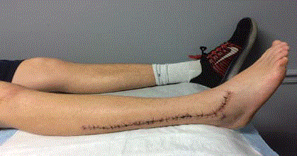
Figure 7
Incision four weeks status-post Watson-Jones lateral ankle stabilization procedure with modification of utilizing the split peroneus longus tendon.
References
- Al-Mohrej O, Al-Kenani N. Chronic ankle instability: Current perspectives. Avicenna J Med. 2016;6(4):103-8.
- Bonnel F, Toullec E, Mabit C, Tourne Y, Sofcot. Chronic ankle instability: Biomechanics and pathomechanics of ligaments injury and associated lesions. Orthop Traumatol Surg Res. 2010;96(4):424-32.
- Bouchard J. The Modified Evans Ankle Stabilization For Correction of Lateral Ankle Instability. The Podiatry Institute. Chapter 12. 1994. p50-58.
- Want W, Xu G. Allograft tendon reconstruction of the anterior talofibular ligament and calcaneofibular ligament in the treatment of chronic ankle instability. BMC Musculoskelet Disord. 2017;18:150.
- Baraza N, Hardy E, Shahban SA. Re-Operation Rates Following Brostrom Repair. JSM Foot and Ankle. 2017;2(1):1019.
- Petrera M, Dwyer T, Theodoropoulos J, Ogilvie-Harris D. Short-to medium-term outcomes after repair for lateral ankle instability with immediate postoperative weightbearing. Am J Sports Med. 2014;42(7):1542-8.
- de Vries JS, Krips R, Sierevelt IN, Blankevoort L, van Dijk CN. Interventions for treating chronic ankle instability. Cochrane Database Syst Rev. 2011;(8): CD004124.
- Karlsson J, Rudholm O, Bergsten T, Faxen E, Styf J. Early range of motion training after ligament reconstruction of the ankle joint. Knee Surg Sports Traumatol Arthrosc. 1995;3(3):173-7.
- Gillespie H, Boucher P. Watson-Jones Repair of Lateral Instability of the Ankle. J Bone Joint Surg Am. 1971;53(5):920-4.
- Hamido F., Ibrahim S, Abo-El-Noor T, Al-Misfer A, Mutiri H, Salem H. Evaluation of the results of Watson Jones tendoesis in chronic lateral instability of the ankle. Foot and Ankle Surgery. 2007;13(2):56-62.
- Lucht U, Vang PS, Termansen NB. Lateral ligament reconstruction of the ankle with a modified Waatson-Jones operation. Acta Orthop Scand. 1981;52(3):363-6.
- Hoy GA, Henderson IJ. Results of Watson-Jones ankle reconstruction for instability. The influence of articular damage. J Bone Joint Surg Br. 1994;76(4):610-3.
- Becker HP, Ebner S, Ebner D, Benesch S, Frossler H, Hayes A, et al. 12-year outcome after modified Watson-Jones tenodesis for ankle instability. Clin Orthop Relat Res. 1999;(358):194-204.