Clinical Image
Tortuous Common Carotid Artery Masquerading as Thyroid Mass
Po-Jun Chen and Hsin-Chien Chen*
Department of Otolaryngology-Head and Neck Surgery, Tri-Service General Hospital, Taiwan
*Corresponding author: Hsin-Chien Chen, Department of Otolaryngology-Head and Neck Surgery, Tri-Service General Hospital, National Defense Medical Center, 325, Section 2, Cheng-Kung Rd, Taipei, 114, Taiwan
Published: 16 Aug, 2018
Cite this article as: Chen P-J, Chen H-C. Tortuous
Common Carotid Artery Masquerading
as Thyroid Mass. Clin Surg. 2018; 3:
2070.
Keywords
Carotid artery; Thyroid; Neck; Tortuosity
Clinical Image
A 58-year-old woman was referred to our hospital for an evaluation of thyroid tumor from local
clinic. The patient had no past medical history. Physical examination revealed a soft and pulsatile
mass over right thyroid region. No gross tumor was noted over nasopharynx, larynx, oropharynx
and hypopharynx. Sono-guided fine needle aspiration biopsy was arranged for a pathological
examination. However, ultrasonography with Doppler Color Flow Image (DCFI) showed a tortuous
Common Carotid Artery (CCA) compressed right thyroid gland. No focal lesion was noted in the
thyroid gland. The procedure of FNA biopsy was cancelled to avoid possible bleeding. Contrastenhanced
Computed Tomography (CT) of the neck further revealed the tortuosity of right CCA and
compression to right thyroid gland (Figure 1). The image of 3D-CT reconstruction showed a reverse
S-shaped tortuosity of right CCA (Figure 2). A clinical diagnosis of
tortuosity of right CCA was made.
The incidence of pulsatile neck mass is relatively rare in the
entity of neck mass [1]. Pulsatile neck masses are categorized into
vascular lesion and non-vascular lesions [1,2]. The differential
diagnosis of vascular lesions includes aneurysm, carotid body tumor,
and tortuosity of carotid artery, arteriovenous fistula, prominent
carotid bulb, strong venous pulsations or dilated jugular bulb. On
the contrary, non-vascular lesions include enlarged lymph nodes,
branchial cleft cyst, thyroid tumor, cystic hygroma, lipoma and
neurogenic tumor. The diagnosis of pulsatile neck mass should
require thorough physical examination, ultrasonography with DCFI,
CT or CT angiography or magnetic resonance imaging to rule out
potential dangerous diseases such as aneurysm [1,3].
The tortuosity of CCA almost occurs in female with mean age
between 50 and 60 year-old and usually presents on the right side
of CCA [2]. Most patients are asymptomatic, but some case reports
present throbbing, dysphagia, dyspnea or painful sensation associated
with the mass and even the symptoms of cerebrovascular insufficiency
due to decrease of blood flow [2,4]. The etiology of tortuosity of CCA
remains unclear [5]. Hypertension and atherosclerotic vascular
disease may contribute to elevation of the aortic arch and subsequent
formation of a buckle in the CCA [2,5]. Visualization of the aortic
knob at the level of the clavicle or higher on a chest X-ray should raise
suspicion for this diagnosis [2].
The management of tortuosity of CCA requires no specific
therapy [2]. Surgical treatment including graft insertion, repositioning
techniques and end-to-end anastomosis after resection of CCA is
performed among the patients with symptoms of cerebrovascular
insufficiency [2]. Because the patient had no neurological symptoms
or impending complications, conservative management was
recommended. This case illustrates the importance of including
carotid artery variation should keep in mind in the differential
diagnosis of pulsatile neck mass before performing a blind fine needle
aspiration biopsy [1,3].
Figure 1
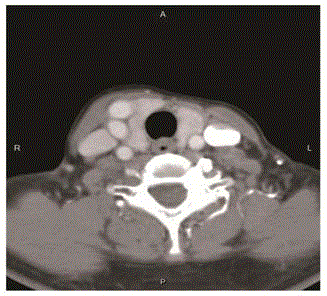
Figure 1
Axial view contrast-enhanced CT of the neck further revealed multiple lumens of right CCA and
overriding to right thyroid gland.
Figure 2
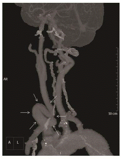
Figure 2
The reconstruction image of 3D-CT showed a reverse S-shaped tortuosity of right CCA.
References
- Takeuchi Y, Numata T, Suzuki H, Konno A, Kaneko T. Differential diagnosis of pulsatile neck masses by Doppler color flow imaging. Ann Otol Rhinol Laryngol. 1995;104(8):633-8.
- Leipzig TJ, Dohrmann GJ. The tortuous or kinked carotid artery: pathogenesis and clinical considerations. A historical review. Surg Neurol. 1986;25:478-86.
- Godin MS, Rice JC, Kerstein MD. Tortuosity of the right common carotid artery simulating aneurysm. South Med J. 1988;81:1382-5.
- Yildiz S, Cece H, Karayol S, Ziylan Z. Concurrence of the tortuosity of bilateral common and left internal carotid arteries in a case with common origin of the innominate trunk and left common carotid artery. Surg Radiol Anat. 2010;32:797-9.
- Del Corso L, Moruzzo D, Conte B, Agelli M, Romanelli AM, Pastine F, et al. Tortuosity, kinking, and coiling of the carotid artery: expression of atherosclerosis or aging? Angiology. 1998;49(5):361-71.