Case Report
A Rare Case of Choledochal Cyst Connecting Intra- and Extra-Hepatic Duct
Shu-Jung Yang1, Yi Chen2, You-Hsin Chiu3, Shen-Yang Huang1,4 and Chia-Man Chou1,4*
1Department of Surgery, Taichung Veterans General Hospital, Republic of China, Taiwan
2School of Post-baccalaureate Chinese Medicine, Republic of China, Taiwan
3Department of Radiology, Taichung Veterans General Hospital, Republic of China, Taiwan
4Department of Medicine, National Yang-Ming University, Republic of China, Taiwan
*Corresponding author: Chia-Man Chou, Division of Pediatric Surgery, Department of Surgery, Taichung Veterans General Hospital, National Yang-Ming University, No. 1650, Sec. 4, Taiwan Boulevard, Taichung 40705, Taiwan, Republic of China
Published: 14 Jul, 2018
Cite this article as: Yang S-J, Chen Y, Chiu Y-H, Huang
S-Y, Chou C-M. A Rare Case of
Choledochal Cyst Connecting Intra- and
Extra-Hepatic Duct. Clin Surg. 2018; 3:
2024.
Abstract
Choledochal cysts are rare congenital dilatations of the extra and/or intrahepatic bile ducts. This
disease often presents non-specific symptoms that perplex early diagnosis. We report a rare
anatomical location of a choledochal cyst in a 5-year-old female patient, and it is a variant of the
classical types of the hepatic duct cysts. Awareness of this type of variation in choledochal cysts
would aid preoperative diagnosis, more accurate interpretation of imaging results and surgical
planning.
Keywords: Choledochal cyst; Hepatic duct; Larparoscopic cyst
Introduction
Choledochal cysts are rare congenital dilatations of the extra and/or intrahepatic bile ducts found primarily in children and estimated of much higher incidence in Asia, where it reaches approximated 1:1000, as compared to Western population [1,2]. A choledochal cyst increases the risk of malignant transformation up to 10% and patients may still be exposed at higher risk for biliary malignancies even after surgical resection [1,3]. The most commonly used classification system updated by Todani et al. [4] described 5 broad types of choledochal cysts. The most commonly reported is type I, which includes cystic, focal or fusiform dilatations of the common bile duct. The clinical presentations are often non-specific, such as nausea, vomiting, abdominal pain and jaundice. Nevertheless, image techniques are essential to aid the diagnosis [5]. Larparoscopic cyst excision and hepaticojejunostomy for children have become a popular treatment protocol [6].
Case Presentation
A 5-year-old girl presented several episodes of right upper quadrant abdominal pain after an
upper airway infection. She denied any fever, nausea/vomiting, abdominal cramping pain, teacolor
urine or diarrhea during these episodes. Initially, she received survey at another hospital,
where choledochal cyst was diagnosed. Medical staff there suggested further Magnetic Resonance
Cholangiopancreatography (MRCP) and surgical survey. Her parents hesitated and came to our
hospital for the second opinion. At presentation to our pediatric ward, her red blood cell count was
5.31 × 10⁶/μL (normal 4.28 to 5.05 × 10⁶/μL), hemoglobin 10.6 g/dL (normal 11.6 g/dL to 13.7g/dL),
Mean Cell Volume (MCV) 64.2 fL (normal 74.9 fL to 84.6 fL), and activated partial thromboplastin
time (APTT) 35.1 sec (normal 24.3 to 32.7 sec). Her white blood cell count, total bilirubin, direct
bilirubin, Alanine Transaminase (ALT), Alanine Aspartate Phosphatase (AST), Gamma-Glutamyl
Transpeptidase (γ-GT), amylase and creatinine were all within normal limits.
Pre-operative abdominal ultrasound and MRCP (Figure 1) showed a cystic lesion near the
common bile duct, 1.45 cm × 1.29 cm in size, and Todani type 1 or 2 choledochal cyst was considered.
During surgical exploration, a cyst lesion was found at the junction of intra- and extra-hepatic duct
(Figure 2), which was confined within the common hepatic segment. This finding was confirmed by
intraoperative cholangiography (Figure 3). The dilatation part of the cyst included part of left and
right hepatic duct, as well as the common hepatic duct (Figure 4A). After cholecystectomy, the cyst
was excised at about 1 cm below bifurcation point (Figure 4B). Reconstruction was carried out with
a Roux-en-Y end-to-side hepaticojejunostomy. Seven days after surgery, the girl was discharged
and followed up with normal daily activities 3 months later. Histological analysis confirmed that
the resected specimen contain thickened fibrotic wall with chronic
inflammation and intact overlying biliary type epithelium. The cyst
contained no stones, and the cyst wall was continuous with the
common hepatic duct.
Figure 1
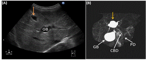
Figure 1
Images of abdominal ultrasound (A) and MRCP (B). The solid
arrows pointed out the cyst.
GB: Gallbladder; CBD: Common Bile Duct; PD: Pancreatic Duct.
Figure 2
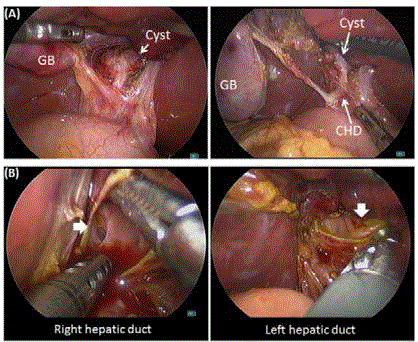
Figure 2
(A) Laparoscopic view found a fusiform dilatation over the
bifurcation of the Common Hepatic Duct (CHD). (B) The cyst was opened to
identify the orifices of right and left hepatic ducts.
Discussion
More severe complications of choledochal cyst include pancreatitis, cholecystitis, liver cirrhosis and cholangiocarcinoma [7]. Surgical treatment and reconstruction is varied according to different types of choledochal cyst. In 2008, Calvo-Ponce et al. [8] introduced an extra variant type in addition to Todani’s modified classification of biliary duct cysts according to their findings in a fifty year-old female patient, which is similar to this pediatric case. This is an extremely rare anatomical location for a choledochal cyst, which could be the cause of the unique clinical manifestation of our patient. The bile flow may be disturbed by the altered route, which leads to intermittent right upper quadrant abdominal pain without any fever or nausea. The gross examination and imaging of the cyst is quite misdirecting as that of type II choledochal cyst. However, under laparoscopic view, there is no saccular diverticulum from the common bile duct, but a fusiform dilation sitting at the convergence of intra- and extrahepatic ducts. Awareness of this variant type in choledochal cysts would aid pre-operative diagnosis, more accurate interpretation of imaging results and surgical planning.
Figure 3
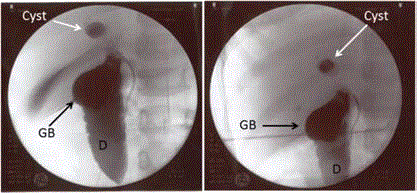
Figure 3
Intra-operative cholangiography showed the cyst position. D:
Duodenum.
Figure 4
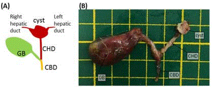
Figure 4
Schematic drawing of the relative location of the choledochal cyst
(A) and the resected choledochal cyst, gallbladder, and biliary ducts (B). GB:
Gallbladder; CHD: Common Hepatic Duct; CBD: Common Bile Duct.
Acknowledgement
This work was supported by Taichung Veterans General Hospital, Taichung, Taiwan (Project No. TCVGH-1075401C).
References
- Soares KC, Kim Y, Spolverato G, Maithel S, Bauer TW, Marques H, et al. Presentation and Clinical Outcomes of Choledochal Cysts in Children and Adults: A Multi-institutional Analysis. JAMA Surg. 2015;150(6):577-84.
- Visser BC, Suh I, Way LW, Kang SM. Congenital choledochal cysts in adults. Arch Surg. 2004;139(8):855-60.
- Ronnekleiv-Kelly SM, Soares KC, Ejaz A, Pawlik TM. Management of choledochal cysts. Curr Opin Gastroenterol. 2016;32(3):225-31.
- Todani T, Watanabe Y, Narusue M, Tabuchi K, Okajima K. Congenital bile duct cysts: Classification, operative procedures, and review of thirty-seven cases including cancer arising from choledochal cyst. Am J Surg. 1977;134(2):263-9.
- Stringer MD, Dhawan A, Davenport M, Mieli-Vergani G, Mowat AP, Howard ER. Choledochal cysts: lessons from a 20 year experience. Arch Dis Child. 1995;73(6):528-31.
- Liem NT, Pham HD, Dung le A, Son TN, Vu HM. Early and intermediate outcomes of laparoscopic surgery for choledochal cysts with 400 patients. J Laparoendosc Adv Surg Tech A. 2012;22(6):599-603.
- Wiseman K, Buczkowski AK, Chung SW, Francoeur J, Schaeffer D, Scudamore CH. Epidemiology, presentation, diagnosis, and outcomes of choledochal cysts in adults in an urban environment. Am J Surg. 2005;189(5):527-31.
- Calvo-Ponce JA, Reyes-Richa RV, Rodriguez Zentner HA. Cyst of the common hepatic duct: treatment and proposal for a modification of Todani's classification. Ann Hepatol. 2008;7(1):80-2.