Case Report
Surgical Indication of Inner Ear Malformation associated with Bacterial Meningitis
Yumiko Hamano1, Kina Kase1,2, Ayano Tetsuka1, Mari Dias1, Kenichi Nakamura1, Toru Sasaki1 and Makoto Ito1,3*
1Department of Otolaryngology, Jichi Medical University, Tochigi, Japan
2Department of Otolaryngology-Head and Neck Surgery, Kanazawa University, Ishikawa, Japan
3Department of Pediatric Otolaryngology, Jichi Medical University, Jichi Children’s Medical Center Tochigi, Tochigi, Japan
*Corresponding author: Makoto Ito, Department of Pediatric Otolaryngology, Jichi Medical University, Jichi Children’s Medical Center Tochigi, 329-0498, Japan
Published: 13 Jul, 2018
Cite this article as: Hamano Y, Kase K, Tetsuka A, Dias M,
Nakamura K, Sasaki T, et al. Surgical
Indication of Inner Ear Malformation
associated with Bacterial Meningitis.
Clin Surg. 2018; 3: 2012.
Abstract
Bacterial meningitis in children is a life-threatening infection of the central nervous system that
is mostly associated with inner ear malformations and Cerebrospinal Fluid (CSF) leakages. With
bacterial meningitis, inflammation may spread to the healthy ears and cause bilateral deafness in
some cases. In this paper, two cases of congenital inner ear malformations are presented. In Case
1, inflammation of the bacterial meningitis spread to the healthy ears and cause bilateral deafness
and the patient received cochlear implant surgery. In Case 2, there was no history of meningitis
or any findings of CSF leakage on CT, but the patient received exploratory surgery and repair of
the potential risk of CSF leakage. In recent years, inner ear malformations have been increasingly
diagnosed after newborn hearing screening, without any signs of meningitis. Early identification of
the specific type of inner ear malformation and determining the associated risk of meningitis are
vitally important. In cases of inner ear malformations with a high risk of CSF leakage and bacterial
meningitis, surgical repair of CSF leakage should be considered before bacterial meningitis develops.
Keywords: Cerebrospinal fluid (CSF); Bacterial meningitis; Computed tomography (CT); Newborn hearing screening (NHS)
Introduction
Bacterial meningitis in childhood is a life-threatening infection of the central nervous system
that is mostly associated with inner ear malformations and Cerebrospinal Fluid (CSF) leaks. Up
to 33% of cases of repeated meningitis in children are caused by otolaryngological etiologies [1].
Bacterial meningitis is not only potentially life-threatening, but serious complications, such as brain
dysfunction, can occur. In addition, inner ear inflammation associated with meningitis may cause
severe bilateral hearing loss (total deafness) [2].
In recent years, inner ear malformations have been increasingly diagnosed with temporal
bone Computed Tomography (CT) after Newborn Hearing Screening (NHS), without any signs of
meningitis. Although it is obvious that not all cases of inner ear malformation require prophylactic
surgical closure of CSF leakage to prevent bacterial meningitis, early identification of the cases at
high risk of bacterial meningitis is required after NHS. In this paper, two cases of congenital inner
ear malformations are presented, and surgical repair of CSF leaks is considered.
Case Presentation
Case 1
A young boy, 5years and 4months old, was brought to our emergency department because he had
high fever and his consciousness had become cloudy. He had severe neck stiffness and hypertonia
of the upper limbs. Blood tests showed neutropenia. On cerebrospinal fluid examination, increasing
number of neutrophils in the cerebrospinal fluid was observed, and the cerebrospinal fluid sugar
level was decreased. Computed Tomography (CT) and Magnetic Resonance Imaging (MRI) showed
brain edema. Temporal bone CT showed bilateral congenital inner ear malformations, incomplete
partition Type I (IP-1) (Figure 1a and b). In addition, a soft tissue shadow was found in the
tympanum and mastoid cavity that suggested a CSF leak of the right ear (Figure 1a).
In fact, this patient had already been diagnosed as having bilateral inner ear malformations
at the age of 2 years at another hospital, and a hearing aid had been
fitted for the contralateral left ear. The patient underwent a newborn
hearing test at birth and was judged as refer; he was then seen by
another doctor at the age of 4 months to undergo a complete hearing
test. ABR showed severe hearing loss of the right ear and intermediate
hearing loss of the left ear. He started using a hearing aid for the left
ear. At the age of 2 years, he underwent CT examination and was
diagnosed as having bilateral congenital inner ear malformations
(Figure 1c and d). Partial soft tissue shadow that suggested fluid
collection was found in the tympanum and mastoid cavity of the right
ear. Since he had never had a history of meningitis, watchful waiting
was selected. After the diagnosis of inner ear malformations at the age
of two, he developed sudden bacterial meningitis at the age of 5 years
and 4 months.
The patient was immediately admitted to the ICU and given
intravenous antibiotics (Ampicillin: ABPC 300 mg/kg + Ceftriaxone:
CTRX 114 mg/kg). On the second day of hospitalization, he showed
mild hemiparesis and facial paralysis. Since Streptococcus pneumonia
was detected on CSF culture, ABPC was increased (400 mg/kg), and
methylprednisolone (mPSL) pulse therapy was added. Intravenous
antibiotics were administered for 14 days, and the meningitis was
cured. During the course of the illness, there were no complaints of
otalgia or tympanic findings suggestive of acute otitis media.
A CSF leak due to the right congenital inner ear malformation was
considered the source of the meningitis infection. The CSF leak was
repaired via the oval window with removal of the stapes of the right
ear. Under general anesthesia, atympanostomy was performed with
resultant significant watery otorrhea. The liquid was sugar-positive
and was considered to be CSF. A small defect at the central part of the
stapes footplate was found, and CSF was discharged from the bone
defect. Stapes was removed, and after the leakage pressure weakened,
multiple pieces of fascia and auricular cartilage were inserted into
the inner ear to repair the CSF leak (Figure 2a and 2b). Spinal fluid
pressure was not lowered by lumbar spinal drainage or mannitol. It
was decided to wait after removal of the whole stapes, and when the
cerebrospinal fluid leak decreased, it was repaired. MRI examination
10 days after surgery confirmed that the inner ear cavity was occluded
with filling materials. There was no recurrence of the CSF leak or of the
meningitis after surgery (Figure 2d arrow). Although the CSF leakage
was stopped by surgery, hearing loss of the opposite ear occurred due
to inner ear inflammation caused by bacterial meningitis. Therefore,
cochlear implant surgery on the opposite ear was performed.
Case 2
A young girl, 3years and 6months old, underwent a newborn
hearing test at birth and was judged as refer; she then underwent a
complete hearing test at 4 months. ABR showed severe hearing loss
in her right ear. During the course, although there were no episodes
of acute otitis media or meningitis, she was brought to our hospital
at the age of 3 years and 6 months for a detailed examination. CT
confirmed a right inner ear malformation (IP-1) (Figure 3c arrow
head). Development of the right mastoid air cells was good, and
they were well-ventilated. Although there was no evidence of a bone
defect of the stapes footplate, a small soft tissue shadow was observed
between the cruses of the stapes (Figure 3c arrow).
Since the right inner ear congenital anomaly was IP-1, it was
explained to her parents that they should pay particular attention
to the symptoms of bacterial meningitis at the onset of acute otitis
media. Since the parents wanted preventive surgery if the risk of
bacterial meningitis was high, it was planned to check the footplate
of the stapes at surgery and perform a definitive CSF leak repair if
necessary. The findings at surgery showed a membranous bulging
between the cruses of the stapes. When this was cut, there was a small
bone defect in the center of the stapes footplate (Figure 4a and 4c
arrows). Although the CSF leakage was not observed from the oval
window, definitive surgical repair was performed by filling the inner
ear with removal of the stapes, similar to Case 1 (Figure 4b).
Figure 1
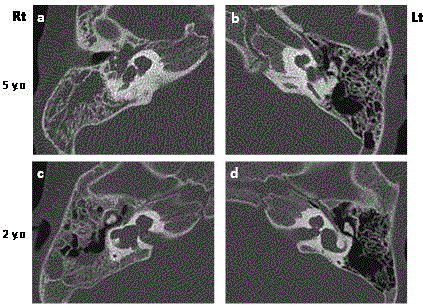
Figure 1
Temporal bone CT shows bilateral congenital inner ear
malformations and incomplete partition Type I (IP-1) on right ear (Figure 1a).
A soft tissue shadow is seen in the right ear tympanum and mastoid cavity
that suggests CSF leak (Figure 1b).
CT scan at the age of 2 years (Figure 1c, 1d) shows partial soft tissue shadow
in the tympanum and mastoid cavity of the right ear (Figure 1c).
Figure 2
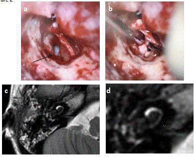
Figure 2
Intra-operative view of Case 1.Stapes was removed (Figure 2a,
arrow), and after the leakage pressure has weakened, multiple pieces of
fascia and cartilage are inserted into the inner ear to repair the CSF leakage
(Figure 2a, 2b). MRI examination 10 days after surgery confirms that the
inner ear cavity (Figure 2c, 2d) is occluded with filling materials (Figure 2d
arrow).
Discussion
Case 1 was a bilateral case of hearing impairment due to inner
ear malformations, while Case 2 was a unilateral case (both IP-I).
Case 1 was diagnosed as having an inner ear malformation on CT at 2
years of age, and a CSF leak in the right ear was suspected. However,
since there was no clinical history of bacterial meningitis, the parents
were advised to follow him carefully. Because the patient developed
bacterial meningitis at 5 years and 4 months, he underwent surgical
repair of the CSF leak by filling the inner ear. The CSF leak stopped
after surgery, but unfortunately, hearing loss of the good hearing
ear due to bacterial meningitis was observed, and cochlear implant
surgery was performed. On the other hand, in Case 2, an inner ear
malformation (IP-I) was diagnosed during examination for unilateral
hearing loss. There was no history of meningitis or any findings
of CSF leak on CT, but the patient underwent exploratory surgery
because IP-I is one of the high-risk inner ear anomalies associated
with bacterial meningitis. Since there was a small bone defect in the
stapes footplate, surgical repair of the potential risk of CSF leak was
performed in Case 2.
Inner ear malformations may involve bone defects of the stapes
footplate. Furthermore, in cases with weakness between the cochlea
and internal auditory canal, CSF leakage is likely to occur from the
small hole of the stapes footplate. Rao et al. [3] reported that 59%
of cases of repeated bacterial meningitis in children were due to
anatomical congenital malformations of the temporal bone. Savva et
al. [4] reported that persistent CSF leaks from inner ear malformations
were observed in 15% to 25% of repeated bacterial meningitis cases
in children. In cases with bacterial meningitis, inflammation may
spread to the healthy ear and cause bilateral deafness in some cases.
Therefore, in cases with inner ear malformations with obvious
cerebrospinal fluid leakage, surgical repair of the CSF leakage should
be performed before the development of meningitis.
Children with certain congenital malformations of the inner ear,
including those with Michel type, cochlear aplasia, common cavity
deformity, cochlear hypoplasia, and IP-I, have a higher incidence of
spontaneous CSF leaks and resulting meningitis [5]. IP-I is one of
the incomplete partition anomalies and accounts for approximately
20% of inner ear malformations [6,7]. In IP-I, there may be a defect
between the cochlea and internal auditory canal, and the CSF
completely fills the cochlea. IP-I may also involve a defective stapes
footplate. Therefore, meningitis can occur in IP-I patients. On the
other hand, there is no fragility of the site separating the internal
auditory canal and the cochlear cavity in IP-II, so that the risk of
cerebrospinal fluid leakage is low. Previously, there were several
reports of repeated bacterial meningitis due to Mondini type inner
ear malformations, but Mondini type strictly conforms to IP-II in the
classification of Sennaroglu [6,7], so the risk of meningitis is low in
Mondini type deformity.
Since β2-transferrin is present only in the CSF, it is very useful
for diagnosing CSF leaks [8]. However, when there is a copious
CSF gusher, care must be taken, because a large amount of CSF
otorrhea can occur into the external auditory canal even with a small
tympanostomy incision. Therefore, in the case of a bulging tympanic
membrane, it is better to consider tympanostomy as an examination
for final confirmation in the operating room.
Muzzi et al. [9] reported in their review that the average diagnostic
delay from the first episode of meningitis was 3.44 ± 3.41 years, the
number of meningitis episodes that occurred before the correct
diagnosis and definitive surgical treatment was 3.27 ± 1.81, and
certain numbers of children died from inner ear malformation-related
meningitis. Newborn hearing screening programs should prompt
a diagnostic work-up in the case of hearing impairment to prevent
inner ear malformation-related meningitis. Early identification of
the specific type of inner ear malformation and determination of
the associated risk of meningitis are of vital importance. Since NHS
provided an opportunity for the early identification of high-risk inner
ear anomalies, temporal bone CT should be recommended to children
with sensorineural hearing loss early in life. In cases with specific
inner ear malformations, in our opinion, prophylactic surgical repair
of CSF leakage should be performed to prevent bacterial meningitis.
Figure 3
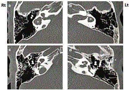
Figure 3
CT confirms right inner ear malformation (incomplete partition
type 1: IP-1) (Figure 3c arrow head) and bilateral dilated vestibules (Figure
3a,3b white asterisk). Development of the right mastoid air cells is good, and
they are well-ventilated. A small soft tissue shadow is observed between the
cruses of the stapes (Figure 3c arrow).
Figure 4
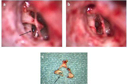
Figure 4
Intra-operative view of Case 2. There is a small bone defect in the
center of the stapes footplate (Figure 4a, 4c arrows). Stapes was removed
(Figure 4 b), filling the inner ear with multiple pieces of fascia and cartilage.
Conclusion
Bacterial meningitis in childhood is not only a life-threatening infection, but also a definitive cause of severe bilateral hearing impairment. In children with the high-risk type of congenital inner ear malformations, such as Michel type, cochlear aplasia, common cavity deformity, cochlear hypoplasia, and IP-I, especially with associated CSF leakage, surgical repair should be considered before bacterial meningitis develops.
Acknowledgement
This study was supported by research grants from the Ministry of Education, Science, Sports, Culture and Technology of Japan (Grant Number 17K11342).
References
- Rupa V, Agarwal I, Rajshekhar V. Congenital perilymph fistula causing recurrent meningitis: lessons learnt from a single-institution case series. Otolaryngol Head Neck Surg. 2014;150(2):285-91.
- Kakigi A, Tomofuji S, Takeda T, Sawada S. Cerebrospinal fluid otorrhea in a patient with congenital inner ear dysplasia. Auris Nasus Larynx. 2001;28(4):357-9.
- Rao AK, Merenda DM, Wetmore SJ. Diagnosis and management of spontaneous cerebrospinal fluid otorrhea. Otol Neurotol. 2005;26(6):1171-5.
- Savva A, Taylor MJ, Beatty CW. Management of cerebrospinal fluid leaks involving the temporal bone: report on 92 patients. Laryngoscope. 2003;113(1):50-6.
- Phelps PD, Lloyd GA. Mondini dysplasia is not associated with meningitis and cerebrospinal fluid fistula. Arch Otolaryngol Head Neck Surg. 1991;117(8):931-2.
- Sennaroglu L, Saatci I. A new classification for cochleovestibular malformations. Laryngoscope. 2002;112:2230-41.
- SennaroÄŸlu L, Bajin MD. Classification and Current Management of Inner Ear Malformations. Balkan Med J. 2017;34(5):397-411.
- McCudden CR, Senior BA, Hainsworth S, Oliveira W, Silverman LM, Bruns DE, et al. Evaluation of high resolution gel β(2)-transferrin for detection of cerebrospinal fluid leak. Clin Chem Lab Med. 2013;51(2):311-5.
- Muzzi E, Battelino S, Gregori M, Pellegrin A, Orzan E. Life-threatening unilateral hearing impairments. Review of the literature on the association between inner ear malformations and meningitis. Int J Pediatr Otorhinolaryngol. 2015;79(12):1969-74.