Clinical Image
Successful Repair with Aorto-Patch Plasty for Supra-Valvular Aortic Stenosis
Eyupserhat Calik, Umit Arslan and Bilgehan Erkut*
Department of Cardiovascular Surgery, Atatürk University, Turkey
*Corresponding author: Bilgehan Erkut, Department of Cardiovascular Surgery, Atatürk University, Erzurum, Turkey
Published: 06 Jun, 2018
Cite this article as: Calik E, Arslan U, Erkut B. Successful
Repair with Aorto-Patch Plasty for
Supra-Valvular Aortic Stenosis. Clin
Surg. 2018; 3: 1983.
Keywords
Supravalvular aortic stenosis; Aortic disease; Surgical therapy
Clinical Image
Supra-valvular aortic stenosis is a rare congenital disease that characterized by narrowing above
the sinotubular junction, and causes left ventricle hypertrophy. A 12-year-old boy was admitted
to our clinic with exercise intolerance, angina, and syncope complaints and with the diagnosis
of supravalvular aortic stenosis. Left ventricle was enlarged in telecardiogram; there had been
electrocardiographic findings of left ventricle hypertrophy. Mean pressure gradient was 70 mmHg
and maximum gradient 112 mmHg in cardiac catheterization. We successfully treated a traditional
diamond shaped patch aortoplasty was used to relieve supra-valvular aortic stenosis. Pressure
gradients were almost disappeared postoperatively with good surgical result. He was discharged
after 9 days.
Many different surgical techniques have been developed to correct supravalvular aortic stenosis
as diamond-shaped and pantaloon-shaped patch techniques, single-patch augmentation of the
non-coronary sinus, extended aortoplasty of the non-coronary and right coronary sinuses using
a pantaloon patch, a three-patch repair of all three aortic sinuses [1,2]. These techniques provides
symmetric reconstruction of the aorta with good postoperative results and no gradient across
aortic valve and aortic valve insufficiency remains, providing excellent long-term relief of localized
supravalvular gradients and preservation of aortic valve competence [3].
Figure 1
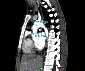
Figure 1
Multiplanar reconstruction image shows the diffusely stenosed ascending aorta in computed
tomographic angiograms.
Figure 2
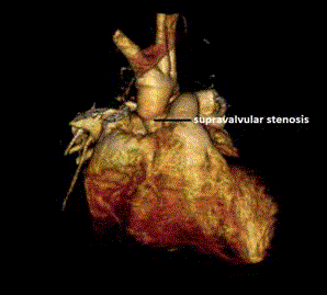
Figure 2
CT angiogram with 3D reconstruction showing the supravalvular aortic stenosis.
Figure 3
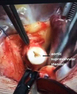
Figure 3
Supravalvular aortic stenosis during resection in operation.
Figure 4
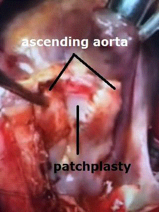
Figure 4
Image during operation of diamond shaped patch aorto-plasty with pericardial patch.
References
- Thistlethwaite PA, Madani MM, Kriett JM, Milhoan K, Jamieson SW. Surgical management of congenital obstruction of the left main coronary artery with supravalvular aortic stenosis. J Thorac Cardiovasc Surg. 2000;120(6):1040-6.
- McElhinney DB, Petrossian E, Tworetzky W, Silverman NH, Hanley FL. Issues and outcomes in the management of supravalvar aortic stenosis. Ann Thorac Surg. 2000;69(2):562-7.
- Mongé MC, Eltayeb OM, Costello JM, Johnson JT, Popescu AR, Rigsby CK, et al. Brom Aortoplasty for Supravalvular Aortic Stenosis. World J Pediatric Congenital Heart Surg. 2018;9(2):139-46.