Case Report
Adrenal Cyst Presenting as Asymptomatic Hydatid Cyst
Fadi Rayya1*, Haya Swilem2, Ruba Zuhri2 and Nour Seilin2
1Department of General Surgery, Al-Assad University Hospital, Syria
2Department of Medicine, Damascus University, Syria
*Corresponding author: Fadi Rayya, Department of General Surgery, University Hospital Damascus, 17-Nissan Street, Damascus, 10769, Syria
Published: 06 Jun, 2018
Cite this article as: Rayya F, Swilem H, Zuhri R, Seilin
N. Adrenal Cyst Presenting as
Asymptomatic Hydatid Cyst. Clin Surg.
2018; 3: 1978.
Abstract
Although it is possible to found hydatid cysts in any organ of the body, especially in the liver and
lungs, it rarely exists in the adrenal gland. We present a rare case of left adrenal hydatid cyst in a 48-
year old woman with an incidental finding and asymptomatic. Nothing was remarkable on physical
examination. Routine laboratory tests showed eosinophils, Bilirubin and alkaline phosphatase in
the normal range. In her medical history, she underwent spinal fusion surgery 4 months ago, during
the follow up after spinal fusion surgery, a simple abdominal X-ray showed a roughly 12 cm round
cyst with peripheral calcification in the left hypochondriac. She underwent surgical resection of the
hydatid cyst.
Keywords: Adrenal cyste; Hydatid cyste
Introduction
Hydatid cyst is a parasitic disease caused by Echinococcus granulosus, there is no specific gender for the disease. It is common in the Mediterranean region, although it is still rare in the North European and American regions. There are two types of parasites in the infection, which causes different diseases, Cystic Echinococcosis (CE) that causes hydatid cysts, where most infections are usually asymptomatic, and Alveolar Echinococcosis (AE) is usually confined to animals and rare in humans which can be fatal when not treated [1]. AE poses a much greater health threat to people than CE [1]. Symptoms take two ways systemic and local symptoms and this depends on the site and organ on which cyst settles [2]. Mebendazole, Albendazole and Praziquantel may be effective in treating. Although, surgical excision of the cyst remains the treatment of choice. However, attention is still paid to this disease as a result of the direct and indirect economic costs it causes by WHO statistics [3].
Case Presentation
A 48-year-old woman with a history of spinal fusion surgery presented to our department for
post-operative follow up. A simple abdominal X-ray was performed and showed a roughly 12 cm
round cyst with peripheral calcification in the left hypochondriac (Figure 1). Her past medical
history was notable for complete thyroidectomy 15 years ago and hemorrhoids surgery 8 years ago.
There was no history of childhood contact with animals. She denied abdominal pain, constipation,
diarrhea, vomiting or weight loss. There was nothing remarkable in physical examination. Vital
signs were stable.
Laboratory studies including Complete Blood Count (CBC) and biochemical profile were
within normal limits.
Multi Slice Computerized Tomography scan (MSCT) of the abdomen revealed a round cyst
measuring (8.2 cm × 7 cm × 8 cm) with peripheral calcification and wall thickening. It was located
among stomach, pancreas and transverse colon without obvious origin (Figure 2). The cyst appeared
inseparable from left adrenal gland which suggested a diagnosis of an adrenal hydatid cyst.
The patient underwent a transabdominal excision of the cyst. She was discharged from hospital
5 days after uneventful postoperative recovery. Histopathologic examination was consistent with
hydatid cyst. Cyst fluid analysis showed numerous inflammatory cells without atypia or irregular
mitotic activity (Figure 3).
Discussion
Adrenal cysts are uncommon; with an incidence 0.06% to 0.18% at autopsy [4]. The first adrenal
hydatid cyst was described in 1670 by Greiselius [5]. Echinococcosis hydatid cysts account for 6%
to 7% of all adrenal cysts [4,6,7]. There is no specific age to adrenal
cysts occurrence, but they are most commonly detected in the fifth
and sixth decades. They usually appear unilateral in 92% of the cases
and do not show a tendency for a particular side [4,6,7]. A review of
27cases is summed up in Table 1. The cases consisted of 12 males and
15 females (1:1), with age range 15 to 80 years (median: 48.9 years).
Furthermore, 13 cases were in left side (48.14%), while 14 cases were
in the right side (51.85%). The size of the cysts ranges from 3 cm to 20
cm (median: 9.4 cm). We found only six cases reported a history of
contact with animals, 4 had a history while two denied it.
Parasitic cysts affecting the adrenals are usually secondary or are
part of disseminated echinococcosis. The primary hydatid disease of
the adrenal gland is rare [6,7]. In our review, we found only 7 cases
(26.92%) of secondary origin and 19 cases (73.08%) of primary origin.
This is mostly because only the primary cysts are reported.
Abdominal hydatid cysts usually present with non-specific
symptoms. Symptoms often vary due to the site, and the size of
the cysts. Most adrenal hydatid cysts are incidentally detected at
sonography or computed tomography [8]. Hypertension is an
uncommon presentation and may result from external compression
of the renal parenchyma, a state known as Page kidney [9]. In our
review, 20 patients suffered from pain (74.07%), six patients from
hypertension (22.22%), and four patients found incidentally (14.8%).
The diagnosis has become easy with the ultrasound and CT,
with the diagnostic sensitivity ranging from 93% to 98% for
ultrasonography and approximately 97% for CT [10,11]. Imaging
features vary in hydatid cyst, depending on the stage of the growth
of the cyst, with/without any complications and the presence of
damaged tissue [10]. Based on ultrasonographic appearance, hydatid
cysts are classified by WHO into five subtypes: type I is a well-defined,
anechoic lesion; type II demonstrates separation of the membrane
characterized as the ‘water lily’ sign; type III contains septa and
intraluminal daughter cysts; type IV is a non-specific solid mass; and
type V is a solid mass with a calcified capsule [12].
On CT, the cyst walls can range in thickness from 2 mm to 1 cm,
authors report that MRI in adrenal hydatid cysts cases is more specific
and useful than CT 6 because it shows the contents of the cyst with
the surrounding membrane. Calcification is seen at radiography in
20% to 30% of hydatid cysts [12], and here we noticed peripheral
calcification around the cyst.
Routine laboratory tests do not show specific results, eosinophilia
is present in 25% of all persons who are infected 1, in our case,
before surgery, eosinophils was in normal range, but after the
surgery the value of eosinophils decreased significantly. Bilirubin or
alkaline phosphatase level may be elevated due to liver involvement.
Leukocytosis may suggest infection of the cyst like our case.
Laboratory tests may demonstrate anemia if there is substantial
intracystic bleeding and leukocystosis if the cyst is infected. In some
cases of hydatid cyste, eosinophilia and positive echinococcal serology
may be present. Their diagnostic value is low, however, and both were
negative in our case. The presence of calcification on plain abdominal
films is strongly suggestive of hydatid cyste or pseudocyst.
The treatment of choice is pericystectomy of the hydatid cyste, or,
if this is not possible, total excision of the adrenal gland.
Management options for hydatid cysts include surgery,
percutaneous treatment, anti-infective drug treatment or observation.
Normally, we do not resort to Albendazole or Mebendazole, as it is
less effective in the single lesion, and can be useful in widespread
diseases; on the other hand, the drug can not affect the calcification
surrounding the cyst as in our patient. Also Albendazole is significantly
more effective than Mebendazole in the treatment of liver cysts 1. All
treatment choices depend on location of cysts, size and existence of
calcification. Therefore, the surgery was the treatment of choice in
our case, because of single lesion, surrounding calcification, large size
and site. In addition, several reports confirmed the effectiveness of
laparoscopic hydatid surgery.
During the follow-up after surgery in 6 months, some studies
suggest a reduction in the rate of immunoglobulin (total and specific
IgE, IgG, IgM, IgA), However, this follow-up is not necessary for all
patients.
Table 1

Table 1
Cases of cystic adrenal echinococcosis in the literature.
Figure 1
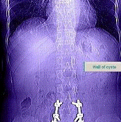
Figure 1
Abdomen X-rays demonstrates cyst with peripheral calcification in
the left hypochondriac.
Figure 2
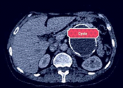
Figure 2
CT shows the adrenal cyste.
Figure 3
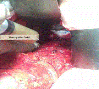
Figure 3
Intraoperative view shows the cystic fluid.
References
- Centers for disease control and prevention. Parasites - Echinococcosis. 2012.
- Duman K, Girgin M, Hamcan S. Uncomplicated hydatid cysts of the liver: Clinical presentation, diagnosis and treatment. J Gastrointest Dig Syst. 2016;6(3):430.
- Benner C, Carabin H, Sánchez-Serrano LP, Budke CM, Carmena D. Analysis of the economic impact of cystic echinococcosis in Spain. Bull World Health Organ. 2010;88(1):49-57.
- de Bree E, Schoretsanitis G, Melissas J, Christodoulakis M, Tsiftsis D. Cysts of the adrenal gland: Diagnosis and management. Int Urol Nephrol. 1998;30(4):369-76.
- Wedmid A, Palese M. Diagnosis and treatment of the adrenal cyst. Curr Urol Rep. 2010;11(1):44-50.
- Abeshouse GA, Goldstein RB, Abeshouse BS. Adrenal cysts; review of the literature and report of three cases. J Urol. 1959;81(6):711-9.
- Akcay MN, Akçay G, Balik AA, Böyük A. Hydatid cysts of the adrenal gland: Review of nine patients. World J Surg. 2004;28(1):97-9.
- Schoretsanitis G, de Bree E, Melissas J, Tsiftsis D. Primary hydatid cyst of the adrenal gland. Scand J Urol Nephrol. 1998;32(1):51-3.
- Tazi F, Ahsaini M, Khalouk A, Mellas S, Stuurman-Wieringa RE, Elfassi, et al. Giant primary adrenal hydatid cyst presenting with arterial hypertension: a case report and review of the literature. J Med Case Reports. 2012;6:46.
- Turgut AT, Akhan O, Bhatt S, Dogra VS. Sonographic spectrum of hydatid disease. Ultrasound Q. 2008;24(1):17-29.
- Wegener OH, Chapter 18 in Whole Body Computed Tomography. 1993, Boston: Blackwell Scientific Publications.
- Group WIW, International classification of ultrasound images in cystic echinococcosis for application in clinical and field epidemiological settings. Acta Tropica. 2003;85(2):253-61.