Case Report
Compound Odontoma-Diagnosis and Treatment in Pedıatrıc Dentıstry: Three Case Reports
Alem Coşgun1, Behiye Sezgin Bolgül1*, Ezgi Meriç1 and Berk Turgay2
1Department of Pedodontics, Mustafa Kemal University, Turkey
2Department of Oral and Maxillo Facial Surgery, Mustafa Kemal University, Turkey
*Corresponding author: Behiye Sezgin Bolgül, Department of Pedodontics, Mustafa Kemal University, Hatay, Turkey
Published: 18 May, 2018
Cite this article as: Coşgun A, Bolgül BS, Meriç E, Turgay
B. Compound Odontoma-Diagnosis and
Treatment in Pedıatrıc Dentıstry: Three
Case Reports. Clin Surg. 2018; 3: 1971.
Abstract
Odontomas are the most common odontogenic tumors. They are considered to be hamartomas
rather than neoplasms, and are composed of natural teeth tissues: enamel, dentin, and cementum
and pulp tissue. They are broadly classified into compound odontoma (small tooth like structures)
and complex odontoma (a conglomeration of dentin, enamel and cementum). Generally,
odontomas have been associated with trauma during primary dentition as well as with inflammatory
and infectious processes, hereditary anomalies (Gardner syndrome, Hermann's syndrome),
odontoblastic hyperactivity and alterations in the genetic components responsible for controlling
dental development. This study presents three cases of odontoma and treatments.
Keywords: Compound odontoma; Child; Tumors
Introduction
Odontomas are the most common odontogenic tumors. They are considered to be hamartomas rather than neoplasms, and are composed of natural teeth tissues: enamel, dentin, cementum and pulp tissue [1]. According to the World Health Organization classification, two distinct types of odontomas are acknowledged: complex and compound odontomaIn complex odontomas, all dental tissues are formed, but appeared without an organized structure. In compound odontomas, all dental tissues are arranged in numerous tooth-like structures known as denticles [2]. Generally these malformations are intraosseous, but occasionally they may erupt into the oral cavity [3,4]. The etiology of the odontoma is unknown. Generally, odontomas have been associated with trauma during primary dentition as well as with inflammatory and infectious processes, odontoblastic hyperactivity and alterations in the genetic components responsible for controlling dental development [5]. Odontomas can also manifest as part of syndromes, such as basal cell nevus syndrome, Gardner syndrome, familial colonic adenomatosis, Tangier disease or Hermann syndrome [6]. Conservative surgical excision is the treatment of choice. Compound and complex odontomas are well encapsulated and easily enucleated from the surrounding bone [7].
Case Presentation
Case 1
13-year-old boy referred to our clinic because of an unerrupted maxillary santral incisor (Figure
1). The lesion was disturbing the eruption of the maxillary central incisor (Figure 2). Lesions removed
surgically (Figure 3). The histopatological examination confirmed that the lesions are compound
odontomas. Following the surgery, the persistant tooth started eruption (Figure 4 and 5).
Case 2
A trauma patient referred to our clinic with a broken central tooth (Figure 6). The tooth was
broken due to a bicycle accident two years ago. He was 11-year-old
boy. The central incisor was root canal treathed. There weren’t any
known syndrome or systemic disease of the patient. We thought that
the etiology of the odontoma might be releated to the trauma so we
removed the odontoma because it might affect the prognosis of the
tooth (Figure 7). Odontomas removed surgically (Figure 8). The
histopatological examination confirmed that the lesion is a compound
odontoma. We restorated the tooth after root canal treatment (Figure
9 and 10).
Case 3
9-year-old boy referred to our clinic because of an unerrupted
maxillary santral incisor (Figure 11). The lesion was disturbing the
eruption of the maxillary central incisor (Figure 12). The lesion was
removed surgically (Figure 13). The histopatological examination
confirmed that the lesion is a compound odontoma. Following the
surgery, the persistant tooth started eruption (Figure 14 and 15).
Figure 1
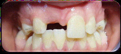
Figure 1
Intraoral view of pre-op.
Figure 2
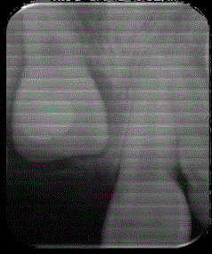
Figure 2
Periapical radiography of pre-op.
Figure 3
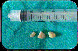
Figure 3
View of odontomas.
Figure 4
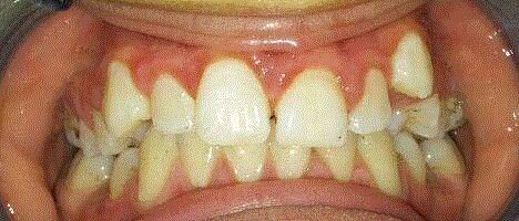
Figure 4
Intraoral view of post-op.
Figure 5
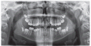
Figure 5
Panoramic radiography of post-op.
Figure 6
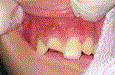
Figure 6
Intraoral view of pre-op.
Figure 7
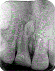
Figure 7
Periapical radiography of pre-op.
Figure 8
Figure 8
View of odontomas.
Figure 9
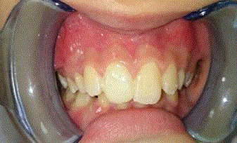
Figure 9
Intraoral view of post-op.
Figure 10
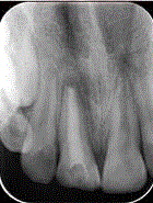
Figure 10
Periapical radiography of post-op.
Figure 11
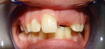
Figure 11
Intraoral view of pre-op.
Figure 12
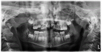
Figure 12
Panoramic radiography of pre-op.
Figure 13
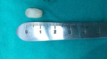
Figure 13
View of odontomas.
Figure 14
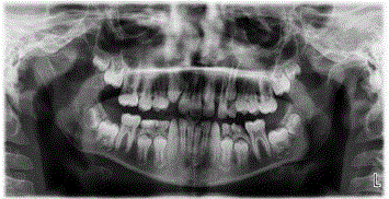
Figure 14
Panoramic radiography of post-op.
Discussion
Odontomas represent the most common type of odontogenic benign jaws tumors among patients younger than 20 years of age [8]. In our cases reported, the incidence age of odontoma is consistent with the literature. Odontomas are slow-growing, asymptomatic neoplasms found in jaws. In about 80% of cases, they are associated with impacted or un erupted teeth. Although they are commonly asymptomatic, clinical indicators of odontoma may include retention of deciduous teeth, non eruption of permanent teeth, pain, expansion of the cortical bone, tooth displacement [9]. Orthodontic treatment may be indicated to correct malocclusion. İn our two cases reported, odontomas are associated with un erupted teeth. The treatment according to the available literature is surgical extraction with complete removal of any associated soft tissues, since the odontoma may interfere with eruption of the permanent tooth displace the adjacent teeth or give rise to a dentigerous cyst [6].
Figure 15
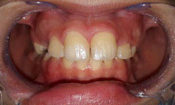
Figure 15
Intraoral view of post-op.
Conclusion
The results achieved indicate that early diagnosis of odontomas in primary dentition is essential in order to prevent later complications, requires less expensive treatment, ensure better prognosis, avoid relapse of the lesion, avoid displacement or devitalisation of adjacent tooth.
References
- Baldawa RS, Khante KC, Kalburge JV, Kasat VO. Orthodontic management of an impacted maxillary incisor due to odontoma. Contemp Clin Dent. 2011;2(1):37.
- Morgan PR. Odontogenic tumors: a review. Periodontol 2000. 2011;57(1):160-76.
- Özcan G, Şekerci AE, Ekizer A, Kara Ö. Erupted Compound Odontoma: Two Case Reports. Turkiye Klinikleri. J Dental Sci Cases. 2015;1(2):126-30.
- Liu J-K, Hsiao C-K, Chen H-A, Tsai M-Y. Orthodontic correction of a mandibular first molar deeply impacted by an odontoma: A case report. Quintessence International. 1997;28(6):381-5.
- Owens B, Schuman N, Mincer H, Turner J, Oliver F. Dental odontomas: a retrospective study of 104 cases. J Clin Pediatr Dent. 1997;21(3):261-4.
- Sánchez OH, Berrocal ML, González JM. Metaanalysis of the epidemiology and clinical manifestations of odontomas. Med Oral Patol Oral Cir Bucal. 2008;13(11):E730-4.
- Amailuk P, Grubor D. Erupted compound odontoma: case report of a 15-year-old Sudanese boy with a history of traditional dental mutilation. Br Dent J. 2008;204(1):11-4.
- Katz R. An analysis of compound and complex odontomas. ASDC journal of dentistry for children. 1989;56(6):445-9.
- Bordini J, Contar CM, Sarot JR, Fernandes Â, Machado MÂN. Multiple compound odontomas in the jaw: case report and analysis of the literature. Journal of Oral and Maxillofacial Surgery. 2008;66(12):2617-20.