Case Report
Lingual Agenesis: A Case Report and Review of Literature
Mark Enverga, Ibrahim Zakhary* and Abraham Khanafer
Department of Oral and Maxillofacial Surgery, University of Detroit Mercy, School of Dentistry, USA
*Corresponding author: Ibrahim Zakhary, Department of Oral and Maxillofacial Surgery, University of Detroit Mercy, School of Dentistry, USA
Published: 09 Mar, 2018
Cite this article as: Enverga M, Zakhary I, Khanafer A.
Lingual Agenesis: A Case Report and
Review of Literature. Clin Surg. 2018;
3: 1933.
Abstract
Aglossia is a rare condition characterized by the complete absence of the tongue. Its etiology is still unknown. The underlying pathophysiology involves disruption of the development of the lateral lingual swellings and tuberculum impar during the second month of gestation. In this case report, a 26-year old African American female with aglossia presented to the Detroit Mercy Oral Surgery Clinic. The patient presented for extraction of tooth #28, which was located within a fused bony plate at the floor of the mandible. This study presents a case of aglossia, as well as a critical review of aglossia in current literature.
Introduction
Aglossia is a rare condition characterized by the complete absence of the tongue. The exact etiology of aglossia is still unknown. Possible etiologic factors during embryogenesis include maternal febrile illness, drug ingestion, hypothyroidism, and cytomegalovirus infection. Heat-induced vascular disruption in the fourth embryonic week and chronic villous sampling performed before 10 weeks of amenorrhea, also called the disruptive vascular hypothesis, may be another possible cause of aglossia [1,2]. The underlying pathophysiology involves disruption of the normal embryogenic development of the lateral lingual swellings and tuberculum impar during the second month of gestation [3]. Most cases of aglossia are associated with other congenital limb malformations and craniofacial abnormalities, such as hypodactyly, adactyly, cleft palate, Pierre Robin sequence, Hanhart syndrome, Moebius syndrome, and facial nerve palsy. Animal studies have shown that the transcription factor Hand2 plays a role in tongue morphogenesis and the development of the limb and branchial arch [2,4]. Situs inversus and thyroid dysfunction are also described to occur with aglossia. An isolated defect limited to the absence of the tongue alone is extremely rare and only a few cases have been described in current literature. Normal tongue development is vital for many functions, such as suckling, swallowing, chewing, and speech. At birth, the tongue is essential for nourishment and the suckling reflex. Newborns with aglossia seldom survive more than three days [3]. In addition, the tongue is associated with the normal growth and position of the mandible, hyoid bone, and teeth. Due to the lack of muscular pressure of the tongue, the establishment of occlusion and the development of the dental arch shape are altered [5]. Multiple cases in literature have described hypertrophy of the floor of the mouth, exaggerated lip movements, constriction of the oropharynx, and hypertrophy of the uvula [6]. Other orofacial alterations reported are the absence of deciduous mandibular teeth and the crowding of permanent mandibular teeth [7,8]. Mandibular hypoplasia, micrognathia, macrosomia, and Angle’s class II malocclusion are also features associated with aglossia [9]. In regards to phonation, most patients have intelligible speech; however, speech impediments are common. The hypertrophic mylohyoid, hyoid bone, and tongue base act as a pseudo-tongue that creates a seal with the posterior palate. The creation of the palatal seal allows for speech and swallowing functions.
Materials and Methods
A systematic PubMed and Medline search was conducted using the key words aglossia, lingual agenesis, and aglossia adactylia and aglossia congenita. Age at presentation, gender, race, comorbidities, limb abnormalities, speech deficits, clinical characteristics, rendered treatment, and treatment outcome were collected, tabulated, and subjected to analysis (Table 1). A 26 year old African American female presented to our Oral Surgery clinic for the extraction of tooth #28 (Figure 1). Medical history revealed asthma, sleep apnea, and sickle cell trait. Patient stated that she used a feeding tube to eat until the age of 10 and a tracheal tube until the age of 7 to assist with breathing. No limb abnormalities were reported. Intraorally, the patient presented with lingual agenesis, a fused mandible, and bony plate in the floor of the mouth, uncharacteristic of typical aglossia cases. Her mandibular teeth are displaced lingually and tooth #28 is located in the center of the bony plate. A panoramic radiograph was taken (Figure 2). Tooth #28 showed extensive decay and diagnosed with symptomatic apical periodontitis. Patient was anesthetized and tooth #28 was removed using straight elevator (Figure 3). To close the defect created following extraction, a lateral repositioned flap was advanced, a collagen membrane was placed, and secured by four (3-0) Chromic Gut sutures.
Figure 1
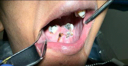
Figure 1
Clinical visualization of tooth #28. Mandibular teeth are lingually
tipped. Tooth #28 located at the center of the bony plate of the mandible.
Figure 2
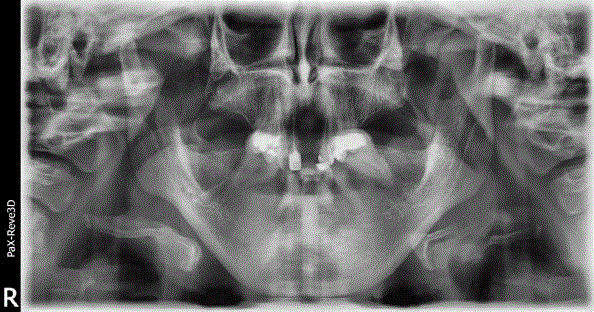
Figure 2
Panoramic radiograph showing fused mandible and bony plate at
the center of the mandible. Tooth #28 is located at the center of the bony
plate.
Figure 3
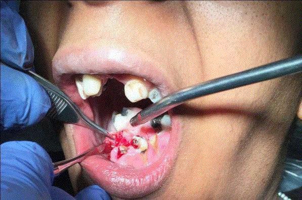
Figure 3
Reflection of the periodontium during the extraction of tooth #28.
Results
Although aglossia is normally incompatible with life, many cases have been reported and described in literature. Jussieu first described congenital aglossia in 1718, and Rosenthal was the first to describe aglossia’s correlation with adactylia in 1932. Our review of literature consisted of 19 case reports of patients with either aglossia or hypoglossia. A 52.6% (10/19) of patients were male and 47.4% (9/19) of patients were female. Most patients in the reviewed literature had some type of limb abnormality or craniofacial malformation. A 47.4% (9/19) of patients presented with limb abnormalities, such as hypodactyly, syndactyly, adactyly, or hypomelia. A 10.5% (2/19) presented with cleft palate and 5.2% (1/19) presented with Moebius Syndrome. Situs inversus was found in 15.8% (3/19) of the cases reviewed. Uncharacteristically, 31.6% (6/19) of patients did not have an associated limb or craniofacial abnormality. A 36.8% (7/19) of patients suffered from a speech impediment. Hypertension, hypothyroidism, and patent ductus arteriosus were comorbidities associated with singular cases of aglossia. 26.3% (5/19) of patients were of Asian descent and 10.5% (2/19) of patients were of Caucasian descent; however, 63.2% (12/19) of cases did not report the patient’s race. Orthopedic distraction osteogenesis was used to treat patients in 21.1% (4/19) of cases, only one of which provided additional speech and hearing therapy. There was no reported treatment in 42.1% (8/19) of cases.
Discussion
The exact etiology of lingual agenesis is still unknown; however,
it is frequently associated with certain limb and craniofacial
malformations. In 63.2% of the reviewed cases, patients with aglossia
presented with a limb abnormality or craniofacial defect. Similar to
the patient presented in our case report, 31.2% of studies did not
observe patient limb or craniofacial abnormalities. Our case report
also describes mandibular fusion and a bony plate at the floor of the
mouth. Characteristically, the reviewed cases reported hypertrophy
of the floor of the mouth, acting as a pseudo-tongue to assist with
speech and swallowing.
Orofacial alterations due to the absence of the tongue ultimately
affect psychological, physical and social development; therefore,
professionals in the areas of nutrition, psychology, speech, general
dentistry, orthodontics, maxillofacial surgery, and implantology are
needed. At birth, the tongue is essential for nourishment and the
suckling reflex. Newborns with aglossia seldom survive more than 3
days. Adjusting the way the child feeds allows for proper nourishment
and growth for newborns and infants [3].
Orthodontic and surgical treatment are used to correct the
malocclusion and orthognathic constriction caused by aglossia.
A 21.1% of the reviewed cases utilized orthopedic extraction
osteogenesis to expand the dental arch. Rapid maxillary expansion,
mandibular expansion, and distraction osteogenesis, followed by
orthodontic treatment early in life have been successfully utilized
to align crowded teeth, provide functional occlusion, and produce
proper esthetics. In some cases, however, these appliances were
unsuccessful because they altered the patient’s naturally modified
swallowing and chewing functions. Because older patients have
shown functional adaptation to most tongue functions, dental arch
expansion and surgical reconstruction of the tongue is not a preferred
treatment [3,7].
Delayed and slurred speeches were the most common problems
during the development of patients with aglossia. Although speech
therapy was conducted in only one of the reviewed cases, speech
therapy is the suggested treatment option. In patients with aglossia, the
hyoid bone and the mandible strongly correlate with the movements
of the pseudo-tongue during speech. Due to the restricted range of
motion of the pseudo-tongue, the mylohyoid and tongue base are
more dependent on the mandible and hyoid for range of motion
during speech. Exaggerated movements of the hyoid and mandible
provide compensatory movements for speech in the absence of a
tongue. Over time, adjusting to the altered range of motion through
speech therapy can allow for speech that is more comprehensible
[2,6-22].
Conclusion
The complete absence of the tongue is an uncommon occurrence.When managing aglossia patients, the intricate complications associated with the absence of a tongue call for a multidisciplinary approach. Early management of lingual agenesis is crucial for the development of feeding, swallowing, and speech. Treatment of aglossia and its accompanying characteristics later in life must be elective due to possible disruption of the adaptive function of the pseudo-tongue.
Table 1

Table 1
Data Collection.
References
- Gruber B, Burton BK. Oromandibular-limb hypogenesis syndrome following chorionic villus sampling. Int J Pediatr Otorhinolaryngol. 1994;29(1):59-63.
- Gupta SR. Isolated aglossia congenita: A rare case of oromandibular limb hypogenesis syndrome type I B. J Oral Maxillofac Pathol. 2012;16(3):414-9.
- Salles F, Anchieta M, Bezerra PC, Torres ML, Queiroz E, Faber J. Complete and isolated congenital aglossia: case report and treatment of sequelae using rapid prototyping models. Oral Surg Oral Med Oral Pathol Oral Radiol Endod. 2008;105(3):e41-7.
- Barron F, Woods C, Kuhn K, Bishop J, Howard MJ, Clouthier DE. Downregulation of Dlx5 and Dlx6 expression by Hand2 is essential for initiation of tongue morphogenesis. Development. 2011;138(11):2249-59.
- Kumar P, Chaubey KK. Aglossia: a case report. J Indian Soc Pedod Prev Dent. 2007;25(1):46-8.
- McMicken BL, Von Berg S, Long Wang AK, Vento-Wilson M, Rogers K. Speech and swallow kinematics of a person with congenital aglossia. Anatomy & Physiology: Current Research 2015;5(176):2161-940.
- Cappellette M, Ferreira da Costa CM, de Nobrega M, de Oliveira WC, Weckx LLM. Oromandibular and limb hypogenesis syndrome: treatment report. Oral surg oral med oral pathol oral radiol. 2013;116(4):e230-6.
- McMicken B, Von Berg S, Iskarous K. Acoustic and Perceptual Description of Vowels in a Speaker with Aglossia. Communication Disorders Quarterly. 2012;34(1):38-46.
- Rasool A, Zaroo MI, Wani AH, Darzi MA, Bashir SA, Bijli AH, et al. Isolated aglossia in a six year old child presenting with impaired speech: a case report. Cases J. 2009;2:7926.
- Ardran GM, Fulford GE, Kemp FH. Aglossia congenita; cineradiographic findings. Arch Dis Child. 1956; 31(159):400-7.
- Ardran GM, Beckett JM, Kemp FH. Aglossia Congenita. Arch Dis Child. 1964;39:389-92.
- Nevin NC, Burrows D, Allen G, Kernohan DC. Aglossia-adactylia syndrome. J Med Genet. 1975;12(1):89-93.
- Mishima K, Sugahara T, Mori Y, Sakuda M. Case report: hypoglossia-hypodactylia syndrome. J Cranio Maxillofac Surg. 1996;24(1):36-9.
- Jang GY, Lee KC, Choung JT, Son CS, Tockgo YC. Congenital aglossia with situs inversus totalis--a case report. J Korean Med Sci. 1997;12(1):55-7.
- Grippaudo FR, Kennedy DC. Oromandibular-limb hypogenesis syndromes: a case of aglossia with an intraoral band. Br J Plast Surg. 1998;51(6):480-3.
- Amor DJ, Craig JE. Situs inversus totalis and congenital hypoglossia. Clin Dysmorphol. 2001;10(1):47-50.
- Cho J, Kim J, Jang K, Kim C, Hahn S, Lee S. Mandibular expansion in a Congenital aglossia patient. THE J Korean Academy Pedtatric Dentistry. 2003;30:673-7.
- Kantaputra P, Tanpaiboon P. Thyroid dysfunction in a patient with aglossia. Am J Med Genet A. 2003;122A(3):274-7.
- Gathwala G, Singh J, Dalal P, Garg A. Hypoglossia-hypodactyly syndrome in a newborn. J Cranio Maxillofac Surg. 2011;39(2):99-101.
- McMicken BL, Kunihiro A, Wang L, Von Berg S, Rogers K. Electropalatography in a Case of Congenital Aglossia. J Commun Disord Deaf Stud Hearing Aids. 2014.
- Bommarito S, Zanato LE, Vieira MM, Angelieri F. Aglossia: Case Report. Int arch otorhinolaryngol. 2016;20(1):87-92.
- Kaissi A, Safi H, Ghachem MB, Hendaoui L, Chehida F. Aglossia-adactylia sequence and Moebius syndrome involvement. African J oral Health. 2005;2:1-2.