Case Report
Retroperitoneal Lymphangioma in Childhood: Is there an Association with Cryptorchidism?
Taro Ikeda*, Takayuki Masuko, Kenjiro Takagi and Toshiki Rikiyama
Department of Surgery and Perinatal Medicine, Saitama Medical Center, Jichi Medical University, Japan
*Corresponding author: Taro Ikeda, Department of Surgery and Perinatal Medicine, Saitama Medical Center, Jichi Medical University, 1-847, Amanuma-cho, Omiya-ku, Saitama- City, Saitama, 330-8503, Japan
Published: 16 Feb, 2018
Cite this article as: Ikeda T, Masuko T, Takagi K, Rikiyama
T. Retroperitoneal Lymphangioma in
Childhood: Is there an Association with
Cryptorchidism?. Clin Surg. 2018; 3:
1917.
Abstract
In this case report, we present a six-month-old boy who undergone a surgical removal of painless mass from the inguinal area which extended to the abdominal cavity to the right side of the bladder.
Introduction
Lymphangiomas are relatively-rare and benign lesions believed to be congenital malformations of the lymphatic anlage rather than true tumors. Retroperitoneal lymphangiomas are considered to be also relatively-rare and account for 1% to 10% of all lymphangiomas. Most retroperitoneal lymphangiomas typically occur in children under 2 years old; however, in some cases, these lesions can be found after a long asymptomatic period due to slow development. There is no typical complication with these lesions and their various symptoms include an acute abdomen complicated by bleeding or infection, palpable tumors and other associated symptoms. In this report, we present a case of retroperitoneal lymphangioma associated with intraperitoneal cryptorchidism.
Case Presentation
A six-month-old boy was brought to our hospital because of a mass in the right inguinal area. He had no previous history of such a mass. Physical examination showed no thoracoabdominal findings. A painless mass, 2 cm in size and soft, was palpable in the inguinal area. On Ultra Sound (US) examination, the lesion was diagnosed as a cystic mass of the cord hydrocele. The patient was examined again because of a change in mass size. Repeat US revealed the cystic mass to have extended along the abdominal cavity to the right side of the bladder. Computed Tomography (CT) was performed because of the possibility of retroperitoneal lymphangioma (Figure 1). CT showed a cystic mass extending from the right side of the bladder to the inguinal canal; a diagnosis of retroperitoneal lymphangioma was thus confirmed. Surgical excision was then scheduled and performed using the inguinal approach after the mass had been detected under a laparoscope. During surgery, a portion of the mass was found in the internal inguinal ring after opening the inguinal canal, and the periphery of the mass was gradually avulsed. The testis was avulsed and separated from the mass, as it was adherent to the wall of the mass just under the internal inguinal ring (Figure 2). Then, the mass was deflated and removed through the internal inguinal ring after its contents had been aspirated. The testis was difficult to pull down to the base of the scrotum due to poor extension of its blood vessels; it was thus fixed at an upper site of the scrotum. His postoperative course remains excellent and follow-up at 12 months showed no lymphangioma recurrence.
Discussion
We present a case of retroperitoneal lymphangioma complicated by cryptorchidism. This case is a rare because there are no reports on associations of these lesions in the medical literature. Retroperitoneal lymphangiomas are considered to be relatively rare and account for 1 to less than 10% of all lymphangioma cases [1-4]. There is no typical complication with these lesions and their various symptoms include an acute abdomen complicated by bleeding or infection, palpable tumors and other associated symptoms [1-8]. Although there are a few reports on lymphangioma extending to the groin, no case of retroperitoneal lymphangioma with cryptorchidism has previously been reported [7,8]. In our case, it was impossible to pull down the testis to the scrotum during surgery when it was not separated from the cystic mass. This was because the testis adhered firmly to a wall of the retroperitoneal lymphangioma. The testis was identified during surgery so that it could be preserved and immediately fixed within the scrotum. No malformations, such as hypoplasia, were identified in the testis, the testicular arteries and veins or in the seminal duct. Also, no process vaginalis was observed; these findings suggest that the retroperitoneal lymphangioma developed/ presented around the same time that the testis would normally have descended, thereby preventing the testis from descending. In general, lymphangiomas are characterized to be an anomaly in the lymphatic system or connective dysplasia that develops at 5 weeks of intrauterine life. The testis, meanwhile, was highly likely to be generated before 28 weeks of intrauterine life when it descends into the scrotum. Thus, in our case, the presence of lymphangioma appears to have played a key role in the occurrence of cryptorchidism.
Figure 1
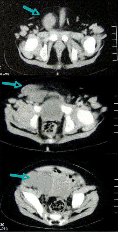
Figure 1
CT showed a cystic mass extending from the right side of the
bladder to the inguinal canal (arrow).
Figure 2
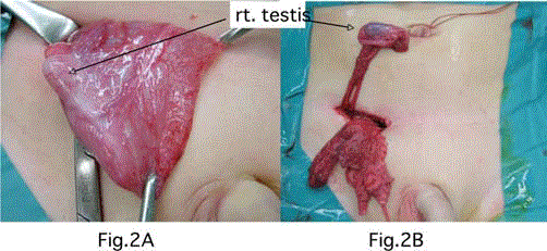
Figure 2
Surgical findings. (A) the mass was found in the internal inguinal
ring after opening the inguinal canal; (B) right testis was avulsed and
separated from the mass, as it was adherent to the wall of the mass just
under the internal inguinal ring.
References
- De Perrot M, Rostan O, Morel P, Le Coultre C. Abdominal lymphangioma in adults and children. Br J Surg. 1998;85(3):395-7.
- Hancock BJ, St-Vil D, Luks FI, Di Lorenzo M, Blanchard H. Complications of lymphangiomas in children. J Pediatr Surg. 1992;27(2):220-6.
- Shankar KR, Roche CJ, Carty HML, Turnock RR. Cystic retroperitoneal lymphangiomas: treatment by image-guided percutaneous catheter drainage and sclerotherapy. Eur Radiol. 2001;11(6):1021-3.
- Waldhausen J, Holterman JM, Tapper D. Identification and surgical management of cystic retroperitoneal lymphangioma in children. Pediatr Surg Int. 1996;11(4):283-5.
- Konen O, Rathaus V, Dlugy E, Freud E, Kessler A, Shapiro M, et al. Childhood abdominal cystic lymphangioma. Pediatr Radiol. 2002;32(2):88-94.
- Hebra A, Brown MF, MiGeehin KM, Ross AJ. Mesenteric, omental, and retroperitoneal cysts in children: a clinical study of 22 cases. South Med J. 1993;86(2):173-6.
- Steyaert H, Guitard J, Moscovici J, Juricic M, Vaysse P, Juskiewenski S. Abdominal lymphangioma in children: benign lesions that can have a proliferative course. J Pediatr Surg. 1996;31(5):677-80.
- Abantanga FA. Groin and scrotal swellings in children aged 5 years and below: a review of 535 cases. Pediatr Surg Int. 2003;19(6):446-50.