Case Report
Adenocarcinoma and Intestinal Duplication of the Ileum: Report of a Case Highlighting the Malignant Potential of an Uncommon Condition
Marcelo A Beltran1,2*
1Department of Surgery, Serena Hospital, Chile
2Department of Clinics, Catholic University of the North, Chile
*Corresponding author: Marcelo A Beltran, Department of Surgery, Serena Hospital, Department of Clinics, Catholic University of the North, La Serena – IV Region, P.O. Box 912, Chile
Published: 12 Feb, 2018
Cite this article as: Beltran MA. Adenocarcinoma and
Intestinal Duplication of the Ileum:
Report of a Case Highlighting the
Malignant Potential of an Uncommon
Condition. Clin Surg. 2018; 3: 1903.
Abstract
Introduction: Gastrointestinal duplications are recognized, however uncommon developmental
abnormalities, and may occur anywhere along the whole gastrointestinal tract, most commonly at
the ileum. Their presentation in adults is an infrequent clinical condition. A recent review found
only 15 cases of ileal duplication in adults published since 1884, with adenocarcinoma in 4 of those
15 cases (27%) highlighting the malignant potential of this uncommon condition.
Case Presentation: A 36-years-old female was admitted to the Emergency Unit. A Computed
Abdominal Tomography (CAT) Scan demonstrated small bowel loops surrounding a tubular
cystic structure which was compressing the adjacent bowel. Surgical exploration revealed multiple
malignant implants covering the visceral and parietal peritoneum and infiltrating completely the
omentum. At the ileal mesentery a tubular cystic whitish tumor was found. We unroofed the
cyst and resected as much wall as we could, however a complete resection of the tumor was not
considered. Thirty-four days after the operation the patient died. The histopathology of the cystic
wall was compatible with the architecture of the intestinal wall extensively infiltrated by a mucinous
adenocarcinoma moderately differentiated; a mucosal lining in parts atrophic and in parts infiltrated
or replaced by adenocarcinoma was also observed.
Conclusion: This uncommon malignancy developing in an even more uncommon developmental
condition such as ileal duplication highlights the importance of diagnosis and appropriate treatment
in the infancy because the threatening malignant potential when presenting late in life.
Keywords: Intestinal duplication; Ileal duplication in adults; Small bowel adenocarcinoma
Introduction
Primary adenocarcinoma of the small intestine is rare, and accounts for 1% to 5% of all gastrointestinal tumors [1,2]. This neoplasm presents a challenge to the clinician due to its rarity combined with non-specific signs and symptoms that delay prompt diagnosis and treatment making its prognosis dismal with 5-year survival rates ranging from 20% to 30% [1-3]. Frequently, these tumors are found at emergency surgery for complications such as intestinal obstruction, perforation or hemorrhage, with a survival of less than a year in all cases [4,5]. Gastrointestinal duplications are recognized but uncommon developmental abnormalities, and may occur anywhere along the whole gastrointestinal tract, more commonly at the ileum. These are usually diagnosed and surgically treated in children, making their presentation in adults an infrequent and curious clinical condition [6,7]. A recent review found only 15 cases of ileal duplication in adults published since 1884 [8]. The same article reports the development of adenocarcinoma in 4 of those 15 cases (27%) and 1 case complicated with squamous cell carcinoma (7%), highlighting the malignant potential of this uncommon condition.
Case Presentation
A 36-years-old female without any previously known diseases, was admitted to the Emergency Unit of our institution with the presumptive diagnosis of acute abdomen and secondary peritonitis. She complained of abdominal pain starting seven days before associated to abdominal distension, nausea and vomiting. On physical examination the blood pressure was 131/76, with a cardiac frequency of 126 beats per minute and 38.9 °C rectal temperature. The skin was pale and diaphoretic. The abdomen was distended, dull at percussion, and bowel sounds were absent at auscultation. The palpation was painful with rebound sensitivity present mainly over the left lower abdominal quadrant. Suspecting a gynecologic condition we performed a gynecological ultrasonography which was normal; however during the exam the gynecologist noticed an abnormal cystic structure adjacent to the small bowel. Subsequently a Computed Abdominal Tomography (CAT) Scan was performed; it demonstrated some small bowel loops surrounding a tubular cystic structure which was compressing the adjacent bowel, and peritoneal free fluid (Figure 1A). The white blood cell count was raised (25,700 x mm3) and the C-reactive protein was 356.06 mg/l. With the diagnosis of a complicated intestinal cystic tumor the patient underwent emergency surgery. We opened the abdominal wall with a midline incision over and below the umbilicus. The peritoneal cavity was filled with 5 liters of serous fluid that was aspirated; we collected 10 milliliters for cytological study. The exploration revealed multiple malignant implants covering the visceral and parietal peritoneum and infiltrating completely the omentum. At the ileal mesentery we found a tubular cystic whitish tumor measuring 12 cm of diameter and 15 cm of length (Figure 1B). Some loops of ileum were surrounding and adherent to this tumor. We bluntly dissected the ileal loops of the tumor and opened the cyst, it was filled with a dark serous fluid and we sent a sample to cytology. We unroofed the cyst and resected as much wall as we could, however a complete resection of the tumor was not considered due to the extensive peritoneal dissemination observed. The procedure was concluded and the postoperative recuperation was uneventful, the patient was sent home 7 days after the surgery in relatively good condition and tolerating oral alimentation. Thirty-four days after the operation the patient died. The cytological samples taken from the peritoneal and cystic fluids were positive for malignant cells. The histopathology of the cystic wall was compatible with the architecture of the intestinal wall extensively infiltrated by a mucinous adenocarcinoma moderately differentiated; a mucosal lining in parts atrophic and in parts infiltrated or replaced by adenocarcinoma was observed (Figure 2A). A well structured muscular layer was recognized, and the myenteric nervous plexus was also identified (Figure 2B) which together with the anatomical localization of the tumor confirmed the diagnosis of malignant degeneration from ileal duplication.
Figure 1A
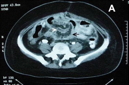
Figure 1A
Wound appearance at the time of diagnosis.
Figure 1B
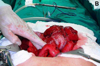
Figure 1B
Ileal loops can be appreciated surrounding and adhering to
the malignant cystic duplication. There were many visceral and parietal
peritoneal implants.
Figure 2A
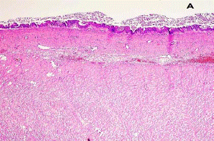
Figure 2A
Intestinal wall where it can be recognized mucosa with atrophic
vellosities, areas of infiltrated adenocarcinoma, and well developed muscular
wall (HE 10x).
Figure 2B
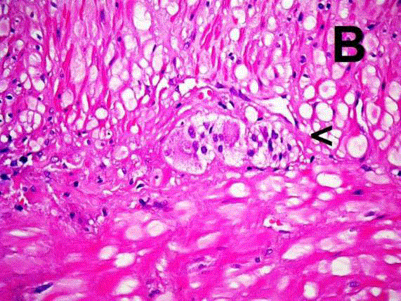
Figure 2B
Muscular wall where some myenteric plexus were recognized
(arrowhead) (HE 40x).
Discussion
The few anecdotic reports on malignant degeneration of ileal
duplications in adults highlight the important pathological potential
of these rare congenital malformations when they are not diagnosed
and treated during the infancy, and posteriorly presenting in adult
life. According to the available literature, in the adult population,
these conditions presents as an acute abdomen requiring emergency
surgery, just as occurred with our patient [8-12]. Complications
described in adults included intestinal obstruction, perforation,
infarction, hemorrhage from ectopic gastric mucosa, and pancreatitis
from heterotopic pancreas [7,8,12]. To these habitually described
complications we must add malignant degeneration, which as we
have seen, constitutes a rare but serious and mortal complication.
The cause of intestinal duplications is unknown, however a number of theories, mainly vascular, have been proposed. Fifty percent of
all gastrointestinal duplications occur in the small bowel, with the
majority of them (>70%) in the ileum where they can assume tubular
or spherical cystic forms [7]. Small bowel duplications are located
on the mesenteric border and share a common blood supply with
the adjacent bowel. In 1998 Long et al. [13] proposed the current
classification of intestinal duplications, according to this classification;
Type 1 duplications are adjacent and lateral to the mesentery of
the bowel and their blood supply is parallel and independent from
the blood supply of the adjacent bowel, Type 2 duplications are
located between the two peritoneal layers of the mesentery and their
irrigation is dependent from the adjacent bowel blood supply. The
cystic malignant duplication of our patient; was a Type 1 duplication.
Preoperative diagnosis of malignant ileal duplication with
ultrasonography and CAT scan was made only in one case before
[12], and was suspected in our case as well, on which we also used
ultrasonography and CAT scan to make the presumptive diagnosis.
Ultrasonographical characteristics of malignant ileal duplications
were described as a two-layered wall cyst adjacent to bowel; the inner
echogenic layer represents mucosa and the outer hypoechoic layer
represents muscle, a hyperechoic mass arising from the inner layer
(mucosa) and projecting into the lumen can be seen and is interpreted
as a tumor. CAT scan findings are useful to confirm the presence
of the cystic double-layered structure and the presence of tumor
in the lumen [12,14]. However, even though these characteristics
were described in this patient and the radiologist and us, suspected
the preoperative diagnosis of a malignant intestinal duplication,
we performed and open exploration through an extensive midline
laparotomy. Laparoscopic exploration is a validated and useful
approach in patients with suspected malignant intestinal obstruction;
nonetheless open exploration is also a safe and validated approach in
these patients.
According to previous reports the treatment of malignant
degeneration of ileal duplications must be resection of the
compromised segment and primary anastomosis; apparently
with this technique they have achieved excellent outcomes [8-12].
However most of these publications give just little detail regarding the
outcome of their patients. What is clear from this small experience is
that all patients presented with some complication of their tumors
and were operated on emergency grounds. Only in 2 cases, including
the present case, the diagnosis was preoperatively suspected based
on radiological findings. Three authors reported the diameter of the
duplication; all were larger than 10 cm of diameter. The segment of
ileum compromised was not reported in 2 cases, in the other 3 there
was 2 duplications located adjacent to the middle ileum and 1 at the
distal ileum. The survival of the patient was reported only in 1 article
and was of over a year, our patient barely survived 1 month, she had very disseminated disease and, consequently her outcome was
expected (Table 1). The aim of the surgical treatment of small bowel
cancer is complete resection with curative intention, which can be
frequently achieved (75% cases) and influences the prognosis [5]. This
fact is probably similar for patients with malignant degeneration of
ileal duplications. However, to the traditionally delayed diagnosis of
small bowel cancer, we must add the infrequent presentation of ileal
duplications in adults, making this rare condition a very dangerous
scenario.
Table 1

Table 1
General characteristics of adenocarcinoma arising in ileal duplication.
References
- Lioe TF, Biggart JD. Primary adenocarcinoma of the jejunum and ileum: Clinicopathological review of 25 cases. J Clin Pathol. 1990;43(7):533-6.
- Chaiyasate K, Jain AK, Cheung LY, Jacobs MJ, Mittal VK. Prognostic factors in primary adenocarcinoma of the small intestine: 13-year single institution experience. World J Surg Oncol. 2008;6:12-17.
- Dabaja BS, Suki D, Pro B, Bonnen M, Ajani J. Adenocarcinoma of the small bowel Presentation, prognostic factors, and outcome of 217 patients. Cancer. 2004;101(3):518-26.
- Beltran MA, Cruces KS. Primary tumors of jejunum and ileum as a cause of intestinal obstruction: A case control study. Int J Surg. 2007;5(3):183-91.
- Wu TJ, Yeh CN, Chao TC, Jan YY, Chen MF. Prognostic factors of primary small bowel adenocarcinoma: Univariate and multivariate analysis. World J Surg. 2006;30(3):391-8.
- Bower RJ, Sleber WK, Klesewetter WB. Alimentary tract duplications in children. Ann Surg. 1978;188(5):669-74.
- Hocking M, Young DG. Duplications of the alimentary tract. Br J Surg. 1981;68(2):92-6.
- Babu MS, Raza M. Adenocarcinoma in an ileal duplication. J Assoc Physicians India. 2008;56:119-20.
- Johnson JA, Poole GV. Ileal duplication in adults: Presentation and treatment. Arch Surg. 1994;129(6):659-61.
- Devos B, Schreurs L, Duponselle E, Hendrix T, Van Dijck H, Van Vuchelen J. [Adenocarcinoma in a cystic duplication of the ileum]. Acta Chir Belg. 1987;87(4):235-8.
- Ribaux C, Meyer P. [Adenocarcinoma in an ileal duplication]. Ann Pathol. 1995;15(6):443-5.
- Tew K, Soans BK, Millar EA. Adenocarcinoma in an ileal duplication cyst: Ultrasound and computed tomography findings. Australas Radiol. 2000;44(2):228-31.
- Long L, Zhang JZ, Wang YX. Vascular classification of small intestinal duplications: Experience with 80 cases. J Pediatr Surg. 1998;33(8):1243-5.
- Macpherson RI. Gastrointestinal tract duplications: Clinical, pathologic, etiologic, and radiologic considerations. Radiographics. 1993;13(5):1063-80.