Research Article
Efficacy of Portable Ultrasound to Detect Pneumothorax Post-Lung Resection
Farah Mohammad*, Adhnan Mohamed, Ilan Rubinfeld, Efstathios Karamanos, Karen Byers,
Keith Killu and Zane Hammoud
Department of Thoracic Surgery and General Surgery, Henry Ford Hospital, USA
*Corresponding author: Farah Mohammad, Department of Thoracic Surgery and General Surgery, Department of General Surgery, Henry Ford Hospital, 2799 West Grand Boulevard, Detroit, MI 48202, USA
Published: 12 Feb, 2018
Cite this article as: Mohammad F, Mohamed A, Rubinfeld
I, Karamanos E, Byers K, Killu K, et
al. Efficacy of Portable Ultrasound
to Detect Pneumothorax Post-Lung
Resection. Clin Surg. 2018; 3: 1901.
Abstract
Background: The role of bedside ultrasonography in detection of Pneumothorax in the acute care
setting is well established. However, its role in the diagnosis of Pneumothorax following chest tube
removal post-lung resection has yet to be elucidated. Our aim was to assess the efficacy of portable
ultrasound in the detection of Pneumothorax following chest tube removal post-lung resection.
Methods: The study was approved by the institutional review board and all patients gave informed
consent prior to enrollment. Patients underwent bedside transthoracic ultrasonography and chest
radiography after an intraoperatively placed chest tube for lung resection was removed. Chest
radiography was the standard in diagnosis of Pneumothorax post-chest tube removal.
Results: A total of 78 patients were included in the study. Chest radiography detected Pneumothorax
in 38 patients (49%). Of the 78 patients, Ultrasonography (US) detected Pneumothorax in 32 of
these patients. With CXR as our standard, our sensitivity and specificity for ultrasound was 84% and
60%, respectively. The positive and negative predictive values were 67% and 80% respectively. Only
6 patients were “false negative”, i.e. negative ultrasound but ultimately positive CXR, none of whom
required further intervention.
Conclusion: Our study demonstrates that portable sonography is efficacious in the detection of
Pneumothorax after chest tube removal post-lung resection. This suggests that sonography may
replace routine Chest X-Ray (CXR), thus leading to reduced overall costs and radiation exposure.
Further studies are required to further refine the role of portable ultrasound post lung resection.
Keywords: Pneumothorax; Ultrasound; Perioperative care
Introduction
Lung resection is the most commonly performed procedure in thoracic surgery. After lung resection, air in the pleural space, i.e. pneumothorax may occur as a result of the introduction of air from the atmosphere or from lung parenchyma, e.g. staple line. In order to evacuate the post lung resection pneumothorax, chest tubes are routinely placed intraoperatively [1]. In standard practice, a Chest X-Ray (CXR) is obtained daily to monitor for occurrence of pneumothorax as one major indication. Furthermore, once it is deemed that there is no further need for the chest tube, it is removed. A CXR is then routinely performed, again to rule out the occurrence of pneumothorax as a major indication, for it is feared that an undetected pneumothorax, even if clinically silent, may lead to major morbidity and possibly mortality in patients who may at baseline have limited pulmonary reserve. Routine CXRs add co stand radiation risks to the patient [2]. Transthoracic ultrasound plays a significant role in the diagnosis and evaluation of a wide range of thoracic pathologies, including peripheral parenchymal, pleural and chest wall diseases [3]. Multiple studies have shown ultrasonography to be more sensitive and specific in the diagnosis of pneumothorax compared to CXR in the setting of emergency medicine and critical care [4-6]. However, its role in thoracic surgery has yet to be elucidated. We hypothesized that transthoracic ultrasound may be an alternative to CXR for detection of a pneumothorax post lung resection. We sought to compare the two modalities in order to determine whether ultrasound may be equivalent to standard CXR for the detection of pneumothorax post-lung resection. Lung ultrasound is a useful modality in detecting or ruling out pneumothorax. It depends on many artifacts and signs to achieve this. When starting a lung ultrasound exam, one should try to obtain a view with the “Bat sign” by using the B mode on the ultrasound machine (Figure 1) [3]. This is a sign formed by two rib shadows and the pleural line in between, resembling a bat flying towards you. Examining the pleural line movement created by the visceral against the parietal pleura and its presence rules out the presence of pneumothorax. This is called the “Lung sliding sign” [3]. Confirming the lung sliding can be done also by using the M mode on the ultrasound machine. The M mode identifies the structures in motion over time. The movement of the pleural line will create a different artifact compared to the chest wall, and this is called the “Seashore sign”, and its presence rules out the presence of pneumothorax (Figure 2) [3]. The absence of pleural movement will create no difference in the artifact between the pleural line and the chest wall, and that can be identified as the “Stratosphere sign” [3]. The “Lung point” sign is created when a localized transition from the intrapleural air to the intra-parenchymal air happens [7]. Can be seen on the B mode as well as on the M mode as a transition point. This has 100% specificity for pneumothorax [7].
Table 1

Table 1
Patients' Demographics and Clinical Characteristics.
Table 2
Table 2
Sensitivity, specificity, NPV and PPV of U/S on diagnosis of
pneumothorax after chest tube removal*.
Table 3
Table 3
Comparison in the detection of pneumothorax CXR vs. US.
Patients and Methods
The study was approved by the institutional review board of
Henry Ford Hospital. Patients were enrolled from May 2010 to
March 2014. All patients gave informed consent prior to enrollment.
All patients under went lung resection by either wedge resections or
lobectomy. The indications for lung resection included malignancy,
undiagnosed lung nodules or bullous disease. Methods of resection
varied from conventional thoracotomy, axillary thoracotomy, Video-
Assisted Thoracic Surgery (VATS) or robotic assisted procedures.
Pleural fluid drainage with one or more chest tubes was performed in
all of these patients. Post-operatively, a CXR was performed routinely
to monitor the status of the lung and pleural space. Once there was no
need for further drainage based on clinical assessment of the patient
and the surgeon’s decision, the chest tube was removed. After removal
of the chest tube, a 2-view CXR is routinely obtained in our practice.
Transthoracic ultrasonography
All enrolled patients underwent bedside thoracic ultrasound
performed by a resident, fellow, or physician assistant. Patients were
positioned either supine or with the head of the bed at 30 degrees,
depending on the patient’s comfort. The bedside ultrasound machine
used in this study was the GE Logic (Wauwatosa, WI, USA). The
ultrasound transducer used was a linear transducer (12 MHz) and
was placed at multiple points on the patient’s chest. After applying gel
to the transducer face, the transducer was placed on the anterior chest
wall initially with the indicator of the transducer marker pointing
cephaled. This position will allow two rib shadows to be identified,
and the pleural line between them, identifying the “bat sign”. The lung
point sign was also used to identify the presence of pneumothorax, if
present. A pneumothorax was ruled out in the presence of the “Lung
sliding” sign on B mode or the “seashore” sign in M mode in the
anterior chest region [8,9]. The performing medical professional was
blinded to the results of CXR until the ultrasound was completely
interpreted. Routine CXR accompanied with US were done within
two hours of one another.
Statistical analysis
Demographic data collected included gender, age, race as well
as the post-removal occurrence of pneumothorax. Data are given
as percentages. The sensitivity, specificity, positive and negative
predictive values of transthoracic ultrasound in the diagnosis of
pneumothorax was calculated, with CXR used as the standard. Data
was analyzed in Foundation for Statistical Computing, Vienna,
Austria.
Figure 1
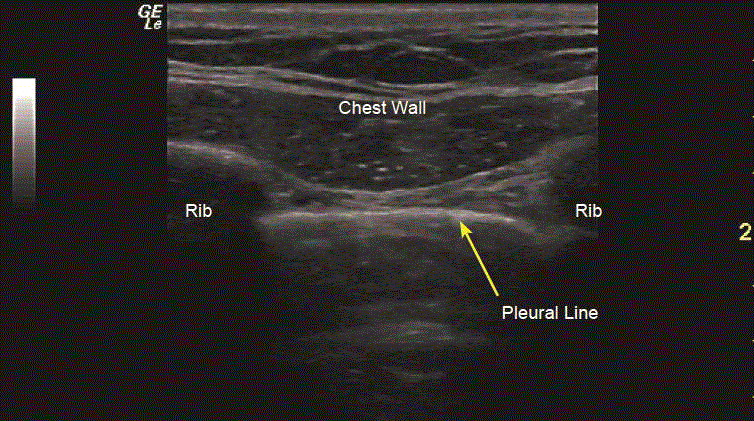
Figure 1
The "Bat Sign".
Results
A total of 78 patients (36 females and 42 males) were enrolled. Median age was 64 years (range 43-84 years) as depicted in Table 1. CXR confirmed a pneumothorax in 38 out of 78 patients (49%). Ultrasonography detected pneumothorax in 32 of these 78 patients. Only six patients were “false negative”, i.e. negative ultrasound but ultimately positive CXR, none of whom required further intervention, as shown in Table 2. With CXR as our standard, our sensitivity and specificity for ultrasound was 84% and 60% respectively. The positive and negative predictive values were 67% and 80% respectively.
Figure 2
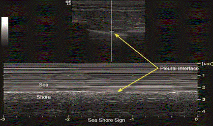
Figure 2
The "Seashore Sign”.
Comment
The routine use of postoperative CXR in cardiothoracic surgical
patients has been challenged [10-12]. CXRs are done after chest tube
removal to rule out complications such as pneumothorax, pleural
effusions or hemothorax. Eisenberg and Khabbaz report the incidence
of pneumothorax after chest tube removal in cardiac surgery patients
to be 9.3% [13]. They are generally not life threatening if small or
moderate in size, but a delay in diagnosis can lead to respiratory
compromise, especially in patients with limited lung reserve. When
a pneumothorax is detected, CXRs are used serially to monitor the
progression or resolution of a pneumothorax in the postoperative
care of a patient. The cumulative cost of all the CXRs done can be
substantial [14-17]. The associated considerable cost and exposure
to radiation has led to investigations of alternative techniques for
exclusion of post-interventional pneumothorax.
The availability of portable ultrasound has raised the interest
and popularity of transthoracic ultrasound in the past decade.
Wernecke et al. [18] in 1987 reported the first use of ultrasound
to detect pneumothorax. The use of transthoracic ultrasound has
been well studied in the diagnosis of thoracic injury in the setting
of trauma [19]. However, there is scant literature in assessing its
role in the diagnosis of iatrogenic pneumothorax in the thoracic
surgery patient [20]. Several studies report ultrasound to be more
sensitive than CXR with a specificity of up to 100% in the diagnosis
of pneumothorax after computed tomography guided biopsy [21,22].
Furthermore, CXR can be unreliable leading to a misdiagnosis rate of
30% [23]. Most basic ultrasound machines can be used for thoracic
applications. Portable machines allow the performing physician to
interpret the results immediately. In the chest, air tends to rise to
the least dependent area. In a supine patient, this corresponds to the
apical region, i.e. midclavicular region, between the second and fourth
intercostals spaces [3]. The majority of significant pneumothoraces
have been shown to be identifiable in this position in trauma patients
[24,25]. With the probe in the sagittal position, the “Bat Sign” can
be identified. The two layers of the pleura can be seen sliding across
one another between the setworibs [23]| forming the lung sliding sign.
Identification of sliding pleural lines effectively rules out the presence
of pneumothorax in the majority of patients [9,26]. The negative
predicted value of this technique has been reported to be between
99.2% to 100% [4,5,27]. The absence of sliding sign, however, may
not be reliable in certain conditions, such as acute respiratory distress
syndrome, pulmonary fibrosis, at elect as is and pleural adhesions
[5,28]. Another sign to rule out pneumothorax is the Seashore sign
on the M Mode. Absence of the seashore sign or the presence of the
barcode sign in M mode [8,9] can be due to pneumothorax. The Lung
Point sign has a 100% sensitivity and specificity in the detection of
pneumothorax when present. Compared with CXR, transthoracic
ultrasound offers some major advantages. In the vast majority of cases,
it can be performed in 2 min to 15 min [6,19]. It also allows instant
diagnosis, decreases costs, and eliminates the radiation risks and
unnecessary transport of the patient [22]. Furthermore, transthoracic
ultrasound may obviate the need for serial CXRs to follow possible
progression of pneumothorax once detected, further reducing
cost and radiation exposure. All operators in this study had only
undergone basic ultrasound skill training as part of their curriculum.
This highlights the benefit of ultrasound as any healthcare provider
with basic ultrasound skills can perform it [29,30]. Our study has
several limitations. Transthoracic ultrasound, while expeditious and
cost effective, cannot quantify pneumothorax. In the majority of cases
however, quantification is not a clinically relevant issue. Ultrasound
is also operator dependent, and detection of pneumothorax can be
variable from user to user. Our study also had a small number of
patients. However, our early experience led us to posit a great deal of
confidence in the technique. Our patients also had underlying lung
disorders, as well as recent surgery, which may makes liding sign not
reliable as an indicator in ruling out pneumothorax. Again, this did
not appear to greatly hinder our ability to obtain reliable results.
Conclusion
Our study suggests that ultrasound may be an effective imaging modality to rule out pneumothorax post lung resection. Advantages include the potential to reduce cost and radiation exposure compared to the standard CXR. Further studies are required to refine the role of portable ultrasound post-lung resection.
References
- Miller KS, Sahn SA. Chest tubes. Indications, technique, management and complications. Chest. 1987;91(2):258-64.
- Fong Y, Whalen GF, Hariri RJ, Barie PS. Utility of routine chest radiographs in the surgical intensive care unit. A prospective study. Arch Surg. 1995;130(7):764-8.
- Husain LF, Hagopian L, Wayman D, Baker WE, Carmody KA. Sonographic diagnosis of pneumothorax. J Emerg Trauma Shock. 2012;5(1):76-81.
- Lichtenstein DA, Menu Y. A bedside ultrasound sign ruling out pneumothorax in the critically ill. Lung sliding. Chest. 1995;108(5):1345-8.
- Lichtenstein D, Mezière G, Biderman P, Gepner A. The comet-tail artifact: an ultrasound sign ruling out pneumothorax. Intensive Care Med. 1999;25(4):383-8.
- Dulchavsky SA, Schwarz KL, Kirkpatrick AW, Billica RD, Williams DR, Diebel LN, et al. Prospective evaluation of thoracic ultrasound in the detection of pneumothorax. J Trauma. 2001;50(2):201-5.
- Lichtenstein D, Mezière G, Biderman P, Gepner A. The "lung point": an ultrasound sign specific to pneumothorax. Intensive Care Med. 2000;26(10):1434-40.
- Barillari A, Kiuru S. Detection of spontaneous pneumothorax with chest ultrasound in the emergency department. Intern Emerg Med. 2010;5(3):253-5.
- Johnson A. Emergency department diagnosis of pneumothorax using goal-directed ultrasound. Acad Emerg Med. 2009;16(12):1379-80.
- Hornick PI, Harris P, Cousins C, Taylor KM, Keogh BE. Assessment of the value of the immediate postoperative chest radiograph after cardiac operation. Ann Thorac Surg. 1995;59(5):1150-3.
- Rao PS, Abid Q, Khan KJ, Meikle RJ, Natarajan KM, Morritt GN, et al. Evaluation of routine postoperative chest X-rays in the management of the cardiac surgical patient. Eur J Cardiothorac Surg. 1997;12(5):724-9.
- Graham RJ, Meziane MA, Rice TW, Agasthian T, Christie N, Gaebelein K, et al. Postoperative portable chest radiographs: optimum use in thoracic surgery. J Thorac Cardiovasc Surg. 1998;115(1):45-50.
- Eisenberg RL, Khabbaz KR. Are chest radiographs routinely indicated after chest tube removal following cardiac surgery? AJR Am J Roentgenol. 2011;197(1):122-4.
- Gray P, Sullivan G, Ostryzniuk P, McEwen TA, Rigby M, Roberts DE. Value of postprocedural chest radiographs in the adult intensive care unit. Crit Care Med. 1992;20(11):1513-8.
- Swensen SJ, Peters SG, LeRoy AJ, Gay PC, Sykes MW, Trastek VF. Radiology in the intensive-care unit. Mayo Clin Proc. 1991;66(4):396-410.
- Bekemeyer WB, Crapo RO, Calhoon S, Cannon CY, Clayton PD. Efficacy of chest radiography in a respiratory intensive care unit. A prospective study. Chest. 1985;88(5):691-6.
- Strain DS, Kinasewitz GT, Vereen LE, George RB. Value of routine daily chest x-rays in the medical intensive care unit. Crit Care Med. 1985;13(7):534-6.
- Wernecke K, Galanski M, Peters PE, Hansen J. Pneumothorax: evaluation by ultrasound--preliminary results. J Thorac Imaging. 1987;2(2):76-8.
- Kirkpatrick AW, Sirois M, Laupland KB, Liu D, Rowan K, Ball CG, et al. Hand-held thoracic sonography for detecting post-traumatic pneumothoraces: the Extended Focused Assessment with Sonography for Trauma (EFAST). J Trauma. 2004;57(2):288-95.
- Saucier S, Motyka C, Killu K. Ultrasonography versus chest radiography after chest tube removal for the detection of pneumothorax. AACN Adv Crit Care. 2010;21(1):34-8.
- Goodman TR, Traill ZC, Phillips AJ, Berger J, Gleeson FV. Ultrasound detection of pneumothorax. Clin Radiol. 1999;54(11):736-9.
- Sartori S, Tombesi P, Trevisani L, Nielsen I, Tassinari D, Abbasciano V. Accuracy of transthoracic sonography in detection of pneumothorax after sonographically guided lung biopsy: prospective comparison with chest radiography. AJR Am J Roentgenol. 2007;188(1):37-41.
- Jalli R, Sefidbakht S, Jafari SH. Value of ultrasound in diagnosis of pneumothorax: a prospective study. Emerg Radiol. 2013;20(2):131-4.
- Tocino IM, Miller MH, Fairfax WR. Distribution of pneumothorax in the supine and semirecumbent critically ill adult. AJR Am J Roentgenol. 1985;144(5):901-5.
- Ball CG, Kirkpatrick AW, Laupland KB, Fox DL, Litvinchuk S, Dyer DM, et al. Factors related to the failure of radiographic recognition of occult posttraumatic pneumothoraces. Am J Surg. 2005;189(5):541-6.
- Targhetta R, Bourgeois JM, Chavagneux R, Balmes P. Diagnosis of pneumothorax by ultrasound immediately after ultrasonically guided aspiration biopsy. Chest. 1992;101(3):855-6.
- Blaivas M, Lyon M, Duggal S. A prospective comparison of supine chest radiography and bedside ultrasound for the diagnosis of traumatic pneumothorax. Acad Emerg Med. 2005;12(9):844-9.
- Ball CG, Kirkpatrick AW, Feliciano DV. The occult pneumothorax: what have we learned? Can J Surg. 2009;52(5):E173-9.
- Noble VE, Lamhaut L, Capp R, Bosson N, Liteplo A, Marx JS, et al. Evaluation of a thoracic ultrasound training module for the detection of pneumothorax and pulmonary edema by prehospital physician care providers. BMC Med Educ. 2009;9:3.
- Monti JD, Younggren B, Blankenship R. Ultrasound detection of pneumothorax with minimally trained sonographers: a preliminary study. J Spec Oper Med. 2009;9(1):43-6.