Case Report
Orthognathic Surgery: A Case Report
Eda Naifoğlu1, Mehmet Emre Yurttutan1, Dt. Can Arslan2, Ayşe Tuba Altuğ2 and Ayşegül Mine
Tüzüner Öncül1*
1Department of Dentistry Oral and Maxillofacial Surgery, Ankara University, Turkey
2Department of Dentistry Orthodontics, Ankara University, Turkey
*Corresponding author: Aysegul Mine Tuzuner Oncul, Department of Dentistry Oral and Maxillofacial Surgery, Ankara University, Turkey
Published: 24 Jan, 2018
Cite this article as: Naifoğlu E, Yurttutan ME, Arslan Dt.C,
Altuğ AT, Öncül AMT. Orthognathic
Surgery: A Case Report: A Case
Report. Clin Surg. 2018; 3: 1883.
Abstract
Orthognathic surgery is a procedure that combines orthodontia and maxillofacial surgery to align
skeletal problems of the jaws. Though there are different methods of osteotomy, now a day’s Lefort I
and bilateral Sagittal split ramus osteotomy are the common ones.
A 16 years old male patient referred to our clinic complaint of anterior open bite. Clinical
examination revealed an Angle Class III malocclusion and 7 mm open bite. According to Steiner’s
lateral cephalometric norms the patient had a high angle profile. Orthognathic surgery was preferred
as the treatment choice. Our treatment started with fixed orthodontic appliance. 13 months later he
underwent bimaxillar orthognathic surgery. Jaws remained in intermaxillary fixation for 2 weeks
with intermaxillary elastics. 7 months after surgery fixed orthodontic appliance was removed.
Introduction
Orthognathic comes from Greek terms. Ortho means “straight, in order” [1] and gnatho means jaw [2]. Orthognathic surgery is a procedure that combines orthodontia and maxillofacial surgery to align the maxilla and the mandible to correct dental and skeletal position of maxilla and mandible, improve Temporomandibular joint function and oropharyngeal airway [3,4]. Speaking and masticatory difficulties, dental crowding with skeletal class II or III malocclusion, open bite, congenital defects, retrognathia, prognathia, difficulty in closing lips effortlessly and sleep apnea are frequent indications for orthognathic surgery [4]. Le Fort I and bilateral sagittal split ramus osteotomy are the most common methods to correct this Dentofacial deformities.
Case Presentation
A 16 years old male patient who was referred to Ankara University Faculty of Dentistry Department of Orthodontics had a main complaint of anterior open bite. Due to this problem the patient had problems with chewing and articulation. Clinical examination revealed an Angle Class III malocclusion with 7 mm open bite. Pre-treatment radiological evaluation according to Steiner’s lateral cephalometric norms showed SNA angle 75°, SNB 75°, ANB 0°, GoGn/SN 41°, which indicated that the patient had a high angle profile. Skeletal maturation level of the patient was Ru according to the hand-wrist radiograph; therefore orthognathic surgery was preferred as the treatment choice. Prior to surgery the patient undergone orthodontic treatment including teeth levelling, alignment and decompensation phases done with fixed orthodontic appliances. (Edgewise prescription – slot 0,018’’) He was ready for surgery 13 months later.
Surgical Procedure
The operation performed under general anesthesia. Nasal endotracheal intubation is applied
to check occlusion during surgery. Local anesthesia also injected (%4 articaine and 1/100.000
epinephrine.) A vertical incision on midline and a circum vestibular incision were made with
no. 15 lancet. The circum vestibular incision must be between mesial sides of upper first molars.
Mucoperiosteal flap was removed to expose anterior and lateral walls of maxilla, infraorbital nerve
and nasal cavity. Nasal floor was dissected. Osteotomy was carried out with reciprocating saw from
pterygomaxillary junction tonasal incisure. The osteotomy must be 5 mm away from apices of the
teeth. Straight, lateral nasal, pterygoid and septum osteotoms was used in order. Down fracture
was carried out with tessier spreaders at left and right anterior buttresses. To avoid nasal septum
deviation, inferior bone of the nasal cavity was skived and 2 mm nasal septum cartilage was cut out.
The first acrylic plaque, prepared by orthodontists before surgery, guided to intermaxillary fixation
for maxilla’s new position. The maxilla fixed with 1.2 mm titanium miniplates and 2 mm diameter
monocortical screws. Musculus levator labii superior alaeque nasi sutured with 2.0 prolene. The alar base cinch suture was used to avoid the unfavorable increase in nasal width. The mucosal wound was sutured with 3.0 vicryl [5,6].
Local anesthesia was injected for alveolar inferior nerve block in
both sides. The incision was made with no. 15 lancet, starting from
the half way of the ramus just lingual of the external oblique ridge to mesial of 2nd molar inferiorly. Mucoperiosteal flap was removed to
expose mandibular bone. After stripping the insertion of temporalis
muscle soft tissues on the anterior ramus retracted with fork retractor.
Obwegeser type channel retractor placed to buccal side. On medial
mandible, flap carefully removed to identify foramen alveolar is
inferior. After the nerve is identified Heinnehen retractor is placed to
retract and observe the nerve during osteotomy. A vertical osteotomy
was carried out with lindemandrillon the lingual side of mandible, at
the level of lingual close to the foramen and parallel with the occlusal
plane. The osteotomy continued on superior side of mandible, only
in cortical layer with a straight fissure burs. This osteotomy follows
the external oblique ridge, smoothly turns to vertical and ends at
mandibular inferior border [7,8]. Various osteotoms were used only
on corners of osteotomy line. Then two straight osteotoms used to
stretch the proximal and distal fragments of mandible. Spreader
is placed and gently opened until the mandible splitted up while
spongious bone is dissected and inferior alveolar nerve removed to
distal segment with a thin periost elevator. Intermaxillary fixation
carried out under the guidance of second acrylic plaque. The mandible
fixed in new position with 2 mm diameter bicortical screws on both
sides. The intermaxillar fixation removed and the mucosal wound
was sutured with 3.0 vicryl. The day after surgery, intermaxillary
fixation was applied for two weeks to protect the jaws new position
against the muscle forces. 7 months later fixed orthodontic appliance
was removed.
Figure 1
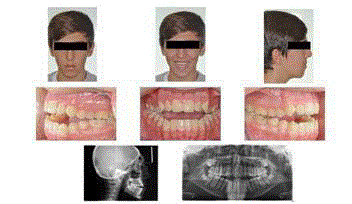
Figure 1
Pre-treatment orthodontic records of the patient.
Figure 2
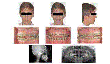
Figure 2
Pre-surgical orthodontic records of the patient.
Figure 3
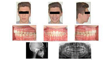
Figure 3
Post-surgical orthodontic records of the patient.
References
- Ortho. Stedman’s Online Medical Dictionary.
- Gnatho. Stedman’s Online Medical Dictionary.
- Sousa CS, Turrini RNT. Complications in orthognathic surgery: A comprehensive review. J Oral Maxillofac Surgery, Med Pathol. 2012;24(2):67-74.
- Robinson RC, Holm RL. Orthognathic Surgery for Patients with Maxillofacial Deformities. AORN J. 2010;92(1):28-52.
- Swennen G, Colle F, De May A, Malevez C. Maxillary distraction in cleft lip palate patients: a review of six cases. J Craniofac Surg. 1999;10(2):117-122.
- Dailey RA, Dierks E, Wilkins J, Wobig JL. LeFort I orbitotomy: a new approach to the inferonasal orbital apex. Ophthal Plast Reconstr Surg. 1998;14(1):27-31.
- Smith GI, Brennan PA, Oh SS, Markus AF. Modification of the Hunsuck sagittal split osteotomy using a nerve hook. Technical note. J Cranio-Maxillofacial Surg. 2002;30(5):292-94.
- Böckmann R, Meyns J, Dik E, Kessler P. The Modifications of the Sagittal Ramus Split Osteotomy. Plast Reconstr Surg Glob Open. 2014;2(12):271.