Case Report
Acute Sinusitis and Implant Failure Following Novel Minimally Invasive Hydraulic Sinus Lift: A Case Report
Ardekian L1*, Levit L2 and Michael L3
1Department of Oral and Maxillofacial, Atidim Medical Center, Israel
2Department of Lipid, Hadassah Medical Center, Jerusalem, Israel
3Department of Prosthodontics, Hebrew University - Hadassah School of Dental Medicine, Jerusalem, Israel
*Corresponding author: Leon Ardekian, Department of Oral and Maxillofacial, Atidim Medical Center, 11 Moshe Levy st. Rishon Le-Tzion,7565828, Israel
Published: 24 Jan, 2018
Cite this article as: Ardekian L, Levit L, Michael L. Acute
Sinusitis and Implant Failure Following
Novel Minimally Invasive Hydraulic
Sinus Lift: A Case Report. Clin Surg.
2018; 3: 1882.
Abstract
The last decade has presented us with a variety of innovative minimally invasive methods for
sinus lifting. One described method is the iRaise, a novel implant device based on hydraulic sinus
membrane elevation. Till now, all reported cases involving this method have been successful,
and no major complications were observed. The current article presents a case of a 45 year-old
healthy woman, who developed acute sinusitis following an iRaise sinus lift. Conservative antibiotic
treatment has failed, and the patient was treated under general anesthesia. The sinus was accessed
by the conventional Caldewell-Luk approach, and designated implant and graft material were
removed. To our knowledge, this is the first case report describing acute sinusitis following the
iRaise method. This case report suggests strong association between the iRaise method and acute
sinusitis, and reviews alternative techniques to the iRaise device.
Keywords: Acute Sinusitis; Caldwell-Luk Procedure; Implant Failure; Hydraulic Membrane
Elevation; Minimally Invasive Sinus Lift
Introduction
Dental implant therapy in the posterior maxilla is a challenging procedure for both general
dentists and maxillofacial surgeons. Posterior maxilla is characterized by "fine" trabecular bone;
with either thin porous cortical bone or no cortical crestal bone with very light density. Additionally,
loss of teeth in the posterior maxilla might lead to sinus pneumatization and reduction of available
bone volume required for conventional implantation. These two factors of reduced bone quality and
quantity might result in insufficient bone support of dental implants [1,2]. In order to overcome
these anatomic limitations, sinus augmentation is usually required for vertical bone height
reconstruction. The sinus lift enables satisfactory osseointegration and long term survival of longer
and wider implants [3].
The sinus augmentation technique was first presented by Tatum [4]. Autogenous cancellous
bone from the lateral iliac crest was used as graft material, adopting a modified Caldwell-Luc
procedure to approach the maxillary sinus [5]. Tatum's traditional method of sinus augmentation
is also called: "open sinus lift" procedure and has been widely used and investigated, and has been
established as an accepted standard for treatment of edentulous maxilla.
Open sinus lift enables direct access and visualization of the whole sinus. However, this
method suffers from many shortcomings. Anatomic considerations may limit the applicability.
Patients usually suffer from periprocedural discomfort: swelling, discoloration, disability and pain,
hematomas and nosebleed. Also, the procedure requires surgical expertise, has a demanding learning
curve and is time and resource consuming. Various complications of open sinus lift were reported
in the literature as follows: obstruction of antronasal foramen, bleeding, infection, infraorbital nerve
laceration, acute maxillary sinusitis, wound dehiscence, and Schneider an membrane perforations,
with consecutive scattering of the grafting material in the sinus cavity [6,7]. Though the open sinus
lift method has proved its worth and effectiveness over the years, it is significantly invasive and
traumatic to the patient, and has stirred up the need for other less invasive methods.
In 1994 Summers has described the osteotome sinus floor approach and ridge expansion
through the alveolar ridge [8]. The sinus lift approach was modified to a less invasive technique,
aiming to achieve higher success rate and fewer complications compared to those introduced by
open sinus lift technique. It is also called: "closed sinus lift". The rational of this technique was the controlled compaction of soft maxillary bone by mallet, conservation
of the residual osseous tissue in the alveolar bone and improvement
of bone density around the implants. When bone compaction is not
sufficient by itself, the osteotome technique enables the clinician to
introduce bone graft material into the sinus through the osteotomy
site, without the lateral window approach. However, this method was
still traumatic to the patient and was limited by residual ridge height
of 5 mm or more [3]. Moreover; it was limited in its ability to raise
the sinus floor. Therefore, the closed sinus lift is a limited approach
that does not necessarily meet with the achievements of the open
approach.
Consequently, during the last decade sinus grafting by minimally
invasive approach has evolved and has been used by a growing
number of clinicians, as shown by an increasing body of scientific
literature. Several modifications of the surgical techniques of closed
sinus lift have been carried out in order to improve the predictability
of clinical outcomes and, at the same time, reduce the amount of
trauma to the patient. Various types of allografts, xenografts and
alloplastic materials have been used as bone substitutes to simplify
the grafting phase, enabling immediate implant placement and
minimizing the patient's discomfort [9].
Hydraulic sinus condensation through osteotomy site was first
described by Chen and Cha at 2005 [10]. Initially, pinhole access to
the sinus is performed by a small round bars. Constant hydraulic
pressure from the hand piece during osteotomy drilling inflates the
Shneiderian membrane through the pinhole at the sinus floor. Once
the membrane is loosened, a bone graft mixture is packed and pushed
using a small sinus condenser. The osteotomy site is enlarged by an
implant drill and the bone graft is condensed again by secondary lift.
At the end of the sinus augmentation, the osteotomy site is ready for
immediate implantation. This minimally invasive sinus lift procedure
had favorable results in a single-center study, but has never been
widely accepted [10,11].
At 2005 Soltan and Smiler suggested anthral membrane sinus
elevation through the lateral window by open sinus lift access [12].
Kfir et al. [7] described a minimally invasive version for anthral
membrane balloon elevation executed via the osteotomy site without
the open access (MIAMBE). Bone grafting was also performed via the
osteotomy site and implant fixation was performed at the same sitting.
The procedure was executed in 24 patients. No major complications
were reported. One patient had rupture of balloon and membrane,
and implant rejection two weeks after procedure. Another patient
had a minor nosebleed. 100% of implants exposed at 6-8 months were
rehabilitated. The authors concluded that this method requires much
abbreviated learning curve, carries excellent procedural success, has
low complication rates and yields very satisfactory long-term results
[7].
A later multicenter study [13] included 112 patients. Of 123
MIAMBE procedures, 119 were successful. Three procedures were
aborted due to membrane tear and successfully retreated at second
attempt. One patient had an infection and oroanthral fistula at 4
weeks after the sinus lift, which required implant removal. The authors
did not specify which anatomic location the infection was present
at, and whether or not the patient had developed signs of sinusitis.
Another study [14] reviewed 34 cases of antral balloon elevation
in 34 patients. Two patients developed acute maxillary sinusitis,
which was defined by the authors as only a minor complication. One
patient had a sinus membrane perforation, which was successfully
managed intraoperatively. The authors did not note if the patient with
membrane tear was the one who has developed the sinusitis. All bone
grafts were stable and integrated well with the implants.
At 2014 Nahlieli introduced the dynamic implant valve approach
(DIVA) [15]. The implant was developed with an internal port and
a sealing screw, and might serve for end scoping direct observation and as a drug delivery system via its corono-apical channel. After
introducing the implant into the posterior maxilla at the osteotomy
site, the internal screw is removed. Bleeding from inside the implant
indicates fracture of the sinus floor, which is followed by saline
irrigation via the internal port. The integrity of the Schneiderian
membrane may be evaluated by an endoscope or by visual observation
of the saline level movement at the coronal opening of the implant,
according to the respiratory movement of the membrane. Careful
saline irrigation and slow ratcheting of the implant separates
the membrane by hydro dissection. A prospective observational
uncontrolled study [16] assessed DIVA implant during follow
up period of up to 60 months. In 94.5% of cases the implantation
was totally successful from objective CBCT, clinical and subjective
patients' viewpoints. No sinus complications or intraoperative
membrane perforation were reported. The authors concluded that
this minimally invasive sinus lift method simplifies the surgery and
secures the optimal dental implant placement.
At 2014 Better et al. [17] has presented a novel procedure and
device, designed as closed sinus lift, using dedicated dental implant
– the iRaise. The implant contains an internal channel, which allows
simultaneous hydraulic sinus membrane elevation and placement
of flowable bone graft. The procedure is considered as minimally
invasive and enables sinus bone augmentation and implant placement
at the same time.
Sinusitis is considered to be one of the major complications and
drawbacks of open sinus lift [18]. The reported incidence of sinus
complications is as lower than 1% to as high as 3.9%. Increased
incidence of complications has been especially observed when the
history before surgery included factors that favored sinusitis [19].
As opposed to these reports; minimally invasive sinus lift appears
to be a safer method with rare complications. The consequences of
postoperative acute sinusitis are severe and might commonly cause
implant and graft failures.
Figure 1
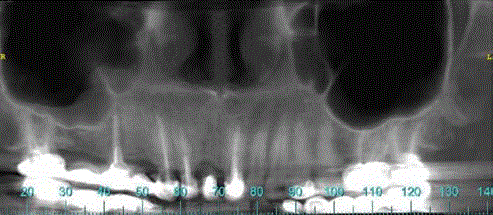
Figure 1
Preoperative cone beam computed tomography (CBCT) scan.
Figure 2
Figure 2
Five weeks postoperative medical computed tomography (MDCT)
scan.
Figure 2(A): Coronal section through the iRaise implant device.
Figure 2(B)
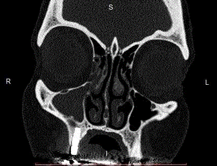
Figure 2(B)
Coronal section through conventional implant anterior to the
iRaise, showing good bone support of the conventional implant.
Figure 2(C)
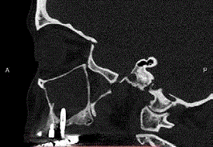
Figure 2(C)
Sagittal section.
Figure 2(D)
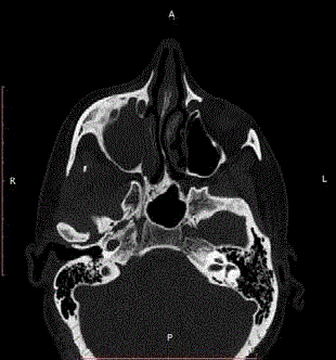
Figure 2(D)
Axial section, bone window.
Figure 2(E)
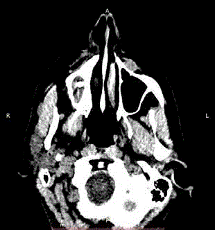
Figure 2(E)
Axial section, soft tissue window. The iRaise implant is
protruding into the infected sinus, no bone formation is demonstrated.
Case Presentation
A 45 year old healthy non-smoker female was referred to a
private clinic in 2016 for rehabilitation of posterior atrophic maxilla
using dental implants. The patient presented with a failing five units
porcelain fused to metal bridge in the upper right quadrant. The
bridge utilized three abutment teeth: canine, second premolar and
the second molar. The first molar and the first premolar were the
pontics. CT scan was obtained, and revealed a pneumotized right
maxilla and a subanthral residual bone of 5 mm (Figure 1). Clinical
and radiographic examination revealed no signs of sinusitis. No
past history of sinus pathologies was present. Therefore, sinus lift
was necessary to support dental implants. It was agreed to perform
a minimally invasive hydraulic sinus floor elevation and bone
augmentation, according to the iRaise technique, as described by
Better et al. [17] (Maxillent, Israel). Risks, benefits, and alternatives of
the proposed procedure were described to the patient, and the patient
signed an informed consent.
Amoxicilline clavulanate 1,750 mg was administered
prophylactically 1 hr preoperatively (Augmentin, SmithKline
Beecham, Brentford, Middlesex, U.K). The patient performed a
mouth wash for 1 minute with chlorhexidine gluconate 0.2% solution
prior to surgery. A full thickness mucoperiosteal flap was reflected.
The implantation site was calculated according to the preoperative
CT scan of the area. The osteotomy site was marked with a small
round bur and then was widened with the full drilling sequence,
according to the company's drilling protocol (Maxillent, Israel). The
hard cortical bone of the sinus cortex was identified and abraded,
using a diamond-tipped cortex drill in order to avoid perforation
of the membrane. A 4.2 mm-diameter iRaise implant was partially
inserted at the site of maxillary right first molar. The tube connector
was then assembled. A saline syringe was connected to the designated
tubing port of the implant and sterile saline solution was injected into
the sinus and aspirated. Saline slightly mixed with blood appeared
in the syringe, indicating proper positioning of the sinus lift implant
device and indicating no perforation of the Schniederian membrane.
Two ml of synthetic bone graft were slowly injected into the sinus
(MBCP gel, Biomatlante, France). The tube connector was removed
and the implant was fully inserted. Implant length was 14.5 mm. An
additional three conventional implants were placed at sites of the
canine, first and second premolar. The surgical flaps were sutured and
primary wound closure was obtained. There were no intraoperative
incidents and the procedure was accomplished uneventful. Postoperative
antibiotic coverage was prescribed with amoxicilline
clavulanate 875 mg twice daily for 7 days.
One month post-operatively the patient visited the clinic
complaining about pain, headache and discomfort at the right inner
side of the mouth. Redness and swelling of the mucosa adjacent
to the implantation site and purulent discharge were present. A
copious irrigation was performed and amoxicilline clavulanate 875 mg was prescribed twice daily for 7 days. The patient showed no
improvement in response to this treatment. CT scan showed total
unilateral opacity of the right maxillary and ethmoidal sinuses, and
diagnosis of acute maxillary sinusitis was confirmed (Figure 2). The
infection was involving the whole maxillary and ethmoid sinuses, and
was not limited to the bone graft. Under general anesthesia the right
sinus was approached by Caldwell-Luc procedure. The iRaise implant
device and the remnants of the graft material were removed from the
sinus together with the infected mucosa. The other three conventional
implants had good bone support and were stable, therefore they
were left intact. Three months postoperatively the patient was free
of symptoms. On clinical examination, normal sinus function and
drainage were restored.
Discussion
Surgical complications after sinus lifts are rarely reported and
their influences have been investigated to a lesser extent [20]. This
article reports a case of acute sinusitis after minimally invasive
sinus lift approach. The reported incidence of acute sinusitis after
open sinus lift appears to be low. Exceptionally few case reports of
acute maxillary sinusitis after open sinus lift were published [21-
23]. In the case of closed sinus lift, only one report [24] presented
an acute maxillary sinusitis following internal sinus lift using the
osteotome technique. However, the patient in that report had a
pre-existing chronic maxillary sinusitis. The authors concluded that
maxillary sinusitis is an inevitable complication of maxillary sinus
augmentation in patients with a history of maxillary sinus disease.
Moreover, to our knowledge, case reports discussing about acute
maxillary sinusitis after sinus lift, by any minimally invasive method,
have not been reported in the literature to date.
When sinusitis does occur, the clinical management of this major
complication is very traumatic for the patient. Except for per oral or
intravenous antibiotic therapy, most often surgical intervention is
inevitable in order to remove the offending source of the infection.
The surgical management can range from debridement and drainage
to removal of grafting material, the infected sinus mucosa and, in some
cases, the removal of implants placed adjacent to the graft. Moreover,
the infected sinus is accessed occasionally by the extensive Caldwell-
Luc procedure under general anesthesia. All of these procedures may
lead to significant pain and discomfort, prolonged overall treatment
and require recurred appointments [3].
To date, there were three studies evaluating the use of the iRaise
sinus lift system, that was published in the indexed literature [17,25-
26]. The first study (2014) was a prospective preliminary study,
evaluating 23 sinus lift procedures performed in 18 patients' cohort
followed for up to 13.1 months [17]. The second study (2016) was
a retrospective analysis of 64 procedures performed in 62 patients
followed for up to 45 months [25]. The third study (2017) was a
prospective case series study; evaluating clinical and radiological
outcomes in a sample of 18 consecutive patients followed for up to 14
month [26]. All studies reported that all procedures were completed
successfully. No intraoperative or postoperative adverse events were
observed, such as membrane tears or sinusitis. Overall implant
survival rate was 100% in both studies. In the retrospective study,
which followed the patients for a longer period, a 100% of prosthetic
cumulative survival rate was also reported [25].
Patients' perception of recovery after minimally invasive iRaise
sinus lift was also evaluated in a prospective pilot study [27].
Patients were assessed by "health-related quality of life" (HRQOL)
questionnaire. The questionnaire was designed to assess the patient's
perception of recovery in four main areas: oral function, general
activity, other symptoms and pain. The questionnaire was completed
each day for consecutive 7 postoperative days by telephone visit.
Patients reported very little discomfort to 'not at all'. Most of them
returned to work on postoperative day 1. The study concluded that
patients undergoing sinus augmentation using the iRaise device can
expect to experience minimum discomfort and immediate return to
everyday activity.
The sinus lift approach with the iRaise implant device harbors
many possible advantages over other minimally invasive techniques.
Closed Trans crestal hydraulic Schneiderian membrane elevation
and simultaneous bone graft augmentation can be accomplished
using a dedicated dental implant, which allows for reducing operative
treatment time, risk of complications, and overall patient discomfort
[25]. This approach mainly differs from previously described
minimally invasive techniques as the Schneiderian membrane
elevation and the bone graft are both performed through the implant
fixture. In addition, the bone grafting is performed with a flowable
material, which can be simply manipulated. The iRaise is a similar
technique to the DIVA in that both of them utilize the implant as
a delivery channel of the graft material. However, DIVA enables
the clinician to visualize the terminal location of the implant apex
by endoscope, securing the Shneiderian membrane from tearing.
Moreover, in order to achieve access to the internal channel of the
iRaise device, a tubing port should be connected to the lateral entry
point of the implant. This need for exact spatial location might
introduce complexity to this technically sensitive procedure.
The sinus membrane elevation requires delicate manipulation
in order to avoid membrane perforation and tears. Intraoperatively,
tearing or perforation of the Schneiderian membrane is the most
common complication of open sinus lift. The reported incidence of
sinus membrane perforation ranges from 7% to 56% of cases [28,29].
One prospective observational uncontrolled study reported the
incidence of membrane perforation to be as high as 44%. [20] Several
authors reported no correlation between Shniederian membrane
perforation and development of maxillary sinusitis [6-30]. On the
other hand, Nolan et al. [31] performed retrospective evaluation on
359 augmented sinuses and found that graft failure was statistically
higher in sinuses in which the membrane was perforated during the
surgery. Finally, Sakkas et al. (2016) concluded that tearing of the
Schneiderian membrane does not affect the success rate of implants
[32]. Despite the perforation, in most of those cases, the sinuses
were repaired and the sinus augmentation completed without other
complications [20].
There are various factors that can cause infection after maxillary
sinus elevation. For example, there could be a loss of stability of the
grafted bone, a pre-existing sinus condition, a membrane perforation,
diabetes mellitus, poor early-infection management, smoking, and
other factors [23]. The main concerns related to the iRaise approach are
the absence of direct visualization of the sinus cavity and Schneiderian
membrane, the limited amount of bone augmentation achieved,
and the high risk of the Schneiderian membrane perforation due to
irregular shape of the sinus floor and attachments caused by scars
[33]. Under absence of direct visualization, the flowable graft material
might be erroneously injected into the sinus. Floating bone graft
materials are known to cause infection after maxillary sinus elevation [34]. Even though membrane perforations are controversially related
to sinusitis, possible membrane perforation might be the cause for
acute sinusitis in our patient. Another possible cause for sinus graft
failure and sinusitis is a pre-existing sinus infection from dental origin.
The right second premolar presented with periapical pathology and
concomitant sinus thickening as shown on preoperative panoramic
cone beam CT scan. This reason is less likely because the patient
was thoroughly examined preoperatively and sinus pathologies were
excluded. The incidence of sinusitis may be reduced by adhering to
clinical recommendations to reduce the incidence of postoperative
complications [35].
Finally, our article presents a major complication after minimally
invasive iraises sinus lift, which is becoming extensively used by general
dentists. Therefore, it has relevance to global dental community.
General practitioners and maxillofacial surgeons as well, should be
familiar with possible unexpected occurrence of acute sinusitis after
minimally invasive sinus lift. More evidence and reports are required
in order to elucidate a more definitive association between the iRaise
sinus lift and acute sinusitis. The current case demonstrates us that
a clinician should be competent in addressing and managing major
sinus complications that might arise after minimally invasive sinus
lifts.
Conclusion
Minimally invasive sinus lift by the novel iRaise implant device has been reported as a safe and reliable technique. Acute sinusitis is a possible complication, which has to be managed immediately in order to reduce complications like parasinusitis, osteomyelitis of the maxillary bone, or the spreading of the infection to adjacent anatomical spaces. To minimize the risk, more evidence is required regarding the safety of this procedure. Moreover, clinical studies are needed to identify the potential factors involved in the occurrence of sinusitis after minimally invasive sinus lift.
References
- Misch CE. Bone character: second vital implant criteria. Dent today. 1988;7:39-40.
- Ribeiro-Rotta RF, Lindh C, Pereira AC, Rohlin M. Ambiguity in bone tissue characteristics as presented in studies on dental implant planning and placement: a systematic review. Clin Oral Implants Res. 2011;22(8):789-801.
- Stern A, Green J. Sinus lifts procedures: an overview of current techniques. Dent Clin North Am. 2012;56(1):219-33.
- Tatum H. Maxillary and sinus implant reconstructions. Dent Clin North Am. 1986;30(2):207-29.
- Boyne PJ, James RA. Grafting of the maxillary sinus floor with autogenous marrow and bone. J Oral Surg. 1980;38(8):613-6.
- Chirilă L, Rotaru C, Filipov I, Săndulescu M. Management of acute maxillary sinusitis after sinus bone grafting procedures with simultaneous dental implants placement - a retrospective study. BMC Infect Dis. 2016;16:94.
- Kfir E, Kfir V, Mijiritsky E, Rafaeloff R, Kaluski E. Minimally invasive antral membrane balloon elevation followed by maxillary bone augmentation and implant fixation. J Oral Implantol. 2006;32(1):26-33.
- Summers RB. A new concept in maxillary implant surgery: the osteotome technique. Compendium. 1994;15(2):152,154-6.
- Del Fabbro M, Rosano G, Taschieri S. Implant survival rates after maxillary sinus augmentation. Eur J Oral Sci. 2008;116(6):497-506.
- Chen L, Cha J. An 8-year retrospective study: 1,100 patients receiving 1,557 implants using the minimally invasive hydraulic sinus condensing technique. J Periodontol. 2005;76(3):482-91.
- Chen L, Cha J, Chen HC. Two different clinical indications using hydraulic sinus condensing (HSC) technique: ten years follow-up. Dent Implantol Update. 2009;20(5):33-8.
- Soltan M, Smiler DG. Antral membrane balloon elevation. J Oral Implantol. 2005;31(2):85-90.
- Kfir E, Goldstein M, Yerushalmi I, Rafaelov R, Mazor Z, Kfir V, et al. Minimally invasive antral membrane balloon elevation - results of a multicenter registry. Clin Implant Dent Relat Res. 2009;11:83-91.
- Rao GS, Reddy SK. Antral balloon sinus elevation and grafting prior to dental implant placement: review of 34 cases. Int J Oral Maxillofac Implants. 2014;29(2):414-8.
- Nahlieli O. Dynamic implants valve approach for dental implant procedures. Chin J Dent Res. 2014;17(1):15-21.
- Nahlieli O, Zagury A, Michaeli E, Bruck N, Nahlieli DD, Casap N. Four-years' experience with dynamic implants with internal port for minimally invasive sinus elevation. Quintessence Int. 2016;47(8):669-75.
- Better H, Slavescu D, Barbu H, Cochran DL, Chaushu G. Minimally invasive sinus lift implant device: a multicenter safety and efficacy trial preliminary results. Clin Implant Dent Relat Res. 2014;16(4):520-6.
- Timmenga NM, Raghoebar GM, Liem RS, van Weissenbruch R, Manson WL, Vissink A. Effects of maxillary sinus floor elevation surgery on maxillary sinus physiology. Eur J Oral Sci. 2003;111(3):189-97.
- Moreno Vazquez JC, Gonzalez de Rivera AS, Gil HS, Mifsut RS. Complication rate in 200 consecutive sinus lift procedures: guidelines for prevention and treatment. J Oral Maxillofac Surg. 2014;72(5):892-901.
- Schwartz-Arad D, Herzberg R, Dolev E. The prevalence of surgical complications of the sinus graft procedure and their impact on implant survival. J Periodontol. 2004;75(4):511-6.
- Beltramini GA, Laganà FC, Giannì AB, Baj A. Maxillary sinusitis after sinus lift due to Gemella morbillorum: antibiotic and surgical treatment. J Craniofac Surg. 2013;24(3):275-6.
- Mahler D, Levin L, Zigdon H, Machtei EE. The "dome phenomenon" associated with maxillary sinus augmentation. Clin Implant Dent Relat Res. 2009;11:46-51.
- Almaghrabi BA, Hatton MN, Andreana S, Hoeplinger MA. Treatment of severe sinus infection after sinus lift procedure: a case report. Implant Dent. 2011;20(6):430-3.
- Alkan A, Celebi N, Baş B. Acute maxillary sinusitis associated with internal sinus lifting: report of a case. Eur J Dent. 2008;2(1):69-72.
- Tallarico M, Better H, De Riu G, Meloni SM. A novel implant system dedicate to hydraulic Schneiderian membrane elevation and simultaneously bone graft augmentation: An up-to 45 months retrospective clinical study. J Craniomaxillofac Surg. 2016;44(8):1089-94.
- Tallarico M, Meloni SM, Xhanari E. Minimally Invasive Sinus Augmentation Procedure Using a Dedicated Hydraulic Sinus Lift Implant Device: A Prospective Case Series Study on Clinical, Radiologic, and Patient-Centered Outcomes. Int J Periodontics Restorative Dent. 2017;37(1):125-35.
- Better H, Slavescu D, Barbu H, Cochran DL, Chaushu G. Patient’s perceptions of recovery after maxillary sinus augmentation with a minimally invasive implant device. Quintessence Int. 2014;45(9):779-87.
- Barone A, Santini S, Sbordone L, Crespi R, Covani U. A clinical study of the outcomes and complications associated with maxillary sinus augmentation. Int J Oral Maxillofac Implants. 2006;21(1):81-5.
- Ewers R. Maxilla sinus grafting with marine algae derived bone forming material: a clinical report of long-term results. J Oral Maxillofac Surg. 2005;63(12):1712-23.
- Kasabah S, Krug J, Simůnek A, Lecaro MC. Can we predict maxillary sinus mucosa perforation? Acta Medica (Hradec Kralove). 2003;46(1):19-23.
- Nolan PJ, Freeman K, Kraut RA. Correlation between Schneiderian membrane perforation and sinus lift graft outcome: a retrospective evaluation of 359 augmented sinus. J Oral Maxillofac Surg. 2014;72(1):47-52.
- Sakkas A, Konstantinidis I, Winter K, Schramm A, Wilde F. Effect of Schneiderian membrane perforation on sinus lift graft outcome using two different donor sites: a retrospective study of 105 maxillary sinus elevation procedures. GMS Interdiscip Plast Reconstr Surg DGPW. 2016;5:11.
- Călin C, Petre A, Drafta S. Osteotome-mediated sinus floor elevation: a systematic review and meta-analysis. Int J Oral Maxillofac Implants. 2014;29(3):558-76.
- Park JS, Kim BC, Choi B, Lee J. Facial skin fistula as a postoperative complication related to maxillary sinus grafting: A case report. Quintessence Int. 2015;46(2):145-8.
- Testori T, Drago L, Wallace SS. Prevention and treatment of postoperative infections after sinus elevation surgery: clinical consensus and recommendations. Int J Dent 2012.