Clinical Image
Temporalis and Pterygoid Enhancement in Trigeminal Neuropathy
Jing Yeo CJ* and R Glenn Smith
Houston Methodist Neurological Institute, USA
*Corresponding author: Crystal Jing Jing Yeo, Houston Methodist Neurological Institute, USA
Published: 18 Dec, 2017
Cite this article as: Jing Yeo CJ, Smith RG. Temporalis and
Pterygoid Enhancement in Trigeminal
Neuropathy. Clin Surg. 2017; 2: 1827.
Clinical Image
A 24-year old teacher developed left tongue ulcer, jaw pain and difficulty opening her mouth. She was seen by a dental surgeon who diagnosed temporal-mandibular joint dysfunction and removed 4 wisdom teeth without relief. She had decreased sensation over the right face and left jaw deviation on mouth-opening. Contrast-enhanced MRI showed asymmetric involution and enhancement of the right temporalis (right panel) and pterygoid (left panel) muscles (Figure 1). Workup was negative for inflammatory and neoplastic processes and detected HSV IgM. Blood flow to denervated muscle is increased, with increased capillary concentration [1]. Extracellular space becomes more prominent as muscle atrophies. These factors cause acutely or sub-acutely denervated skeletal muscle to enhance. Masticator muscle enhances in subacute denervation of the trigeminal nerve [2]. MRI can be used as an ancillary test to support denervation and distinguish between trigeminal neuropathy and or maxillary surgical issues as a cause for facial pain.
Figure 1
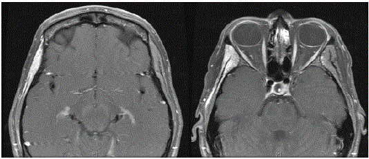
Figure 1
Enhancement of Right Temporalis and Right Pterygoid Muscles of Mastication.
References
- Davis SB, Mathews VP, Williams DW 3rd. Masticator muscle enhancement in subacute denervation atrophy. AJNR Am J Neuroradiol. 1995;16(6):1292-4.
- Fischbein NJ, Kaplan MJ, Jackler RK, Dillon WP. MR imaging in two cases of subacute denervation change in the muscles of facial expression. AJNR Am J Neuroradiol. 2001;22(5):880-4.