Case Report
Catamenial Pneumothorax: Presentation of an Uncommon Pathology: Review of Topic
Rui Haddad1*, Caterin Arévalo2 and David Nigri3
1Department of Thoracic Surgery, Hospital Samaritano, Rio de Janeiro, Brazil
2Resident of General Surgery, Universidad delSinú, Cartagena de Indias, Colombia
3Department of Pulmonology and Bronchoscopy, Hospital Samaritano, Rio de Janeiro, Brazil
*Corresponding author: Rui Haddad, Department of Thoracic Surgery, Rua Barão de Lucena 48- Room 3 Botafogo, CEP: 22260-020 Rio – RJ, Brazil
Published: 06 Dec, 2017
Cite this article as: Haddad R, Arévalo C, Nigri D.
Catamenial Pneumothorax:
Presentation of an Uncommon
Pathology: Review of Topic. Clin Surg.
2017; 2: 1801.
Abstract
The catamenial pneumothorax is defined as the accumulation of air in the pleural cavity that
appears in women infrequently and spontaneously with various clinical presentations. Actually,
it is considered as an extremely rare entity with few cases described in the literature, that is the
reason why the etiology is still discussed. However, a strong association with thoracic endometriosis
syndrome has been found. We want to emphasize how the importance of conducting a diagnosis
and having a timely management would improve the quality of life of the patient and give a better
prognosis of the disease.
Thus, a case report of a 38-year-old female patient who was receiving hormone therapy as a
treatment for abdominal endometriosis and repetitive pneumothorax was presented. In the videoassisted
thoracoscopy we saw diaphragmatic lesions and pneumothorax during the perioperative
and postoperative period. Emphasize the importance of a detailed inspection of each intrathoracic
organ during the surgical procedure, we also showed how the intraoperative pleurodesis, the
placement of a mesh on the diaphragm and the continuity of the hormonal treatment, seems to be
an effective therapy to prevent recurrences and have a better control of the disease.
Keywords: Catamenial pneumothorax; Endometriosis; Thoracic endometriosis syndrome;
Pneumothorax
Introduction
The first description of the recurrent catamenial pneumothorax performed by Maurer et al. [1],
some cases have been described in the world literature, which has allowed to improve their diagnosis,
however, there is still no consensus on the best standard therapeutic attitude of the disease [2].
Endometriosis has been characterized as one of the most important and predisposing factors in
the development of this pathology. We describe the history of a woman with recurrent pneumothorax
caused by pelvic endometriosis with thoracic implants and discuss the different mechanisms and
current therapies for the management of the main complications of this pathology.
Case Presentation
A 38-year-old, non-smoker, presented clinical picture of chest pain and dyspnea of
evolution, with a history of multiple episodes of right pneumothorax. Carrier of pelvic endo3m0 edtaryios soisf
in hormonal therapy and antecedent of bladder and left ureteral obstruction, improved with double
J catheter placement. It was evaluated by our thoracic surgery department, who initially gave us
expectant management of pneumothorax. Given pre and postoperative recurrent respiratory
symptoms, he was submitted to drainage and prosthesis placement by assisted video-assisted
thoracoscopy, evidencing extensive pleural endometriosis with multiple diaphragmatic pores (Figure
1), in tendon centers, pleural micronodules, and scars at the apex. Due to the findings, apicoectomy
of the right upper lobe, diaphragmatic plasty with polyethynylglycol mesh placement covering the
diaphragm defects (Figure 2), pleural biopsy, pleural drainage with a pig-tail catheter, and pleurodesis
with 2% iodopovidone were performed. The pathology report confirmed endometriosis throughout
the specimen withdrawn. He tolerated the procedure well and continued with hormonal therapy.
However, during the postoperative period it presents persistent air leakage through the drain. Due
to the persistence of fistula, she is hospitalized for new examinations and conduct determination.
Patient without respiratory symptoms or pain, normal pulmonary auscultation, with studies of normal radiological images and self-limitation of the fistula. He is discharged with a CT scan at 1 month, which is normal and without
recurrence of symptoms. Due to the persistence of pelvic symptoms,
5 months after thoracoscopic surgery, the patient underwent an ultralow
anterior rectal resection with mechanical anastomosis through a
highly complex robotic surgery (Figure 3), after placement of a chest
tube, which was removed on the third day without problems in the
postoperative period.
Therefore, it is demonstrated the importance of the
interdisciplinary management of the patient by the whole group of
general surgeons, thoracic, urologists, proctologists and gynecologists.
Figure 1
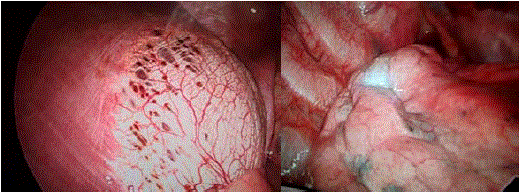
Figure 1
Multiple diaphragmatic fenestrations without active bleeding (two views).
Figure 2
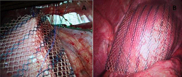
Figure 2
Assisted Video-assisted thoracoscopy with: (A). Placement of mesh in right hemidiaphragm. (B). Positioning of the material.
Figure3
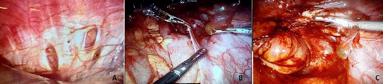
Figure 3
Review of Diaphragm with intra-abdominal chamber lens. Decreased injuries. (B and C). Initiation of adhesion release and dissection of intra-pelvic
tissue planes for subsequent ultra-low anterior rectal resection. Evidence of residual endometriosis in multiple intra abdominal organs.
Figure 4
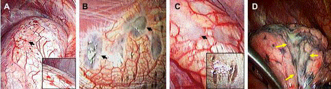
Figure 4
The American Society of Reproductive Medicine classifies the diaphragmatic lesions found in videothoracoscopic surgery according to its macroscopic
appearance in: Rojas (A), black (B), white (C) and cicatricial lesions of the visceral pleural surface of the lung D) [11].
Discussion
Because the frequency of the appearance of endometriosis
in women is in the reproductive age, the incidence of catamenial
pneumothorax associated with it is low, 5% to 15% [3-5], however, in
our case the initial age of Occurrence of the disease, coincided with
that reported (older than 40 years) in previous studies [6].
Also, the rate of complicated endometriosis in this type of
pneumothorax varies from 25% to 60%, requiring inspection of the
pelvic cavity by laparoscopy or laparotomy, in most cases [7].
Endometriosis that involves the chest cavity or lungs
usually presents with catamenial symptoms of pneumothorax,
hemothorax or hemoptysis. Karpel et al. [2] reviewed 84 cases of
pulmonary endometriosis, revealing the dominance of right-sided
pneumothorax in a minority of patients who had concurrent pelvic
endometriosis. Also, in a low percentage of pelvic or diaphragmatic
cases, diaphragmatic fenestrations isolated or associated with the
endometrial focus have been observed and diagnosed [8].
The theory of Sampson et al. [9], proposed to explain the
relationship between these two phenomena, is that the retrograde
menstrual bleeding that appears with columnar cells wrapped
by a stromal with decidual reaction, causes ectopic implants of
endometriosis, compatible with the appearance of collections of
pleural fluid in the Diaphragmatic right side for physiological reasons
of effusion and movement of peritoneal fluids. In addition, the most
frequent extrapelvic location of endometriosis is the respiratory
system [10].
Generally, pulmonary symptoms and cyclic occurrence are
sufficient for the diagnosis of pulmonary endometriosis, and biopsy
is not usually mandatory. However, intrathoracic lesions have
been described during surgery, in which the diaphragm has been
identified and classified according to criteria of the American Society
of Reproductive Medicine (Figure 4), which is important because
its knowledge, together with the correlation of the histopathological
findings, allow a more accurate diagnosis of the types of disease [11].
In addition, patients with abdominal endometriosis and thoracic
implants show predominantly posterior classic sentinel lesions, which
cannot be easily seen by the umbilical port of laparoscopic surgery,
thus reinforcing the recommendation for meticulous examination
of the diaphragm at that level given the high Risk of secondary
pneumothorax and recurrence [12]. However, Muller and Nelems
[2] performed a review, in which they determined that recurrent
catamenial pneumothorax can occur without thoracic endometriosis,
due to the passage of air from the genital tract to the peritoneum
and thence to the chest, also involving some cases of congenital
diaphragmatic defects. Regarding treatment, multiple therapies have
been described to allow definitive control of the disease. These should
be performed individually by assessing the patient's age, desire for
fertility, recurrence, and quality of life with symptoms [2]. Therefore,
if the patient wishes to preserve her fertility, hormone suppression
therapy has been consolidated as the best option, in which the
analogues of gonadotropin hormone through the rupture of ovarian
steroidogenesis, induce the resolution of endometriosis (Pulmonary
or pelvic) [13] and decreased recurrence. Therefore, we emphasize
that the importance of multidisciplinary management among the
gynecologist, urologist, proctologist and general and thoracic surgeon
has also demonstrated the depreciation in the recurrence rates of
Disease, showing better postoperative results [11]. In summary,
according to the literature found and our experience, the ideal
management of thoracic endometriosis is apicectomy, placement
of prosthetic material in the affected areas within the thorax and
hormone therapy [12,14-17].
Conclusion
Although catamenial pneumothorax has been studied as an infrequent disease with multiple recurrences, each patient should be analyzed integrally and individually to correctly determine the aetiology of the pathology, using tools such as new classifications correlated to histological studies, which not only improve the Diagnosis, but, correct with the treatment of women with endometriosis. Currently, worldwide literature shows how the treatment of the combination of continuous hormonal therapy and a thorough review of the pleural, pulmonary and diaphragmatic alterations of endometriosis provides a decrease in recurrence and improves prognosis of the disease.
References
- Maurer ER, Schaal JA, Mendez FL. Chronic recurring spontaneous pneumothorax due to endometriosis of the diaphragm. J Am Med Assoc. 1958;168(15):2013-4.
- Slabbynck H, Laureys M, Impens N, DeVroey P, Schandevyl W. Recurring catamenial pneumothorax treated with a Gn-RH analogue. Chest. 1991;100(3):851.
- Giudice LC, Kao LC. Endometriosis. Lancet. 2004;364(9447):1789-99.
- Azizad-Pinto P, Clarke D. Thoracic endometriosis syndrome: case report and review of the literature. Perm J. 2014;18(3):61-5.
- Alifano M, Trisolini R, Cancellieri A, Regnard JF. Thoracic endometriosis: Current knowledge. Annals of Thoracic Surgery. 2006;761-9.
- Tettey M, Edwin F, Aniteye E, Seffah J, Tamatey M, Ofosu-Appiah E, et al. Thoracic endometriosis syndrome. West Afr J Med. 2013;32(4):302-6.
- Joseph J, Sahn SA. Thoracic endometriosis syndrome: New observations from an analysis of 110 cases. Am J Med. 1996;100(2):164-70.
- Van Schil PE, Vercauteren SR, Vermeire PA, Nackaerts YH, Van Marck EA. Catamenial pneumothorax caused by thoracic endometriosis. Ann Thorac Surg. 1996;62(2):585-6.
- Sampson JA. Metastatic or embolic endometriosis, due to the menstrual dissemination of endometrial tissue into the venous circulation. Am J Pathol. 1927;3(2):93-110.43.
- Joseph J, Reed CE, Sahn SA. Thoracic endometriosis: Recurrence following hysterectomy with bilateral salpingo-oophorectomy and successful treatment with talc pleurodesis. Chest. 1994;106(6):1894-6.
- Kumakiri J, Kumakiri Y, Miyamoto H, Kikuchi I, Arakawa A, Kitade M, et al. Gynecologic Evaluation of Catamenial Pneumothorax Associated with Endometriosis. J Minim Invasive Gynecol. 2010;17(5):593-9.
- Rousset-Jablonski C, Alifano M, Plu-Bureau G, Camilleri-Broet S, Rousset P, Regnard JF, et al. Catamenial pneumothorax and endometriosis-related pneumothorax: Clinical features and risk factors. Hum Reprod. 2011;26(9):2322-9.
- L'huillier JP, Salat-Baroux J. A patient with pulmonary endometriosis. Rev Pneumol Clin. 2002;58(4 Pt 1):233-6.
- Nezhat C, Main J, Paka C, Nezhat A, Beygui RE. Multidisciplinary Treatment for Thoracic and Abdominopelvic Endometriosis. JSLS J Soc Laparoendosc Surg. 2014;18(3):e2014.00312.
- Korom S, Canyurt H, Missbach A, Schneiter D, Kurrer MO, Haller U, et al. Catamenial pneumothorax revisited: Clinical approach and systematic review of the literature. J Thorac Cardiovasc Surg. 2004;128(4):502-8.
- Vercellini P, Viganò P, Somigliana E, Fedele L. Endometriosis: pathogenesis and treatment. Nat Rev Endocrinol. 2013;10(5):261-75.
- Nair SS, Nayar J. Thoracic Endometriosis Syndrome: A Veritable Pandora's Box. J Clin Diagnostic Res. 2016;10(4):QR04-08.