Clinical Image
Melanoma of the Esophagus: Endoscopic Findings of Superficial and Advanced Tumors
Kazuo Koyanagi1 and Soji Ozawa2*
1>/Department of Esophageal Surgery, National Cancer Center Hospital, Tokyo, Japan
2Department of Gastroenterological Surgery, Tokai University School of Medicine, Isehara, Japan
*Corresponding author: Soji Ozawa, Department of Gastroenterological Surgery, Tokai University School of Medicine, Shimokasuya, Isehara, 259-1193, Japan
Published: 30 Nov, 2017
Cite this article as: Koyanagi K, Ozawa S. Melanoma of
the Esophagus: Endoscopic Findings of
Superficial and Advanced Tumors. Clin
Surg. 2017; 2: 1785.
Keywords
Melanoma; Esophageal cancer
Clinical Image
We present two cases of representative endoscopic findings of melanoma of the esophagus
(Figure 1) shows a superficial type of esophageal melanoma. A slightly elevated black pigmentation
covering the esophageal wall entirely at 21 cm to 28 cm from the incisors is visible. Thoracoscopic
and laparoscopic esophagectomy and cervical anastomosis using a gastric conduit were performed.
Pathological examination and immunohistochemistry staining of the resisted specimens showed
a melanoma of the esophagus with the depth of T1b-SM3 and no regional lymph node metastasis
(Figure 2) shows a protruding advanced tumor surrounded by a superficial pigmentation of
melanoma. Thoracoscopic and laparoscopic esophagectomy and a cervical anastomosis using a
gastric conduit were performed. Pathological examination and immunohistochemistry staining
showed melanoma of the esophagus with a depth of pT4a (adventitia) and regional lymph node
metastasis.
Melanoma generally occurs on the skin of the whole body as a black pigmentation and rarely
occurs in the digestive tract. In a previous study, Makuuchi et al. reported that the incidence of
melanoma of the esophagus was 0.3% among all histological types of esophageal cancers in Japan
and the prognosis was extremely poor [1]. Despite recent improvements in the outcomes of patients with melanoma based on the advances in molecular targeted drugs [1,2], surgical resection remains the mainstay of treatment strategies
and neoadjuvant or adjuvant therapies have not yet been established
for patients with esophageal melanoma. A multimodal therapeutic
strategy is urgently needed to treat this desperate disease [3].
Figure 1

Figure 1
Superficial esophageal melanoma. A slightly elevated black pigmentation is visible at 21‒28 cm from
the incisors.
Figure 2
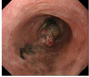
Figure 2
Advanced esophageal melanoma. Aprotruding tumor surrounded by a superficial pigmentation is
visible.
References
- Makuuchi H, Takubo K, Yanagisawa A, Yamamoto S. Esophageal malignant melanoma: analysis of 134 cases collected by the Japan Esophageal Society. Esophagus. 2015;12(2):158-69.
- Tapalian SL, Sznol M, McDermott DF, Kluger HM, Carvajal RD, Sharfman WH, et al. Survival, durable tumor remission, and long-term safety in patients with advanced melanoma receiving nivolumab. J Clin Oncol. 2014;32(10):1020-30.
- Weber J, Mandala M, Del Vecchio M, Gogas HJ, Arance AM, Cowey CL, et al. Adjuvant nivolumab versus ipilimumab in resected stage III or IV melanoma. N Engl J Med. 2017;377:1824-35.