Editorial
Recurrent Adnexal Gland Carcinoma of the Orbit
Heather Fenley*, Chelsea Obourn and Nicholas Purdy
Department of Otolaryngology Head and Neck Surgery, Geisinger Medical Center, Danville, PA, USA
*Corresponding author: Heather Fenley, Department of Otolaryngology – Head and Neck Surgery, Geisinger Medical Center 100 N. Academy Avenue Danville, Pennsylvania, USA
Published: 29 Nov, 2017
Cite this article as: Fenley H, Obourn C, Purdy N.
Recurrent Adnexal Gland Carcinoma of
the Orbit. Clin Surg. 2017; 2: 1773.
Abstract
Primary orbital adenocarcinoma is a rare disease with few reported cases. Symptom onset is typically late, often resulting in diagnosis at advanced stages. It often originates from the lacrimal gland, but may also arise from Adnexal structures. We report the case of a 62 year old man with recurrent Adnexal gland carcinoma of the orbit who presented 4 years following initial treatment with external beam radiation. MRI at presentation showed a right per orbital mass with extension into the orbit. Head and Neck Tumor Board recommendations were for surgical resection including orbital exenteration with free flap reconstruction. Preoperative PET/CT imaging showed low level uptake at the primary site without evidence of metastases. Intraoperatively, widespread subcutaneous extension of the tumor and metastatic cervical adenopathy unexpectedly necessitated a more aggressive surgical procedure than originally planned. Final pathology showed a high-grade orbital Adnexal adenocarcinoma, with per neural and lymph vascular invasion, and metastases to 8 of 41 cervical lymph nodes. Postoperatively, the patient underwent adjuvant chemotherapy and external beam radiation and has done well. This case highlights the importance of maintaining a high index of suspicion and awareness of disease progression when treating primary orbital adenocarcinoma.
Editorial
Primary orbital adenocarcinoma is a rare disease with few reported cases. The natural disease
course is slow growth and late symptom onset [1-4]. It often originates from the lacrimal gland,
but may also arise from Adnexal structures. Adnexal tumors are of sweat gland origin, and have a
propensity for regional metastasis and local recurrence [1]. It most commonly affects those in the 5th
and 6th decades of life, though there are cases reported in the pediatric population as well [2]. There
is a slight male over female predilection. We report a case of recurrent Adnexal gland carcinoma of
the orbit. A 62 year old man presented with 4 months of right upper eyelid swelling. His history was
significant for treatment of right orbital adenocarcinoma with radiation therapy 4 years prior. On
exam, there was firm swelling of the upper eyelid with partial ptosis. Vision was intact. There was no
concerning adenopathy. Subsequent excisional biopsy of the orbital fat was positive for recurrent
adenocarcinoma (Figure 1).
The patient was evaluated at Head and Neck Multidisciplinary Tumor Board was
recommendations were made for surgical resection, including exenteration, with radial forearm
free flap reconstruction. Preoperative PET/CT showed low level uptake at the primary site without
evidence of metastases; however, widespread subcutaneous extension of the primary tumor was
observed intraoperatively. Multiple re-excisions were required to achieve negative margins,
resulting in a larger than anticipated facial defect (Figure 2). A pathologic appearing digastrics node
was identified during neck exploration for vessel anastomosis, which showed metastatic disease on
frozen section necessitating a right neck dissection prior to completing reconstruction.
Surgical pathology showed a high-grade adenocarcinoma measured at 3.5 cm, with apocrine
differentiation, arising from the orbital Adnexal glands. Per neural and lymph vascular invasion
were present. The globe was unaffected. Metastasis was present in 8 of 41 cervical lymph nodes. The
patient recovered well from surgery and completed adjuvant therapy with Xeloda and XRT. Twenty
two months after completing adjuvant treatment, the patient presented with multiple subcutaneous
nodules in the right neck which were biopsied proving metastatic disease. Based on molecular
profiling of the tumor, he was started on Casodex, to which he has responded well thus far. This case
highlights the difficulties in diagnosing and adequately treating orbital Adnexal carcinoma. This
patient’s disease appeared limited initially, though it was much more extensive intraoperatively and
aggressive pathologically. Resection was difficult due to wide subcutaneous spread, and microscopic
disease was far more extensive than the patient’s presentation suggested. Additionally there was
metastasis to multiple cervical lymph nodes on the affected side which were not suspected on PET. Shintaku describe a similar presentation in which case bony metastasis was also present at the time of diagnosis [3].
Early disease diagnosis is important to prevent local spread and
distant metastasis. Due to a lack of distinguishing clinical features, it
is important for practitioners to have a high index of suspicion when a
patient presents with unknown or unexplained orbital findings [1-4].
The current standard of treatment is surgical resection, although no
consensus exists regarding the necessity of exenteration. Determining
the presence of regional spread also is controversial, as advocates for
both sentinel lymph node biopsy and radical neck dissection exist in
the literature [2-4]. Utilization of Moh’s micrographic surgery for
resection of adenocarcinoma in the head and neck may be beneficial
[5]. Latorre et al. [2] describe its use in treating an orbital Adnexal
adenocarcinoma in a 14 year old patient, sparing the globe and
achieving good cosmetic result.
Complete resection often requires the combined effort of multiple
surgical specialties, though prognosis remains poor even with adequate
resection in the case of recurrence. Multimodal treatment including
radiation and chemotherapy may help prevent recurrence, though
long term outcomes are not well documented due to disease rarity.
Regardless of disease presentation, a multidisciplinary approach to
treatment is paramount, as is close monitoring for recurrence.
Figure 1
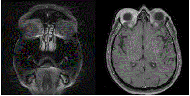
Figure 1
TI weighted MRI images, Coronal (left) and axial (right), showing
right periorbital soft tissue enhancement representing tumor.
Figure 2
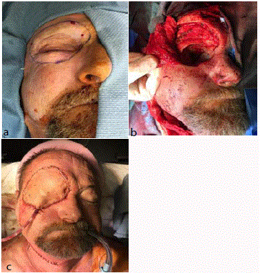
Figure 2
Intraoperative photos showing swelling over the right superior orbit
representing tumor mass and the outline of the planned area of resection (a).
Facial defect following complete resection of tumor and exenteration (b), and
the end result following completion of radial forearm free flap reconstruction
and right modified radial neck dissection (c).
References
- Latorre A, Alghothani L, Lambert D, Jatana KR, Peters S, Foster J, et al. Mucin-producing Malignant Tumor of Lower Eyelid Presenting in a 14-year old Patient. J Clin Aesthet Dermatol. 2012; 5(4): 44-7.
- Zhang Leilei, Ge S, Shengfang Fan, Xianqun. A brief review of different types of sweat-gland carcinomas in the eyelid and orbit. Onco Targets Ther. 2013;6:331-40.
- Shintaku M, Tsuta K, Yoshida H, Tsubura A, Nakashima Y, Noda K. Apocrine adenocarcinoma of the eyelid with aggressive biological behav-ior: a report of a case. Pathol Int. 2001;52(2):169-73.
- Akcay, Emine Kalkan, et al. Apocrine adenocarcinoma of the right eyelid and apocrine adeno-carcinoma of the left maxillary sinus. Can J Ophthalmol. 2008; 43(5): 609-10.
- Wildemore John K, Lee Jason B, Humphreys Tatyana R. Mohs Surgery for Malignant Eccrine Neoplasms. Dermatol Surg. 2004;30 (12):1574-9.