Case Report
Giant Coronary Sinus Associated with Early Prosthetic Valve Failure of the Right Heart
Yoshiaki Saito, Kozo Fukui, Kazuyuki Daitoku and Ikuo Fukuda*
Department of Thoracic and Cardiovascular Surgery, Hirosaki University School of Medicine, Aomori, Japan
*Corresponding author: Ikuo Fukuda, Department of Cardiovascular Surgery, Hirosaki University School of Medicine, Zaifucho, Hirosaki, Aomori 036-8562, Japan
Published: 23 Nov, 2017
Cite this article as: Saito Y, Fukui K, Daitoku K, Fukuda I.
Giant Coronary Sinus Associated with
Early Prosthetic Valve Failure of the
Right Heart. Clin Surg. 2017; 2: 1761.
Abstract
A 71-year-old woman was admitted to our hospital presenting right heart failure. She had undergone mitral valve replacement and concomitant tricuspid valve replacement 7 years before the admission. Computed tomography at the time of the first operation revealed enlargement of the coronary sinus of 3.8 cm with a persistent left superior vena cava. On the latest admission, a transesophageal echocardiogram revealed prosthetic valve failure on the tricuspid valve position. Computed tomography showed giant coronary sinus with a maximum diameter of 6.2 cm. Retricuspid valve replacements was successfully performed. Severe pannus formation and adhesion of native valves were observed on the right ventricular side of the prosthetic valve. This is the first report describing aneurysmal growth of a coronary sinus caused by a prosthetic valve failure.
Introduction
The Coronary Sinus (CS) corrects venous flows from the heart muscle and opens to right atrium. CS dilatation is sometimes observed in the presence of right atrial pressure overload. We herein report a case of giant CS caused by early prosthetic valve failure on the tricuspid position.
Case Presentation
A 71-year-old woman was admitted to our hospital complaining of shortness of breath and progressive lower leg edema. She had undergone mitral valve replacement with a 27-mm bioprosthetic valve (CEP, Edwards Life sciences Corp, Irvine, CA) for degenerative valve disease and concomitant tricuspid valve replacement with a 29-mm CEP for uncontrollable tricuspid regurgitation 7 years before admission. When performing the primary operation, leaflets of the tricuspid valve were fully preserved in TVR. Computed tomography (CT) at the time of the first operation revealed enlargement of the CS of 3.8 cm (Figure 1A), with a persistent left superior vena cava (PLSVC). On the latest admission, a transesophageal echocardiogram revealed restriction of opening and closing of the bioprosthetic valve in the right heart, indicating prosthetic valve insufficiency. The traced valve area was 1.4 cm2. On Swan-Ganz catheter examination, right atrial pressure was 15 mmHg, and right ventricular pressure was 26/14 mmHg. CT showed an enlarged right atrium associated with a giant CS with a maximum diameter of 6.2 cm (Figure 1B). The patient underwent re-TVR for early prosthetic valve insufficiency. Re-sternotomy was made using an oscillating saw. Adhesion in the mediastinum was carefully exfoliated using the harmonic scalpel. Cardiopulmonary bypass was established through aortic cannulation and bicaval drainage. Additional venous drainage catheter was inserted in the PLSVC. In order to minimize the risk for injuring cardiac wall due to radical exfoliation, vacuum assisted venous drainage technique was employed. After the administration of the crystalloid cardioplegia and aortic cross clamp, cardiac arrest was smoothly obtained. After incising the wall of the right atrium, large coronary sinus was observed. In order to reduce the backflow from the coronary sinus into the right artium, a 16 Fr Foley catheter was inserted into the CS (Figure 2A and 2B). The ex-implanted valve was removed in a careful manner. Severe pannus formation was evident at the outflow aspect. Host tissue overgrowth fused the free margins of leaflets at each commissure (Figure 3). Concrete debridement of subvalvular structure was performed and a 29 mm mechanical valve (ATS medical, INC., Minneapolis, MN) was implanted afterwards. After the operation, explanted valve was carefully inspected. No inconsistencies detected in the X-ray as the wire form is intact. Calcification of the fused leaflets was not also detected. The patient survived, there is no sign of recurrence of prosthetic valve insufficiency and the patient is doing well 2 years after the surgery.
Figure 1
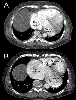
Figure 1A and 1B
(A) Computed tomography (CT) at the time of the first
operation revealed enlargement of the CS of 3.8 cm. (B) CT showed an
enlarged right atrium associated with a giant CS with a maximum diameter
of 6.2 cm.
Figure 2A and 2B
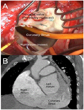
Figure 2A and 2B
(A) In order to reduce the backflow from the coronary
sinus into the right atrium (B) A 16 Fr Foley catheter was inserted into the CS.
Figure 3
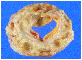
Figure 3
Host tissue overgrowth fused the free margins of leflets at each
commissure.
Discussion
CS dilatation is observed in the presence of PLSVC and right atrial pressure overload [1]. CS dilatation itself is normally benign, though it can cause mechanical compression of left atrium as its diameter grows [3]. Pulmonary hypertension, right ventricular dysfunction, right coronary arteriovenous fistula and arteriovenous hemodialysis fistula have been reported as one cause of CS dilatation in a quite small series of literature [2-5]. Prosthetic valve insufficiency, as presented here has not been described before. Both bioprosthetic valve failure and PLSVC might contribute to CS growth in the presented case. Aneurysmal growth in diameter of CS also has been reported before, thus this fact makes this report a landmark. Owing to recent advances in tricuspid annuloplasty technique, prosthetic valve replacement is seldom required. Of 4,816 tricuspid surgeries in total 19,164 acquired valvular surgery cases arisen in the annual report of 2011 by the Japanese Association for Thoracic Surgery, only 148 isolated and concomitant tricuspid valve replacements (3.1%) were performed in Japan [6]. Long-term survival after TVR is poor with reported 10- year survival rate of 37% to 68% [7-9]. Prosthesis durability in TVR is unclear because only small numbers of TVR cases have been reported to date. Limited reported 10-year and 15-year reoperation-free rate ranges from 75% to 79% and 53% to 63%, respectively [8,9]. Tricuspid valve re-operation is associated with a high mortality rate. Guenther et al. reported a hospital mortality rate of 26.7% [8]. Thus, avoidance of re-operation is of importance. Nakano et al. [9] reported 12 cases of re-TVR cases and of these, 10 cases were caused by native valve attachment and/or pannus formation. This report waken us an idea that resection of valves and sub-valvular apparatus may contribute longer durability of prosthetic valves when performing TVR. In conclusion, CS dilatation was observed under the presence of prosthetic valve stenosis in tricuspid valve position and PLSVC. To the best of our knowledge, this is the first report describing aneurysmal growth of CS caused by a prosthetic valve failure. Resection of tricuspid valves and sub-valvular apparatus may be important when performing TVR.
References
- Kolski BC, Khadivi B, Anawati M, Daniels LB, Demaria AN, Blanchard DG. The dilated coronary sinus: utility of coronary sinus cross-sectional area and eccentricity index in differentiating right atrial pressure overload from persistent left superior vena cava. Echocardiography 2011;28(8):829- 32.
- Gunes Y, Guntekin U, Tuncer M, Kaya Y, Akyol A. Association of coronary sinus diameter with pulmonary hypertension. Echocardiography. 2008;25(9):935-40.
- Gaudino M, Anselmi A, Lombardo A, Riccardo Marano, Gianfederico Possati. Giant coronary sinus aneurysm secondary to right coronary arteriovenous fistula leading to pseudo-mitral stenosis. Arch Med Sci. 2011;7(3):533-5.
- Nathani S, Parakh N, Chaturvedi V, Sanjay Tyagi. Giant coronary sinus. Tex Heart Inst J. 2011;38(3):310-1.
- Wolf M, Scott B. Left-sided high-flow arteriovenous hemodialysis fistula combined with a persistent left superior vena cava causing coronary sinus dilatation. Semin Dial. 2013;26(2):E13-6.
- Amano J, Kuwano H, Yokomise H. Thoracic and cardiovascular surgery in Japan during 2011: Annual report by the Japanese association for thoracic surgery. Gen Thorac Cardiovasc Surg. 2013;61(10):578-607.
- Rizzoli G, De Perini L, Bottio T, Minutolo G, Thiene G, Casarotto D. Prosthetic replacement of the tricuspid valve: biological or mechanical?. Ann Thorac Surg. 1998;66(6):S62-7.
- Guenther T, Noebauer C, Mazzitelli D, Busch R, Tassani-Prell P, Lange R. Tricuspid valve surgery: a thirty-year assessment of early and late outcome. Eur J Cardiothorac Surg. 2008;34(2):402-9.
- Nakano K, Ishibashi-Ueda H, Kobayashi J, Sasako Y, Yagihara T. Tricuspid valve replacement with bioprosthises: long-term results and causes of valve dysfunction. Ann Thorac Surg. 2001;71(1):105-9.