Review Article
Ultrasound Assisted Abdominoplasty
Alfredo Hoyos1* and David E Guarin2
1Department of Plastic, Aesthetic, Maxillofacial, and Hand Surgery, University of San Martin in Bogota, Colombia
2Department of Plastic Surgery, Universidad del Valle, Hospital Universitario del Valle, Colombia
*Corresponding author: Alfredo Hoyos, Department of Plastic, Aesthetic, Maxillofacial and Hand Surgery, University of San Martin in Bogota, Carrera 15# 83–33, Suite 304, Bogotá, Colombia
Published: 23 Nov, 2017
Cite this article as: Hoyos A, Guarin DE. Ultrasound
Assisted Abdominoplasty. Clin Surg.
2017; 2: 1756.
Abstract
The Vaser addition to the abdominoplasty techniques safely allows achieving natural and
aesthetically pleasant outcomes, by means of diminishing trauma of the abdominal wall. A limited
tunneling technique as well as the neoumbilicoplasty is essential part of this approach.
Keywords: Vaser; UAL; High definition; Abdominoplasty
Key Points
1. Current abdominoplasty approaches have the cumulative experience of a century of
procedures; however there are still surgical outcomes that stigmatize the procedure.
2. The ultrasound assisted liposuction is a safe and useful tool to improve the Abdominoplasty
outcomes allowing achieving natural and athletic look.
3. The Neoumbilicoplasty technique allows an optimal placement as well as the possibility of
delaying in cases of vascular risk.
Introduction
Being the third most common surgical cosmetic procedure in the United States [1] is good reason to gather all the available resources, techniques and experience aiming to achieve the best possible outcomes in terms of patient satisfaction, security and aesthetic results. More than a century of surgical advances and understanding of the abdominal wall anatomy [2] allows reducing the trauma, the mortality rates and enhancing the results by including a limited flap dissection, correction of the muscular diastasis, an optimal scar position, umbilical reconstruction and all the contouring advantages of the liposuction techniques [3]. In spite of these facts, there are still surgical outcomes that stigmatize the procedure creating an unnatural abdomen [4] including a poor waistline definition, a visible umbilical scar, inadequate scar position, umbilical color and vertical residual scar. The develop of adjuvant tools to facilitate and enhance the fat extraction can also help in improving the results and consistency of the abdominoplasty [5,6]. The ultrasound technology is one of these energy based tools which has been widely tested and have shown to be a secure and effective method to help the fat extraction [7-9]. This type of energy applied to the fat tissue induces the brake down of the fat cells (emulsification) allowing a less traumatic extraction, lower the blood lost, a faster recovery and relieve the surgeon work [10,11]. This paper discusses the use of the ultrasound technologies combined with the detailed anatomy knowledge to get an accurate and stigma free abdominal contour.
Patient Selection and Classification
The indication for ultrasound assisted Abdominoplasty is determined by a comprehensive physical examination considering the abdominal skin laxity (Matarasso’s [12] classification), skin quality, the striae, fat deposits, scars, the abdominal wall integrity and the relative umbilical position. This approach can be used in deformities with any type of skin laxity associated with fat accumulation with or without muscular diastasis. The ideal patient must be healthy, with a type IV deformity, a body mass index below 33 Kg/m2 and non smoker. Post bariatric patients are not good candidates for this procedure. Previous abdominal and cosmetic procedures are not contraindication for this approach however must be considered to accurately plan the surgery.
Preparation
All patients must be assessed for venous thromboembolism risk using the Caprini score and the adequate preventive measures must be taken. The preoperative set of standard photos are a critical part as well as the time taken to resolve all the doubts related to the procedure, the recovery and all the possible complications that also must be carefully written in the consent.
Figure 1
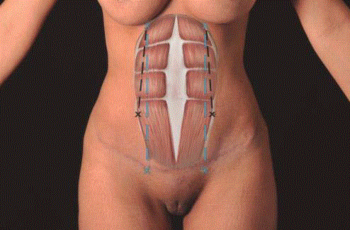
Figure 1
Abdominal marking.
To mark a patient with rectus abdominis diastasis the origin and insertion
of the rectus abdominis is located and a line between them is marked (blue
line).
Figure 2
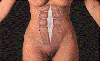
Figure 2
Lipectomy incision.
The horizontal incision is marked by locating the lateral edges of the rectus
abdominis muscle insertion, 2 cm above the pubis after manual ptosis
correction. Then the lateral lines are drawn in an angle of 135 degrees.
Patient Marking
The marking is done with the patient in standing position. Begin by marking the areas with excess of fat tissue. Then the linea alba and the origin and insertion of the rectus abdominis is located and a line between them is marked. The muscular landmarks do not guide this marking when there is diastasis of the rectus abdominis (Figure 1). The areas below the costal margin, the midline and lateral to the rectus abdominis lower insertion are marked for extra liposuction to recreate the superficial anatomy. The lower horizontal incision is marked between the lateral edges of the rectus abdominis muscle insertion, 2 cm above the pubis after manual ptosis correction. Then a lateral line in an angle of 135° (Figure 2).
Operative Technique
The procedure is done in 3-phases: beginning with the
Liposculpture then the Abdominoplasty and finally a neoumbilicoplasty.
Place the patient in prone with the antiembolic
compression device and the prophylactic antibiotic.
Liposculpture of the abdomen. Begin infiltrating the tumescent
solution of 1000 ml of saline plus 10 ml of 1% lidocaine and 1ml of
epinephrine 1:1,000 using an infiltration/extraction ratio of 2:1 to
1.5:1. With the adequate skin care, start the fat emulsification using
the third-generation ultrasound (VASER® - Valeant Pharmaceuticals
North America LLC) with a 3 mm and 3.7 mm grooved probes using
a pulse mode with 70 y 80%. We use powered-assisted liposuction
(POWER X Lipo® - Valeant Pharmaceuticals North America LLC)
to facilitate the extraction. Follow the pre-operative markings beginning in the deep layer and the blending with the intermediate,
and superficial fat layers using 4.6 mm and 3.7 mm cannulas until
reaching the desired flap thickness. In the supra umbilical midline and the negative marked areas a superficial liposuction is performed
using smaller cannulas and achieve the desired etching.
Figure 3
Figure 3
Abdominal flap suture.
The progressive tension sutures the abdominal flap to the midline. From the
xyphoid down to the umbilicus use deep from to enhance the midline. Down
the umbilicus use only superficial continuous stitches.
Figure 4
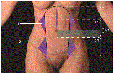
Figure 4
Umbilical position zone.
The umbilicus is placed according to the patient characteristics in a zone
from the midpoint between the xyphoid and the pubis and the lower third.
Figure 5a
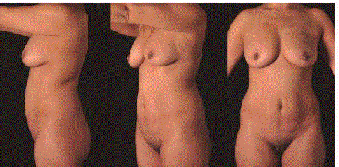
Figure 5a
A 30-year-old female. She had one pregnancy 6 years before de
procedure.
Figure 5b
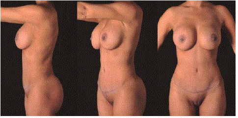
Figure 5b
Ultrasound assisted lipoabdominoplasty. 14 months after
surgery. Observe the long navel-scar distance and the abdomen and
waistline definition that gives a natural looking of the abdomen. Periareolar
breast augmentation as well as 500ml gluteal fat grafting was also done.
Lipectomy
Begin by rising the abdominal flap from the lower incision mark: under the umbilicus in the sub-Scarpa’s layer and in the upper abdomen above the muscular fascia. Use a tunneling technique up to the xiphoid process to preserve the vascular supply as possible. Do not forget to be extremely cautious with the hemostatic control. Locate and mark the medial borders of the rectus abdominis muscles and make the plicature taking into account the point of maximal indentation of the waist to enhance it. Resect the native umbilical stalk and suture the remnant with the surrounding fascia. Use a progressive tension technique to suture the abdominal flap to the midline: 1) profound continuous stitches from the xyphoid down to the umbilicus to enhance the midline in the upper abdomen 2) superficial below the umbilicus Figure 3. Now the skin resection is completed, a closed drainage is placed and the flap is sutured in layers. Extra liposuction over the flap can be done to enhance the definition.
Neoumbilicoplasty
We chose the umbilical position according to the patient’s characteristics and desires. A higher location is used in young and athletic patients while older, non athletic or associated with breast ptosis a lower position is advisable Figure 4 and 5. The procedure can be performed just after the Lipectomy or can be delayed when there is a recognizable risk of flap failure such as congestion, high tension in the closure, large liposuction, secondary procedures and high scar positioning. The delay can be from a week up to 3 months. After choosing the umbilical position, a butterfly-shaped incision is done with 60° in the apex angles creating 4 triangular flaps as shown in Figure 6. Dissect down to the muscular fascia and make a shallow release of the triangular flaps. Secure the three lower flaps with a continuous subcuticular polyglactine stich to the muscular fascia in a spot just below the base of the upper flap which is then loosely sutured to the same spot. Use a gauze splint covered with a topical ointment or antibiotic to induce a round umbilicus shape.
After Care
A loose compression garment and a foam vest are indicated for up to 8 weeks as well as daily drainage. Antithrombotic measures including early mobilization and the required medications are strongly advised. Most patients are left overnight to provide a comprehensive care with an optimal pain control, closely look to the wounds, drainage and nurse care. The drainage is left for the required time to avoid seroma buildup. The postoperative checkups must include a standard photographic record (Figure 5 and 6).
Discussion
A natural and healthy abdominal wall shape and contour is far more complex than the lack of fat, skin redundancy nor laxity. Recreating the natural light and shadows of the skin surface enveloping the underneath muscular anatomy must be the goal of the procedure. The unnatural surgical stigmata [4] is still a common concern that can be addressed by adding the VASER ultrasound technologies to the lipoabdominoplasty. In our experience this technique makes it a safer procedure by means of reduced blood lost, flap trauma and enhancing the outcomes. The umbilical shape, color and position are critical in the abdominal appearance. The vertical umbilical shape is the most common finding on young and attractive abdomen. The untanned area of the buried umbilical scar is frequently an issue that creates a color mismatch with the surrounding skin. Making a neoumbilicoplasty can be an accurate strategy to rightfully create and place the umbilical scar according to the patient characteristics. The ideal position is still a topic lacking of consensus, however we believe that it must be located over an umbilical area rather than an umbilical point. By considering this approach, delaying the umbilical reconstruction is feasible and can help in improving the flap viability in cases such where there is a perfusion risk. Following these considerations when approaching the abdominal contouring let us have consistent and aesthetically pleasant results in a safe way and avoiding the surgical stigma.
Figure 6a
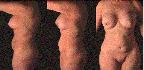
Figure 6a
A 56-year-old female. She had a previous abdominoplasty. Note
the short scar-navel distance.
Figure 6b
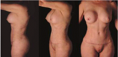
Figure 6b
Ultrasound assisted lipoabdominoplasty before. 1 year after
surgery. Notice the low scar placement and definition of abdomen and
waistline. 300 ml gluteal fat grafting was also done.
References
- American Society of Aesthetic Plastic Surgery. Cosmetic Surgery National Data Bank Statistics. 2016.
- Lancerotto L, Stecco C, Macchi V, Porzionato A, Stecco A, De Caro R. Layers of the abdominal wall: anatomical investigation of subcutaneous tissue & superficial fascia. Surg Radiol Anat. 2011;33(10):835-42.
- Regnault P. The history of abdominal dermolipectomy. Aesthetic Plastic Surgery. 1978;2(1):113-23.
- Lockwood TE. Maximizing aesthetics in lateral-tension abdominoplasty and body lifts. Clinics in Plastic Surgery. 2004;31(4):523-37.
- Saldanha OR, Azevedo SF, Delboni PS, Saldanha Filho OR, Saldanha CB, Uribe LH. Lipoabdominoplasty: The Saldanha Technique. Clin Plast Surg. 2010;37(3):469-81.
- Abramson DL. Ultrasound-Assisted Abdominoplasty: Combining Modalities in a Safe and Effective Technique. Plast Reconstr Surg. 2003;112(3):898-902.
- De Souza Pinto EB, de Souza Pinto Abdala PC, Maciel CM, de Paula Turchiari dos Santos F, de Souza RPM. Liposuction and VASER. Clin Plast Surg. 2006;33(1):107-15.
- Dispaltro FL. Use of ultrasound as an adjunct to lipoplasty. Aesthet Surg J. 2002;22(3):303-4.
- Blondeel PN, Derks D, Roche N, Van Landuyt KH, Monstrey SJ. The effect of ultrasound-assisted liposuction and conventional liposuction on the perforator vessels in the lower abdominal wall. British J Plast Surg. 2003;56(3):266-71.
- Hoyos A, Millard J. VASER-assisted high-definition liposculpture. Aesthet Surg J. 2007;27(6):594-604.
- Hoyos A, Perez M. Arm Dynamic Definition by Liposculpture and Fat Grafting. Aesthet Surg J. 2012;32(8):974-87.
- Matarasso A. Abdominolipoplasty. Clin Plast Surg. 1989;16(2):289-303.