Clinical Image
Ventriculoperitoneal Shunt in the Scrotum? Rare but Possible!
Suhasini Gazula*, A Rajasekhar and N Narender Kumar
Department of Paediatric Surgery, Employees State Insurance Corporation (ESIC) Medical College &
Superspeciality Hospital, Sanathnagar, Hyderabad, India
*Corresponding author: Suhasini Gazula, Department of Paediatric Surgery, Employees State Insurance Corporation (ESIC) Medical College & Superspeciality Hospital, Sanathnagar, Hyderabad, Telangana 500038, India
Published: 16 Nov, 2017
Cite this article as: Gazula S, Rajasekhar A, Narender
Kumar N. Ventriculoperitoneal Shunt in
the Scrotum? Rare but Possible!. Clin
Surg. 2017; 2: 1730.
Clinical Image
A 2-year-old male child with right Ventriculoperitoneal (VP) shunt insertion for obstructive
hydrocephalous at 3 months age presented with left scrotal swelling since 2 months. Examination
revealed a coiled tubular structure in the left scrotum (Figure 1). CT scan showed ventricular end
of the VP shunt in situ with no hydrocephalous; the peritoneal part migrated through a left Patent
Processus Vaginalis (PPV) and was lying coiled in the scrotum but its tip was lying in the peritoneal
cavity (Figure 2 and 3) and hence continuing to drain CSF with no worsening of hydrocephalous.
An open left herniotomy was done during which the PPV was opened (Figure 4), shunt uncoiled,
lower end drainage confirmed and repositioned into the peritoneal cavity through the proximal end
of PPV prior to its ligation (Figure 5).
Shunt migration can occur to the abdominal wall, mediastinum, bladder, and bowel and rarely
to the scrotum via a PPV. Increased intraabdominal pressure by CSF accumulation is proposed as a
causative factor for the prolonged patency of processus vaginalis in children with VP shunts.
Figure 1
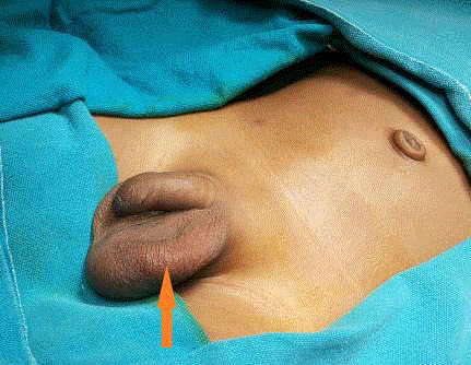
Figure 1
Coiled tubular structure in the left scrotum.
Figure 2
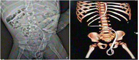
Figure 2 and 3
Left Patent Processus Vaginalis (PPV) and was lying coiled in the scrotum.
Figure 4
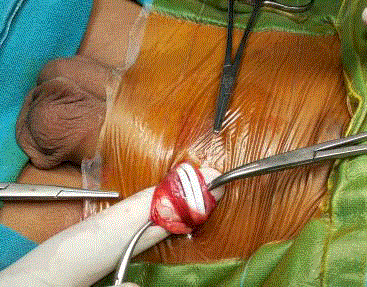
Figure 4
Open left herniotomy was done during which the PPV was opened.
Figure 5
Figure 4
Lower end drainage confirmed and repositioned into the peritoneal
cavity through the proximal end of PPV.