Case Report
A Rare Pulmonary Adenofibroma Mimicking a Metastatic Lesion
Roberto Corzani1*, Cristiana Bellan2, Luca Luzzi1, Marco Ghisalberti1, Tommaso Ligabue1,
Fabiola Meniconi1, Arash Astaneh1, Sascia Pietro Servillo1, Lisa De Leonibus1 and Piero
Paladini1
1Department of Thoracic Surgery, University Hospital of Siena; Siena 53100, Italy
2Department of Pathology, University Hospital of Siena; Siena 53100, Italy
*Corresponding author: Roberto Corzani, Department of Thoracic Surgery, University Hospital of Siena; Siena 53100, Italy
Published: 03 Aug, 2017
Cite this article as: Corzani R, Bellan C, Luzzi L,
Ghisalberti M, Ligabue T, Meniconi F,
et al. A Rare Pulmonary Adenofibroma
Mimicking a Metastatic Lesion. Clin
Surg. 2017; 2: 1691.
Abstract
Pulmonary adenofibroma is a rare benign soft-tissue tumor composed of epithelial and stromal components, both of which are histologically benign. We report a case of pulmonary adenofibroma in a patient with occasional finding of a solitary pulmonary nodule in a CT-scan performed during the staging for a testicular tumor. The therapeutic approach was driven by the positive anamnesis for testicular neoplasm, by the positive PET-scan and most of all by the fact that the lesion was shrinking during chemotherapy and increased in dimension during the follow-up period without adjuvant therapy.
Introduction
Pulmonary adenofibroma is a rare benign soft-tissue tumor composed of epithelial and stromal components, both of which are histologically benign. Rarity of this lesion is confirmed by the fact that only few case reports are available in the literature [1-10]. Radiologically, it appears as a solitary coin lesion: the rarity of this tumor and it’s in usual histologic appearance make it a challenging radiologic diagnosis. In fact, diagnosis is established only by histopathological examination and it needs to be distinguished from other benign and malignant entities with biphasic growth pattern. We report a case of pulmonary adenofibroma in a patient with occasional finding of a solitary pulmonary nodule in a CT-scan performed during the staging for a testicular tumor.
Case Presentation
A 24-year-old man, occasional smoker, was operated for a malignant testicular tumor that the histological examination confirmed to be a germinal tumor (immature teratoma 70%, seminoma in situ 15%, Yolk sac tumor 10% and embrional carcinoma 5%). Pre-operative staging CT-scan, besides the testicular lesion, showed a homogeneous, well-defined, round soft tissue nodule, 8 mm in diameter, localized in the right lower lobe associated with a sub-carinal linfadenopathy (Figure 1). Contrast-enhanced CT showed no enhancement of the lung lesion and PET-scan revealed a mild uptake in the lung lesion (SUV 3.5). After three cycles of chemotherapy with PEB (cysplatin, vepesid and bleomycin) the patient underwent a new CT-scan and PET-scan that showed a dimensional reduction of the pulmonary nodule (4 mm) and the disappearance of the uptake of FDG. These data strengthen the suspicion that the lung lesion could be a metastatic localization of the testicular tumor. CT-scan was performed in follow-up after 12, 15, 18 and 24 months since operation and any modification was detected in the lung nodule, but the last CT-scan performed after 33 months showed a millimetrical increase in size of the suspected metastasis (6 mm, ex 4 mm) even if another PET-scan didn't show any uptake. On the basis of this data we decided to perform a surgical resection of the lung lesion. The patient was placed in a left lateral decubitus position and partial wedge resection of the right lower lobe was performed under video-assisted thoracoscopy (VATS) through CT-guided landmark placement. We inserted a 5-mm, 30-degree thoracoscope through a port in the eighth intercostal space along the middle axillary line; two additional ports were placed in the fifth intercostal space along the right anterior axillary line and in the sixth intercostal space along the right posterior axillary line respectively. We used a 60-mm articulating endothoracic linear cutting device (Endogia iDrive) with 3.5-mm staples to perform a wedge resection where the landmark was placed. There was no evidence of any other abnormalities of the right lung or pleural space. Despite all preoperative data pointed to a metastatic coin lesion, the histopathological examination surprisingly showed a benign lung lesion. On the basis of the macroscopic features, the tumor was a well circumscribed but unencapsulated intraparenchymal peripheral mass, in subpleural location without contact with pleural surface or bronchus, measuring 5 mm in maximum dimension. The cut surface was firm, rubbery, homogenous, tan-pink without areas of hemorrhage and necrosis. The histologic features were characterized by club-shaped papillary structures lined by a layer of simple cuboidal epithelium, sometimes by a tall columnar epithelium at a low power view (Figure 2). The stromal component of papillary structures contains spindle cells with bland cytologic appearance devoid of mitoses or nuclear pleomorphism. Occasionally gland-like spaces were scattered within the lesion. No areas of necrosis or hemorrhage were noted. Bronchial and parenchymal surgical cut margins were free of tumor. Surrounding lung parenchyma was essentially unremarkable. At the immunohistochemical analysis, epithelial cells lining the papillae stained positive for pancytokeratins (CK), EMA, and TTF-1, whereas the stromal component was immune-positive for vimentin and focally for desmin and SMA. Spindle cells were negative for S-100 protein, CD34, CD99 and Bcl-2 (Figure 3 and 4). Based on the histomorphological and immunohistochemical features, the diagnosis of adenofibroma of lung was made.
Figure 1
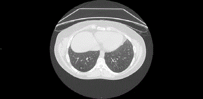
Figure 1
Soft-tissue nodule in the right lower lobe, detected at CT-Scan.
Figure 2
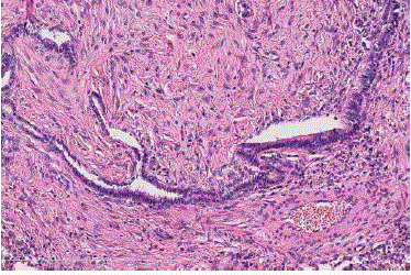
Figure 2
HE: pulmonary adenofibroma with club-like papillae lined by a
single layer of simple epithelium.
Figure3
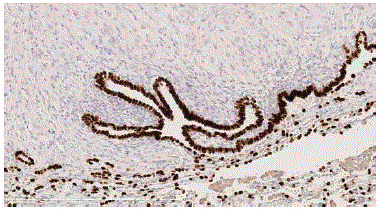
Figure 3
TTF1: Epithelial cells lining the papillae show nuclear positivity for
TTF-1.
Figure 4
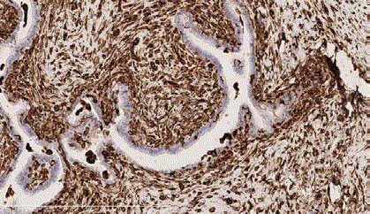
Figure 4
VIM: spindle cells stain positive for vimentin.
Discussion
Pulmonary adenofibroma was first reported by Scarff and Gowar in 1944 [1]. Since then, only few case reports have been added to the literature. Clinical manifestations are vague and nonspecific. It is usually detected incidentally on radiology as a solitary subpleural coin lesion in middle-aged patients with a male ponderance. Histogenesis of this lesion is ambiguous and remains a subject of debate. Scarff and Gowar [1] believed that this tumor probably belongs to the hamartomatous family of the lung lesion. However, Butler and Kleinerman [2] proposed that they were neoplastic rather than developmental, based on the presence of immature cartilage in their lesions. At last, Suster and Moran [3] examined the clinicopathological and immunohistochemical features of two pulmonary adenofibromas and concluded that these tumors may represent an immature form of pulmonary hamartoma. Pulmonary adenofibroma is rarely of clinical significance and surgery is not always required, even if adenofibroma could potentially undergo malignant transformation and metastasize to other organs. Resection was almost always done because of the diagnostic puzzle presented [4-6]. In our case, the therapeutic approach was driven by the positive anamnesis for testicular neoplasm, by the positive PET-scan and most of all by the fact that the lesion was shrinking during chemotherapy and increased in dimension during the follow-up period without adjuvant therapy. The histopathological finding was kind of a surprise. We infer that the response during chemotherapy was due to the fact that, despite the benign nature, adenofibroma shows some low, but significant proliferating activity. In conclusion, pulmonary adenofibroma is a benign lesion of the lung which should enter the scope of differential diagnosis of solitary peripheral lung nodules; the most definitive method for diagnosis in patients with solitary pulmonary nodule is histopathological examination. Limited pulmonary resection using VATS is the most appropriate diagnostic and therapeutic modality in the management of this lesion [7-10].
References
- Scarff RW, Gowar FJS. Fibroadenoma of the lung. J Pathol Bacteriol. 1944;56(2):257-9.
- Butler C, Kleinerman J. Pulmonary hamartoma. Arch Pathol. 1969;88(6):584-92.
- Suster S, Moran CA. Pulmonary adenofibroma: report of two cases of an unusual type of hamartomatous lesion of the lung. Histopathology. 1993;23(6):547-51.
- Cavazza A, Rossi G, De Marco L, Putrino I, Pellegrino S, Piana S. Solitary fibrous pseudopapillary tumor of the lung: pulmonary fibroadenoma and adenofibroma revisited. Pathologica. 2003;95(3):162-6.
- Kumar R, Desai S, Pai T, Pramesh CS, Jambhekar NA. Pulmonary adenofibroma: clinicopathological study of 3 cases of a rare benign lung lesion and review of the literature. Ann Diagn Pathol. 2014;18(4):238-43.
- Vitkovsky T, Zelstman D, Esposito M, Morgenstern N. Pulmonary adenofibroma: cytologic and clinicopathologic features of a rare benign primary lung lesion. Diagn Cytopathol. 2013;41(11):991-6.
- Wang Y, Xiao HL, Jia Y, Chen JH, He Y, Tan QY, et al. Pulmonary adenofibroma in a middle-aged man: report of a case. Surg Today. 2013;43(6):690-3.
- Nicholson AG, Tomashefski JF, Popper Jr H. Hamartoma. In: Travis WD, Brambilla E, Muller-Hermelink HK, Harris CC, editors. World Health Organization classification of tumours. Pathology and genetics of tumours of the lung, pleura, thymus and heart. Lyon: IARC Press; 2004;113-4.
- Varoli F, Vergani C, Caminiti R, Francese M, Gerosa C, Bongini M, et al. Management of solitary pulmonary nodule. Eur J Cardiothorac Surg. 2008;33(3):461-5.
- Akin O, Coskun M. Biliary adenofibroma with malignant transformation and pulmonary metastases: CT findings. AJR Am J Roentgenol. 2002;179(1):280-1.