Case Report
Aneurysmal Bone Cyst of Clavicle in a Child: A Rare Case Report
Suresh Borah1, Hemonta Kr Dutta2* and Manasjyoti Das1
1Department of Orthopaedics, Assam Medical College, India
2Department of Paediatric Surgery, Assam Medical College, India
*Corresponding author: Hemonta Kr Dutta, Department of Paediatric Surgery, Assam Medical College, Dibrugarh, Assam, 786002, India
Published: 20 Oct, 2017
Cite this article as: Borah S, Kr Dutta H, Das M.
Aneurysmal Bone Cyst of Clavicle in a
Child: A Rare Case Report. Clin Surg.
2017; 2: 1685
Abstract
Aneurysmal bone cyst is a benign expanding osteolytic lesion of bones and usually affects teens and
young adults. Metaphyseal end of long bones are usually affected followed by spine and flat bones.
Involvement of clavicle is rare. We report a seven year old child presenting with an aneurysmal bone
cyst of the clavicle which was excised with a bone graft.
Keywords: Aneurysmal bone cyst; Bone tumors; Giant cell tumor
Introduction
Aneurysmal bone cyst (ABC) is a benign but locally destructive tumor of bone. There is presence of spongy or multiloculated blood-filled spaces of variable size separated by connective tissue septa containing trabeculae or osteoid tissue and osteoclast giant cells [1]. It accounts for 1% of all benign bone tumors [2]. Eighty per cent of aneurysmal bone cysts occur in skeletally immature patients who are under age 20 years. There is no sex predilection; the peak incidence is in the second decade of life [2]. Aetiology and pathogenesis of this lesion remains unclear [3]. Aneurysmal bone cysts may involve almost any bone, but the most frequent sites are long tubular bones and vertebrae [4,5]. Most common locations include the proximal humerus, distal femur, proximal tibia, and spine. Among flat bones, the pelvis and scapula are well-known locations. The clavicle is a relatively rare site for this lesion, and not many have been reported in literature [6]. We present a case of aneurysmal bone cyst of lateral end of clavicle in a seven year old girl child.
Patients and Methods
A 7-year-old girl presented in the outpatient department with a swelling over the lateral end of
the right clavicle for the last 6 months. The swelling increased in size gradually and attained the size
of an egg at the time of examination, with smooth surface, mildly tender on palpation and there was
no local rise of temperature. The skin was not adherent to the swelling. Consistency was bony hard
with egg shell cracking like feeling. The margins were distinct and no local lymph nodes were found.
The child had full range of motion at the shoulder without any pain. No other similar swelling was
found in the body. Radiographs (Figure 1) showed a well-defined expansile osteolytic lesion with
thin sclerotic margins arising from the lateral end of the right clavicle. Thin septations could be
seen within the lesion. Blood investigations including ESR, CRP and serum alkaline phosphatise
were within normal limits. The FNAC report was inconclusive. Incisional biopsy was performed
and was sent to two different laboratories; one of which reported as aneurysmal bone cyst and
the other as benign fibro-osseous lesion. Excision of the mass with a portion of normal bone and
fibular transplant with k wire was done (Figure 2A and 2B). The shoulder was immobilized for three
weeks followed by active movement and physiotherapy. The excision biopsy reports confirmed the
case as ABC. The patient was followed up at 3 weeks, 3 months, 6 months and at 1 & 2 years. The
child returned to school after 3 weeks. The patient has full range of movements with no functional
problems.
The implant was removed after one year and the x-rays showed complete union and no signs
of recurrence (Figure 3A and 3B). The child showed full range of movement without any difficulty
(Figure 4).
Discussion
Aneurysmal bone cyst usually presents with pain and swelling over a variable period of time.
Rarely may they present with any compression features of the adjacent structures. In the long bones, they commonly involve the metaphyseal region. But they
rarely cross the joints [5]. Radiographs show an expansile lytic lesion
with blowout or ballooning distension of the periosteum. The mass
is outlined by a thin sheet of periosteal bone. It may also show soap
bubble like appearance [6,7]. CT scan shows multiloculated cystic
nature of the tumor. It may also show fluid levels in the cyst. MRI
depicts the expansile nature and also helps to comment regarding the
nearby structures [8].
The term aneurysmal bone cyst was coined by Jaffe and
Lichenstein [9] in 1942 based upon the radiographic findings. ABC
is a locally aggressive benign tumor and its pathogenesis is uncertain.
It is postulated that it may arise from a local circulatory disturbance
leading to an increased venous pressure and subsequent production
of hemorrhage in the bone [10]. This may explain why there is
low incidence of ABC in clavicle due to low venous pressure here.
Recent studies have shown that most primary ABCs demonstrate at
(16;17) (q22;p13) fusion of the TRE17/CDH11-USP6 oncogene. This
fusion leads to increased cellular cadherin-11 activity that seems to
arrest osteoblastic maturation in a more primitive state. ABC can
be confused with giant cell tumor, chondromyxoid fibroma and
telengiectasic osteosarcoma [11]. GCT commonly appears after
physeal closure. It is less cystic and seldom grows as rapidly as an
ABC. Chondromyxiod fibroma is a rare tumor that generally affects
men in the second and third decade. It is slow growing and most
commonly involve tibia or femur. Its radiological appearance might
be confusing but histological differentiated on the basis of findings of
a mixture of fibrous, myxomatous and chondroid tissue. Distinction
from telangiectatic osteosarcoma is difficult because the conditions
have overlapping clinical and radiologic features. The differentiation
is made from the histologic features. The presence of highly anaplastic
sarcomatous cells with atypical mitoses producing osteoid is highly
diagnostic of osteosarcoma.
There are numerous ways of treating an ABC like curettage,
excision, saucerization, radiotherapy, cryotherapy, vascular occlusion
etc. Complete excision of the tumor remains the best modality of
treatment [12]. However this is associated with functional impairment.
So most commonly curettage and bone grafting is performed [13].
There are reports of spontaneous resolution of ABC [14]. The overall
cure rate is 90% to 95% [15,16]. A younger age, open growth plates,
and a metaphyseal location all have been associated with an increased
risk of recurrence. Embolization of the feeding vessel also serves as an
important treatment by reducing the size of the tumor and allowing
lesser exposure and dissection during surgery. Adjuvant treatment is
done in inaccessible tumors and in conditions where there is potential
risk to damage to nearby structures.
Figure 1
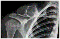
Figure 1
Pre operative X-ray showing the expansile bone mass in the lateral
end of right clavicle.
Figure 2
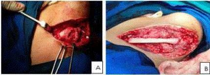
Figure 2
Intra operative photographs showing the tumour (A) and (B) after
excision and bone graft.
Figure3
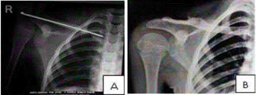
Figure 3
X-rays immediate post-op (A) and 1 year later (B).
Figure 4

Figure 4
Follow up at one year showing complete range of movements.
References
- Schajowicz F. Aneurysmal bone cyst. Histologic Typing of Bone Tumours. Berlin: Springer-Verlag; 1992. p. 37.
- Leithner A, Windhager R, Lang S, Haas OA, Kainberger F, Kotz R. Aneurysmal bone cyst. A population based epidemiologic study and literature review. Clin Orthop Relat Res. 1999;(363):176-9.
- Cottalorda J, Bourelle S. Modern concepts of primary aneurysmal bone cyst. Arch Orthop Trauma Surg. 2007;127(2):105-14.
- Burch S, Hu S, Berven S. Aneurysmal bone cysts of the spine. Neurosurg Clin N Am. 2008;19(1):41-7.
- McCarthy EF, Frassica FJ. Aneurysmal bone cyst. Pathology of Bone and Joint Disorders: With Clinical and Radiographic Correlation. Philadelphia: WB Saunders; 1998. p. 279-84.
- Freiberg AA, Loder RT, Heidelberger KP, Hensinger RN. Aneurysmal bone cysts in young children. J Pediatr Orthop. 1994;14(1):86-91.
- Rosenberg AE, Nielsen GP, Fletcher JA. Aneurysmal bone cyst. In: Fletcher CDM, Unni KK, Mertens F, editors. World Health Organization: classification of tumours: pathology and genetics of tumours of soft tissue and bone. Lyon: International Agency for Research on Cancer Press; 2002. p. 338-9.
- Dabska M, Buraczewski J. Aneurysmal bone cyst. Pathology, clinical course and radiologic appearances. Cancer. 1969;23(2):371-89.
- Jaffe HL, Lichtenstein L. Solitary unicameral bone cyst with emphasis on the roentgen picture, the pathologic appearance and the pathogenesis. Arch Surg. 1942;44(6):1004-25.
- Lau AW, Pringle LM, Quick L, Riquelme DN, Ye Y, Oliveira AM, et al. TRE17/ubiquitin-specific protease 6 (USP6) oncogene translocated in aneurysmal bone cyst blocks osteoblastic maturation via an autocrine mechanism involving bone morphogenetic protein dysregulation. J Biol Chem. 2010;285(47):111-20.
- Lichtenstein L. Aneurysmal bone cyst. A pathological entity commonly mistaken for giant-cell tumor and occasionally for hemangioma and osteogenic sarcoma. Cancer. 1950;3(2):279-89.
- Cole WG. Treatment of aneurysmal bone cysts in childhood. J Pediatr Orthop. 1986;6(3):326-9.
- Malghem J, Maldague B, Esselinckx W, Noel H, De Nayer P, Vincent A. Spontaneous healing of aneurysmal bone cysts. A report of three cases. J Bone Joint Surg Br. 1989;71(4):645-50.
- van Loon CJ, Veth RP, Pruszczynski M, Lemmens JA, van Horn JR. Aneurysmal bone cyst. Long-term results and functional evaluation. Acta Orthop Belg. 1995;61(3):199-204.
- Gibbs CP Jr, Hefele MC, Peabody TD, Montag AG, Aithal V, Simon MA. Aneurysmal bone cyst of the extremities. Factors related to local recurrence after curettage with a high-speed burr. J Bone Joint Surg Am. 1999;81(12):1671-8.
- Papagelopoulos PJ, Currier BL, Shaughnessy WJ, Sim FH, Ebsersold MJ, Bond JR, et al. Aneurysmal bone cyst of the spine. Management and outcome. Spine (Phila Pa 1976). 1998;23(5):621-8.