Case Report
A Rare Complication after Urethroplasty: Epidermoid Inclusion Cyst
Volkan Sarper Erikci*, Merve Dilara Öney and Gökhan Köylüoğlu
Department of Pediatric Surgery, Sağlik Bilimleri University, Katip Çelebi University, Tepecik Training Hospital,
Izmir, Turkey
*Corresponding author: Volkan Sarper Erikci, Department of Pediatric Surgery, Sağlik Bilimleri University, Katip Çelebi University, Tepecik Training Hospital, Kazim Dirik Mah. Mustafa Kemal Cad. Hakkibey Apt. No.: 45 D.10 35100, Bornova-İzmir, Turkey
Published: 11 Oct, 2017
Cite this article as: Erikci VS, Öney MD, Köylüoğlu G. A
Rare Complication after Urethroplasty:
Epidermoid Inclusion Cyst. Clin Surg.
2017; 2: 1671.
Abstract
Epidermoid İnclusion Cysts (EIC) occur as a result of the implantation of the epidermal keratinized squamous epithelial cells and sebaceous glands into the dermis and subcutaneous tissue after trauma and surgical interventions. A 5-year-old boy with a penile EIC who was operated elsewhere at the age of 1 year for an anterior hypospadias with a usage of skin graft covering urethroplasty is presented and discussed with regard to the foregoing literature. In order to avoid psychological and surgical trauma that can be seen after circumcision and hypospadias surgery in children, all the surgical interventions should be performed carefully and during surgical intervention implantation of the epidermis into the dermis and subcutaneous tissue should be avoided.
Introduction
Epidermoid İnclusion Cysts (EIC) occur as a result of the implantation of the epidermal keratinized squamous epithelial cells and sebaceous glands into the dermis and subcutaneous tissue [1,2]. These masses typically present as painless swellings located at the related locations of body. A 5-year-old boy with a penile EIC who was operated for hypospadias with a usage of skin graft covering urethroplasty is presented and discussed in the light of relevant literature.
Case Presntation
A 5-year-old boy was admitted to our clinic with a diagnosis of penile EIC. The patient had been operated at the age of 1 year for an anterior hypospadias with a usage of skin graft covering urethroplasty. Initially, the mass was reported to be small, but later it started to grow rapidly (Figure 1). Under general anesthesia with a vertical insicion on the ventral aspect of the penis the cystic mass was totally excised (Figure 2 and 3). Histopathological examination revealed an epidermal inclusion cyst with a dimension of 2 cm × 1.5 cm × 0.7 cm. The cyst had a capsule at the outer surface and contained keratinized material inside. Postoperative follow-up was uneventful.
Discussion
EICs occur as a result of the implantation of the epidermal keratinized squamous epithelial
cells and sebaceous glands into the dermis and subcutaneous tissue after trauma and surgical
interventions [1,2]. These masses are real cysts containing keratized material and are surrounded by
keratinized squamous epithelial cells. They can be congenital or acquired. Abnormal embryologic
closure of the median raphe is postulated to represent congenital forms of penile EIC [3]. In terms of
acquired etiological factors different theories have been proposed for these masses including penile
surgery and trauma. It is stated that epidermal cells are implanted within a circumscribed space of
the dermis during penile surgical interventions such as circumsicion or hypospadias surgery [2,4,5].
Idiopathic forms of penile EIC have also been described [6]. These masses can be single or multiple
with variable size. Accumulation of epidermal desquamations, secretions and debris in a closed
space leads to formation of a cystic and often painless swelling that gradually increases in size over
time [7]. The cystic mass located at the ventral aspect of the penile shaft in the presented case was
initially reported to be small, but later the family of the child stated that it started to grow rapidly.
Physical examination is all that is needed to diagnose these masses. In doubtful circumstances
ultrasonografy and means of other radiological evaluations may be used to confirm the diagnosis.
The differential diagnosis of penile EICs include urethral diverticula, urethrocutaneous fistula,
dermoid cysts or teratoma. Concerning our case, physical examination of penis was enough to
diagnose EIC and no other diagnostic radiological modalities were used. Complications of EIC
have been reported including rupture and release of keratin that leads to inflammatory reaction, infection, hematoma and rarely carcinomas [8,9]. With regard to our patient, although there is a relatively long time period (4 years)
between the initial hypospadias sugery and surgical excision of penile
EIC, we did not observe any complications in our case during pre- and
postoperative period. Nevertheless, once EIC has been detected at the
penis following trauma or other surgical procedures, surgical excision
of the mass should be a matter of necessity rather than of choice to
avoid above mentioned complications. The management of penile
EIC is complete surgical excision. Meticulous dissection of the mass
is necessary to avoid local recurrences. The capsule of the EIC should
not be violated during surgical excision of the mass otherwise local
implantation of the epidermal keratinized squamous epithelial cells
and sebaceous glands into the dermis may lead to recurrence during
follow-up period. Histopathological examination of the excised mass
is necessary to confirm the diagnosis of EIC. Typically these cystic
masses are lined by keratinized stratified squamous epithelium
containing keratinized sebaceous material inside. Histopathological
examination revealed an EIC with presence of cheesy material at
the cut surface of the mas in our patient. There is no recurrence at 3
months follow-up.
During childhood period, penile EICs are rarely seen and can
mimic other disease states. Surgical treatment, if performed early,
can prevent complications such as infection, hematoma and rarely
carcinomas. Literature on this subject reveals that most patients with
EIC were reported following circumcision [10,11]. An EIC of the penis
after urethroplasty causing an urethro-cutaneous fistula has also been
reported. To our knowledge after hypospadias surgery, there is no
pediatric patient with a EIC not associated with an urethro-cutaneous
fistula. Presented child is probably the first case of EIC following
hypospadias surgery not associated with other complications of
hypospadias. In order to avoid psychological and surgical trauma that
can be seen after circumcision and hypospadias surgery in children,
all the surgical interventions should be performed carefully. During
surgical intervention implantation of the epidermis into the dermis
and subcutaneous tissue should also be avoided. The possibility of
this diagnosis should be kept in mind for the patients with penile
cystic massses and managed accordingly.
Figure 1
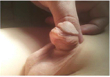
Figure 1
Preoperative appearance of penile EIC.
Figure 2
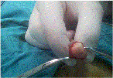
Figure 2
Operative view of EIC during excision.
Figure3
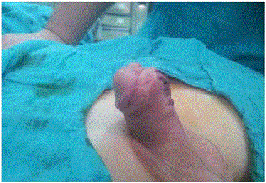
Figure 3
Postoperative view of the penis after removal of EIC.
References
- Okeke LI. Epidermal inclusion cyst as a rare complication of neonatal male circumcision: a case report. J Med Case Rep. 2009;3:7321.
- Suwa M, Takeda M, Bilim V, Takahashi K. Epidermoid cyst of the penis: a case report and review of the literature. Int J Urol. 2000;7(11):431-3.
- Giambanco A, Pensabene M, Giuffré M, Cimador M. Epidermal inclusion cyst of the penis after urethroplasty causing an urethro-cutaneous fistula: a first case report. Ped Med Chir. 2013;35(6):288-9.
- Saini P, Mansoor MN, Jalali S, Sharma A. Penile epidermal inclusion cyst. Indian J Pediatr. 2010;77(7):815-6.
- Park HJ, Park NC, Park SW, Jern TK, Choi KU. Penile epidermal inclusion cyst: a late complication of penile girth enhancement surgery. J Sex Med. 2008;5(9):2238-40.
- Aslan Y, Balci M, Atan A. Idiopathic penile epidermoid cyst in a young patient: three-year follow-up. Eur J Surg Sci. 2011;2(1):16-8.
- Kroll GI, Miller L. Vulvar epithelial inclusion cyst as a late complication of childhood female traditional genital surgery. Am J Obstet Gynecol. 2000;183(2):509-10.
- Somesh G, Sanjeev G, Vijar Kumar J, Bhushan A. A "stone" in the vulva. Sex Transm Inf. 2000;76:319.
- Arizpe SR, Condiani JO. Giant epidermoid cyst: clinical aspect and surgical management. J Dermatol Surg Oncol. 1986;12(7):734-6.
- Ben Chaim J, Livne PM, Binyamini J, Hardak B, Ben-Meir D, Mor Y. Complications of circumcision in Israel: a one year multicenter survey. Isr Med Assoc J. 2005;7(6):368-70.
- Little JS, Keating MA, Rink RC. Median raphe cysts of the genitalia. J Urol. 1992;148(6):1872-3.