Case Report
Indeginous RV-PA Conduit –Future for Intracardia repair of Tetraology of Fallot with Pulmonary Atresia
Suraj Wasudeo Nagre*, Krishnarao Narayanrao Bhosle, Ambrish Khatod and Vignesh
Ravikumar
Department of CVTS, Grant Medical College and Sir J J Group of Hospitals, Mumbai, India
*Corresponding author: Suraj Wasudeo Nagre, Department of CVTS, Grant Medical College and Sir J J group of Hospitals, Byculla, Mumbai, 400008, India
Published: 10 Oct, 2017
Cite this article as: Nagre SW, Bhosle KN, Khatod A,
Ravikumar V. Indeginous RV-PA
Conduit –Future for Intracardia repair
of Tetraology of Fallot with Pulmonary
Atresia. Clin Surg. 2017; 2: 1654.
Abstract
RV-PA conduit like contegra which is a xenograft from bovine IJV, has varied indications from TOF to complex congenital cardiac procedures like noorwood, Ross etc. RV-PA conduit has limited availability also the sizes available are limited. So we used indigenous made RV-PA conduit created from a polyester graft and a biocore valve in a 16 year old male for intracardiac repair of tetralogy of fallot [ICR TOF] with pulmonary atresia post BT shunt. Patient tolerated procedure well and is in regular follow up with RVOT gradient of 10 mm of HG. Indigenous made RV-PA conduit have longer shelf life and can be easily available but minimum size of bioprosthetic valve available was 21 mm hence limitation for use in children and infants. It is better alternative in adults where availability and cost of contegra graft was problem.
Abbreviations
RVPA-Right Ventricle Pulmonary Artery; ICR: Intracardiac Repair; TOF: Tetralogy of Fallot
Introduction
RV-PA conduit like contegra which is a xenograft from bovine IJV, has varied indications from TOF to complex congenital cardiac procedures like noorwood, Ross etc. RV-PA conduit has limited availability also the sizes available are limited. So we used indigenous made RV-PA conduit created from a polyester graft and a biocore valve in an 18 year old male for ICR TOF with pulmonary atresia post BT shunt. Patient tolerated procedure well and is in regular follow up with RVOT gradient of 36 mm of HG. Much complex congenital heart disease requires a conduit with valve. They are available as CONTEGRA and others. But one can use a bioprosthetic valve with poleyster graft as conduit to overcome limitation of size and availability.
Figure 1
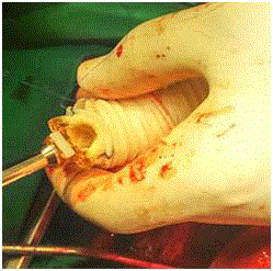
Figure 1
Indigenously made RV-PA conduit first sutured distal end Main
Pulmonary artery with prolene 4-0 continuous manner.
Figure 2
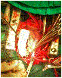
Figure 2
Peroneus brevis tendon after repair with direct tubularization.
Figure 3
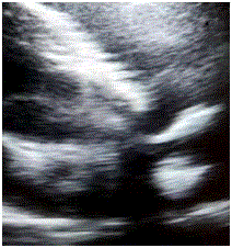
Figure 3
Postopt 2 D Echo showed good flow across the indigenously made
conduit with maximum gradient of 10 mm Hg.
Case Presentation
Sixteen year old male patient admitted with symptoms of NYHA class two dyspnea. Patient was a known case of tetralogy of fallot with pulmonary atresia. Patient had history of left side modified BT shunt done 8 years back with 6 mm PTFE graft. 2D echo was done s/o of complex congenital heart disease with large subaortic vsd of 30 mm with overriding of aorta with pulmonary atresia. MPA 12 mm RPA 7 mm and LPA 9 mm. Bt shunt had flow with gradients of 24 mmhg. Cardiac catch done showed blocked BT shunt, no significant MAPCA and Magoon’s index of 1.4. Patient planned for complete intracardiac repair. Patient body surface area of 1.4/m2. Because of unavailability of Contegra RVPA conduit in India during this period, decision was taken to make artificial RV-PA conduit. Polyester graft of 24 mm and biocore mitral valve of size 21 mm was used. The polyester graft was inturned. The valve was sutured midway to the inturned graft using 2-0 prolene in continuous fashion (Figure 1). RVOT coring was done and VSD was closed with a DVD patch in semicontineous fashion using prolene 4-0 plegdetted suture on CPB. Indigenously prepared RVPA conduit was suture in direction of right ventricle to main pulmonary artery (Figure 2). Patient tolerated procedure well and come off bypass without any problem. Post-opt 2D echo showed good flow across graft with maximum gradient of 10 mm HG (Figure 3). Patient is in regular follow up on antiplatelets with anticoagulant was given initially for 3 months with maintenance of INR between 1 to 2. Then we stopped anticoagulant and continued with antiplatelets. Since one year patient is doing well.
Discussion
The homograft conduit, harvested from human pulmonary artery or aortic tissue, has been the gold standard in Right Ventricle Outflow Tract (RVOT)
reconstruction ever since its inception in 1966 [1,2]. However,
homografts in the pulmonary position suffer from early calcification,
particularly in younger patients, resulting in unavoidable
reintervention [3,4]. To address this problem, the Contegra Bovine
Jugular Vein (BJV) conduit was developed by VenPro Corp. in 1999,
and acquired by Medtronic Inc. in 2001, to supplant the homograft
in RVOT reconstruction. Over the last decade, the Contegra conduit
has gained acceptance from surgeons internationally because of the
availability of an adequate range of sizes (12 mm to 22 mm), the
relatively low cost, and the low reported incidence of calcification.
Breymann et al. [5] reported a significantly lower conduit-related rate
of reoperation after 4 years of follow-up in their Contegra recipients
vs. their homograft patients. Brown et al. [6] declared the Contegra
their 'conduit of choice' in RVOT reconstruction after reporting
excellent early and midterm outcomes [7]. Despite these encouraging
results, other centres have reported cases of Contegra recipients
returning prematurely to the operating room (OR) due to distal
conduit stenosis. Meyns et al. [8] observed severe distal stenosis in
51% of their Contegra recipients at 2 years' follow-up. Gober et al.
[9] reported similar findings of supravalvular stenosis, necessitating
reintervention in 6 of their 38 Contegra recipients after an average
follow-up time of 18 months [10]. In addition, several other groups
have identified proximal aneurysmal dilatation of Contegra conduits
in the setting of distal stenosis-a novel mode of failure infrequently
seen with the homograft or porcine xenografts [11,12]. Consequently,
the debate on the Contegra as the long-term RVOT conduit of choice
has not ended. In our cardiovascular setup we usually use Contegra
bovine valved conduit for surgical correction of severe pulmonic and
infundibular stenosis/atresia. Contegra (Medtronic Inc, Minneapolis,
MN), a biological valved conduit consisting of a zero-pressure
glutaraldehyde preserved heterologous bovine jugular vein with a
trileaflet venous valve with natural sinuses. The claimed advantages
of this conduit are large variety of sizes available, easy tailoring and
suturing, adequate hemodynamics thanks to a favourable effective
orifice area, no need for proximal or distal extension.
The Contegra bovine jugular conduit marketed by Medtronic Inc.
has now been commercially available for nearly a decade in sizes 12
mm to 22 mm, most commonly used only for a substitute for RVOT.
But in year 2015-16 suddenly its importation to India stopped. We
started facing difficult in managing the patients of pulmonary atresia.
To overcome this difficulty we started making indeginous conduits
using polyester graft and bioprosthetic valve. Here we discussed
similar case with good early results. But for final conclusion we has to
use this indigenously made conduit more frequently in more number
of patients and follow the patients for longer duration.
Conclusion
Contegra conduits are costly and have propensity to shrinking and degenerate requiring reoperation sometimes. Indegeneously made RV-PA Conduit is good option in adults when there is unavailability of contegra. It also has longer shelf life and can be prepared from easily available biocore valve. But minimum size biocore valve available is 21mm hence limitation to use in children and infants. Further evaluation is needed in terms of long term results and number of patients. However recent studies in few centres have shown good long term outcome. Manually created RV-PA conduit using artificial grafts become a good alternative in future.
References
- Salem AM. Journal of the Egyptian Society of Cardio-Thoracic Surgery. 2016;24.
- Nagre SW. Surgical removal of embolized atrial septal defect device from pulmonary artery. J Med Case Rep. 2015;4(2):65-9.
- Frank Sellke, Pedro J. del Nido, Scott J. Swanson. Sabiston and Spencer's Surgery of the Chest. Elsevier. 2016.
- Boethig D, Goeler H, Westhoff-Bleck M, Ono M, Daiber A, Haverich A, et al. Evaluation of 188 consecutive homografts implanted in pulmonary position after 20 years. Eur J Cardiothorac Surg. 2007;32(1):133-42.
- Corno AF, Qanadli SD, Sekarski N, Artemisia S, Hurni M, Tozzi P, et al. Bovine valved xenograft in pulmonary position: Medium-term follow-up with excellent hemodynamics and freedom from calcification. Ann Thorac Surg. 2004;78(4):1382-8.
- Boethig D, Thies WR, Hecker H, Breymann T. Mid-term course after pediatric right ventricular outflow tract reconstruction: A comparison of homografts, porcine xenografts and Contegras. Eur J Cardiothorac Surg. 2005;27(1):58-66.
- Brown JW, Ruzmetov M, Rodefeld MD, Vijay P, Darragh RK. Valved bovine jugular vein conduits for right ventricular outflow tract reconstruction in children: an attractive alternative to pulmonary homograft. Ann Thorac Surg. 2006;82(3):909-16.
- Boudjemline Y, Bonnet D, Massih TA, Agnoletti G, Iserin F, Jaubert F, et al. Use of bovine jugular vein to reconstruct the right ventricular outflow tract: early results. J Thorac Cardiovasc Surg. 2003;126(2):490-7.
- Meyns B, Garsse LV, Boshoff D, Eyskens B, Mertens L, Gewillig M, et al. The Contegra conduit in the right ventricular outflow tract induces supravalvular stenosis. J Thorac Cardiovasc Surg. 2004;128(6):834-40.
- Gober V, Berdat P, Pavlovic M, Pfammatter JP, Carrel TP. Adverse mid-term outcome following RVOT reconstruction using the Contegra valved bovine jugular vein. Ann Thorac Surg. 2005;79(72):625-31.
- Bautista-Hernandez V, Kaza AK, Benavidaz OJ, Pigula FA. True aneurysmal dilatation of a contegra conduit after right ventricular outflow tract reconstruction: A novel mechanism of conduit failure. Ann Thorac Surg. 2008;86(6):1976-7.
- Delmo-Walter EM, Alexi-Meskishvilli V, Abdul-Khaliq H, Meyer R, Hetzer R. Aneurysmal dilatation of the Contegra bovine jugular vein conduit after reconstruction of the right ventricular outflow tract. Ann Thorac Surg. 2007;83(2):682-4.