Case Report
Conservative Management of Lower Pole Pelvi-Ureteric Junction Obstruction in Duplicated Collecting System
Carmelo Agostino Di Franco1*, Giovanni Giliberto,1, Daniele Porru1, Tiziano Cebrelli1, Lavinia
Galvagno2 and Bruno Rovereto1
1Department of Urology, University Hospital Foundation IRCCS Policlinico S. Matteo of Pavia, Italy
2English and Spanish Translation and Teaching Service, Enna, Italy
*Corresponding author: Carmelo Agostino Di Franco, Department of Urology, University Hospital Foundation IRCCS Policlinico S. Matteo of Pavia, Italy
Published: 10 Oct, 2017
Cite this article as: Di Franco CA, Giliberto G, Porru D,
Cebrelli T, Galvagno L, Rovereto B.
Conservative Management of Lower
Pole Pelvi-Ureteric Junction Obstruction
in Duplicated Collecting System. Clin
Surg. 2017; 2: 1653.
Abstract
Duplication of collecting system is one of the most common upper urinary tract anomalies, affecting around 15% of the population. In the duplicated system, kidney has two pyelocaliceal systems and it is associated with a single ureter or with a bifid ureter (partial duplication) or, in the case of a complete duplication, with two ureters (double ureters) that drain separately into the urinary bladder. In this last case, usually, ureters follow the “Weigert-Meyer Law” according to which, upper pole ureter drains medially and inferiorly compared to lower pole ureter that drains in bladder in orthotopic position, laterally and superiorly compared to the other one. Partial duplication can be associated with two problems: pelvi-ureteric junction obstruction and retrograde peristalsis of urine in the two ureters as “Yo-Yo phenomenon”. We describe a case of a 20 years-old woman who accessed to our department for right flank pain and fever. CT-scan showed incomplete duplication of right collecting system with hydronephrosis and thinned cortical of lower kidney moiety. Patient was successfully treated in conservative way.
Case Presentation
A 20 years-old female patient accessed to our department for right flank pain and fever. Renal colic or acute pyelonephritis was suspected and the patient performed an abdomen ultrasound showing right hydronephrosis and then abdominal contrast CT-scan. The CT-scan was negative for calculi or renal infectious signs, but revealed incomplete duplication of right collecting system with hydronephrosis and thinned kidney cortical of lower moiety (Figure 1). Blood tests were in normal range, no leucocytosis, creatinine was normal (0,55 mg/dl). Urine culture test was positive for Klebsiella pneumonia and targeted antibiotic therapy was started with rapid fever resolution, but without pain relief. On the basis of CT-scan that showed thinned renal cortical of lower pole. In order to solve painful symptoms, we decided to perform retrograde pyelography and to insert a double J ureteral stent, putting it into the lower kidney system as showed in (Figure 2). In immediate post-operative days, the patient referred resolution of flank pain, so we organized an outpatient dynamic kidney scintigraphy with Tc99m-DTPA (diethylenetriaminepentacetate). Scintigraphy, performed with double J stent, showed a good renal function, normally divided between the two kidneys with a good filtration function of lower right kidney collecting system. However, a couple of weeks after stenting insertion, the patient accessed to our department for flank and abdominal pain. Ultrasound and CT-scan showed slight hydronephrosis and correct insertion of the ureteral stent. Despite analgesic therapy, the patient was intolerant to the stent, therefore in accordance with the patient, we decided to position percutaneous nephrostomy. We obtained fast and good resolution of painful symptoms. After one month, we performed a surgical treatment of latero-lateral interureteropelvic anastomosis; we did not performed reduction of redundant dilated renal pelvis. We inserted a double J 7 Fr ureteral. The patient received adequate analgesic therapy because she had a low pain tolerance. After one month, we removed the ureteral stent with complete resolution of symptoms. A CT-scan performed after 18 months of regular follow-up revealed a moderate dilated right lower pole pelvis (compatible with previous dilatation) with a good filtration and excretion right kidney function (Figure 3).
Discussion
Ureteral duplication is one of the most common renal abnormalities. It can be unilateral or bilateral and sometimes it is associated with other genitourinary tract anomalies [1]. Bifid ureter (or incomplete ureteral duplication) may be associated complete duplication of contralateral ureter,congenital hydronephrosis, Goltz’s syndrome, cephalad-renal ectopia and duplication of pelvis [2]. In two-thirds of the cases, ureteral duplication is incomplete and ureter-ureteral reflux may occur in more than 80% of cases. Severe reflux may be associated with a loss of cortical function in the affected renal segment [3]. Partial duplication is reported to be twice more common in females and on the right side, it is usually diagnosed in young adults and most of the cases show hydronephrosis and recurrent UTI, as in our case. During fetal age, the most common cause of hydronephrosis is represented by pelvi-ureteric obstruction, with 60% to 80% of cases and duplication of renal collecting system is the most common upper tract anomaly with a female: male ratio of 2:1. Embryologically, duplication occurs when two separate ureteric buds arise from a single Wolffian duct (mesonephric duct) [4]. Due to the future lower pole ureter separating from the Wolffian duct earlier, it migrates superiorly and laterally as the urogenital sinus grows and becomes the upper pole moiety [5]. Despite this migration of the upper tract, the insertion maintains the original embryologic relationship inferiorly, and thus the upper pole moiety ureter drains infero-medial to the normal lower moiety ureter (Weigert-Meyer Law). The ectopic insertion often has an ureterocoele that obstructs its own collecting system, and can distort the orthotopic lower pole moiety insertion such that it is prone to reflux [6]. Uretero-ureteral reflux, also known as yo-yo phenomenon, is a common but transitory condition. It is less frequently observed because the images obtained during excretory phase, do not record the event [7]. It should be suspected when there is asymmetric dilatation of ureters. Uretero-ureteral reflux prevents the urinary tract from ever being completely drained and is responsible for flank pain, renal colic symptoms and recurrent urinary tract infections, frequently associated with partial duplication of ureters. In our case, probably, the patient had both reflux, that could explain cyclic and not continuous flank pain and recurrent urinary infections and fever, and pelvi-ureteric junction obstruction showed by contrast CT-scan that revealed hydronephrosis and excretion delay [8]. However, kidney scintigraphy performed with double J stent inside, showed a good renal function. Some authors suggest that the standard surgical treatment of symptomatic ureteral duplication (flank pain, recurrent urinary infections, fever) may be heminephrectomy to reduce the risk of urosepsy in patients with ureteral reflux [9]. In this case, our patient was young (20 years old), renal function was conserved bilaterally, and this suggested us, in accordance with the patient, to perform a conservative surgical treatment. Firstly, we inserted a ureteral double J in lower hydronephrotic collecting system assisting on the one hand to fever resolution but on the other hand to recurrent flank pain with slight upper collecting system dilatation: we think it could be associated with the partial mechanical obstruction due to stent at the ureteral bifurcation point [10]. In fact, after stent removal and positioning of percutaneous nephrostomy, we had rapid pain resolution, confirming our theory. We decided to perform laterolateral inter-ureteropelvic anastomosis obtaining good kidney drainage and complete resolution of pain and recurrent urinary infections.
Figure 1
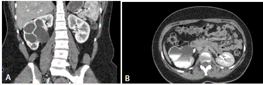
Figure 1
(A and B) Contrast CT-scan shows hydronephrosis of right kidney
lower system with incomplete duplication of right collecting system.
Figure 2
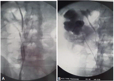
Figure 2
A) Retrograde pyelography showing incomplete duplication (bifid
ureter) with duplication of right collecting system; B) Double J ureteral stent
positioned in right hydronephrotic lower kidney system..
Figure3
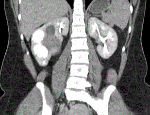
Figure 3
Constrast CT-scan performed after 18 months of follow-up showed
a moderate dilated right lower pole pelvis (compatible with previous dilatation)
with a good right kidney function
Conclusion
Duplication of renal collecting system in adult is usually asymptomatic and its diagnosis is often incidental. When the condition becomes symptomatic, the management can require a different approach to obtain a good clinical and functional outcome.
References
- Tundidor Bermúdez AM. [A bifid ureter with a blind branch]. Arch Esp Urol. 1999;52(7):790-2.
- Das S, Dhar P, Mehra RD. Unilateral isolated bifid ureter: a case report. J Anat Soc India. 2001;50(1):43-4.
- Gündüz K, Günalp I, Erden I. Focal dermal hypoplasia (Goltz's syndrome). Ophthalmic Genet. 1997;18(3):143-9.
- Attia Haider MA. Cephalad renal ectopia, duplication of pelvicalyceal system and patent ductus arteriosus in an adult female. Scand J Urol Nephrol. 1999;33(4):257-9.
- Wu F, Snow B, Taylor A Jr. Potential pitfall of DMSA scintigraphy in patients with ureteral duplication. J Nucl Med. 1986;27(7):1154-6.
- Rege VM, Deshmukh SS, Borwankar SS, Gandhi RK. Blind ending bifid ureter (a case report). J Postgrad Med. 1986;32(4):233-5.
- Gonzalez F, Canning DA, Hyun G, Casale P. Lower pole pelvi-ureteric junction obstruction in duplicated collecting systems. BJU Int. 2006;97(1):161-5.
- Gupta K, Galhotra R, Saggar K. Yo-yo reflux in partial duplication of ureter: A diagnosis on the color and pulse Doppler study. Muller Journal of Medical Sciences and Research. 2013;4(2):116-8.
- Fernbach SK, Feinstein KA, Spencer K, Lindstrom CA. Ureteral Duplication and Its Complications-Scientific Exhibit. Radiographics. 1997;17:109-27.
- Abouassaly R, Gill IS, Kaouk JH. Laparoscopic Upper Pole Partial Nephrectomy for Duplicated Renal Collecting Systems in Adult Patients. Urology. 2007;69:1202-5.