Case Report
Split Central Venous Sampling of Parathyroid Hormone in Non-localizing Parathyroid Tumors
T Tai and DR Maceri*
Department of Otolaryngology – Head and Neck Surgery, Keck School of Medicine of USC, 1975 Zonal Ave, Los
Angeles, CA 90033, USA
*Corresponding author: Dennis R. Maceri, Department of Otolaryngology – Head and Neck Surgery, Keck School of Medicine of USC, 1975 Zonal Ave, Los Angeles, CA 90033, USA
Published: 05 Oct, 2017
Cite this article as: T Tai, DR Maceri. Split Central Venous
Sampling of Parathyroid Hormone in
Non-localizing Parathyroid Tumors. Clin
Surg. 2017; 2: 1647.
Abstract
One- hundred-and-fifteen patients underwent neck exploration for hyperparathyroidism at the Keck School of Medicine, University of Southern California. Twenty-seven of these patients had intraoperative parathyroid hormone levels drawn simultaneously from both internal jugular veins and received a definitive diagnosis of their disease based on tissue pathology. This majority of these diagnoses were comprised of parathyroid adenomas or multiglandular (four-gland) hyperplasia. From this cohort, thirteen cases had parathyroid abnormalities that could not be localized by technectium-99 sestamibi scan, while six patients received imaging results that conflicted between sestamibi scan and high resolution neck ultrasonography or sestamibi scan and 4D CT scan. By utilizing the split venous sampling technique, we demonstrate that the hormone gradient between the left and right internal jugular veins is a statistically valid predictor of the side and the etiology of a parathyroid abnormality. This holds true even in instances where patients receive non-localizing or conflicting preoperative imaging results. The accuracy of this technique improves dramatically as the absolute value of the gradient exceeds 100 pg/ml. Split vein parathyroid hormone sampling is a useful adjunct in guiding parathyroid surgery when conventional imaging modalities are inadequate. Application of this technique provides the surgeon a greater degree of assurance that a minimally invasive approach will result in an appropriate cure.
Introduction
Split Central Venous Sampling of Parathyroid Hormone in Non-localizing Parathyroid Tumors. Parathyroid surgery has improved tremendously since the adoption of intraoperative parathyroid hormone (IO-PTH) monitoring in 1990 by Yves Chapuis and George Irvin as an adjunct to preoperative parathyroid imaging [1,2]. Steady advances in diagnostic radiology and biochemical analysis continue to increase the yield and efficiency of intraoperative PTH assessment so that focused neck exploration can be performed with minimal risk of overlooking residual or multiglandular disease. In its current state, the technetium-99m sestamibi scan accurately localizes hyper functional tissue in 71% - 79% of patients with a single adenoma [3]. Single adenomas account for 85% - 97% of documented hyperparathyroidism – with multi-glandular hyperplasia (2.5% - 15%) and carcinoma (1%) forming the remainder of cases [4]. Coupling sestamibi technology with four-dimensional Computed Tomography (4D CT) and neck ultrasonography demonstrably improves sensitivity to 79% - 95% [5]. Still, there remain instances when preoperative imaging is inconclusive or in conflict amongst the different modalities. The conventional algorithm for parathyroidectomy is outlined in Figure 1. One-sided minimallyinvasive parathyroidectomy is performed when disease is localized preoperatively. Otherwise, patients with inconclusive localization are treated with bilateral exploration of the four parathyroid glands. In a previous study, we demonstrated that intraoperative split Internal Jugular Vein (IJV) PTH values can be used to inform the surgeon of laterality in situations where preoperative imaging is unable to localize parathyroid abnormalities [6]. That study abided by the Mayo protocol, where a >50% reduction compared to baseline peripheral PTH levels 10 or more minutes after careful exploration and resection was considered to be biochemically curative. In the face of nonlocalizing or conflicting preoperative imaging studies, the author maintains the value in employing this split internal jugular vein PTH sampling technique as an aid to determine the etiology and laterality of parathyroid disease. This study evaluates the continued efforts of a single institution to use this method.
Figure 1
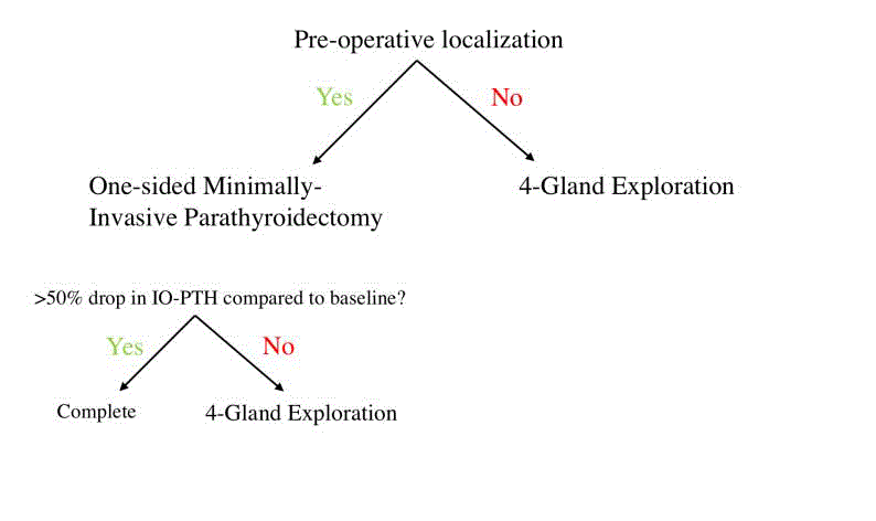
Figure 1
Current algorithm for the surgical approach to parathyroidectomy. One-sided minimally-invasive
parathyroidectomy is only performed in instances of successful pre-operative localization. Successful localization
is defined as localizing sestamibi imaging with coinciding high-resolution ultrasonography for confirmation.
Materials and Methods
Participants
One hundred and fifteen patients underwent neck exploration
for hyperparathyroidism at the Keck School of Medicine of the
University of Southern California between January 2010 to May
2016 (excluding 2012). A cohort of 41 individuals had intraoperative
PTH levels drawn from both jugular veins, which represents the
protocol in question aimed at minimizing the extent of surgery and
determining the ability of increased PTH levels to correlate with the
side of the adenoma. Patients with secondary hyperparathyroidism
(5) or parathyroid cancer (1) were excluded from the study. Patients
without a final pathologic diagnosis (3) and/or without PTH levels
drawn from both left and right internal jugular veins were also
excluded. Revision surgical cases were discounted as well (1). The
medical records of all patients were reviewed retrospectively in
accordance with the regulations and practices formulated by the
Health Sciences Institutional Review Board.
Procedure
Samples drawn for the patient intraoperatively were immediately
analyzed by a technologist with the immunochemiluminesence assay
(Future Diagnostics, Wijchen, The Netherlands). The peripheral vein
sample obtained after induction of general anesthesia served as the
baseline preoperative PTH level. For each patient, the left and right
internal jugular vein samples were compared for gradients. Preoperative
localization studies with technetium-99-sestamibi scintigraphy were
used to evaluate for abnormal parathyroid tissue. These results were
confirmed with high-resolution neck ultrasonography. 4D CT was
used in select patients. In certain cases, sestamibi scans were nonlocalizing.
The split internal jugular vein PTH gradients were used to
direct the initial area of surgical exploration. In all cases, suspicious
gland removal was followed by IO-PTH sampling from a peripheral
vein 10 minutes later. Repeated failure of the PTH levels to drop >50%
from the baseline value after careful unilateral exploration resulted in
a bilateral 4-gland exploration and redrawing of PTH levels until the
criteria was satisfied (Figure 2).
Statistical methods
The p-values for the difference in PTH between the right and left
internal jugular veins were calculated by use of paired sample t tests
stratified by location and pathologic tissue confirmation. The p values
for difference in right and left internal jugular veins by localization and side were calculated with independent sample t tests. Sensitivities and
specificities were generated by comparing gradients which correctly
predicted etiology and side versus those that were incorrect (Figure
3 and 4). P-values for comparing the proportion of non-localizing
studies between this study and the previous study were calculated by
the use of a z-test. Statistical analysis was performed with GraphPad
Prism version 7.0 (GraphPad Software). Statistical significance was
defined as P< 0.05.
Figure 2
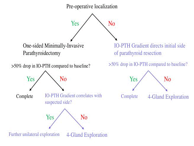
Figure 2
Study algorithm for the surgical approach to parathyroidectomy.
The IO-PTH gradient is used to direct the initial side of parathyroid resection
if preoperative localization is unsuccessful. Furthermore, for patients with
localizing studies that fail to yield the appropriate surgical result, the surgeon
proceeds to perform a bilateral four gland exploration.
Figure3
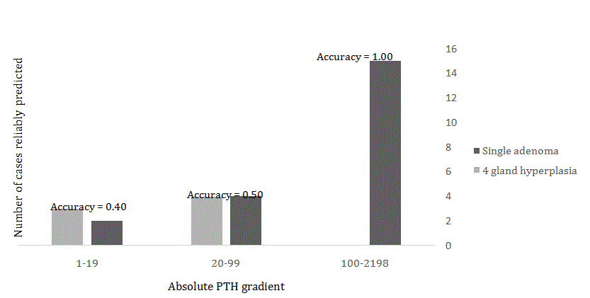
Figure 3
Predictive value of the PTH Gradient. The gradient was calculated
as the difference between the right and left internal jugular split values. The
larger the absolute value of the gradient, the more likely the gradient correctly
predicted the side of the tumor.
Figure 4
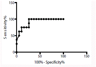
Figure 4
Receiver Operator Characteristics Curve. The area under the
receiver operating characteristics curve for the 27 patients with a final
definitive side diagnosis was 0.911. An area of 1.0 represents a perfectly
predictive test. Therefore, this gradient is significantly better at predicting the
presence of a parathyroid adenoma and its side than chance alone.
Table 1

Table 1
Patients with hyperparathyroidism.
Results
After applying the selection criteria, 27 patients had undergone surgery with bilaterally-drawn internal jugular vein PTH samples and received a definitive pathologic diagnosis to correlate with the intraoperative findings. Thirteen of these patients had nonlocalizing technetium-99-sestamibi scans and 6 cases had conflicting preoperative imaging. Table 1 and 2 illustrates the patients that had localizing or non-localizing scans and the etiology of their disease. Table 3 shows the means and standard deviations of intraoperative internal jugular PTH samples. It demonstrates that left-sided adenomas are significantly more likely to have higher left-sided PTH values while right-sided adenomas are significantly more likely to have high-right sided PTH values. There is no statistically significant relationship between 4-gland hyperplasia patients and their left or right-sided PTH values. Overall, an increase in right-sided IJV PTH values correlated with right-sided adenomas and an increase in leftsided IJV PTH values correlated with left-sided adenomas. Of the 20 patients that had a single adenoma, the PTH gradient was correct in predicting the adenoma’s side in 19 cases. Graph 3 represents the accuracy of this sampling technique: the greater the absolute value of the gradient between the left and right IJ samples, the more likely the gradient was predictive of tumor side. For patients with a PTH gradient 100 and above, both the presence of a unilateral adenoma and its respective side was predicted with 100% accuracy. Below this threshold, the ability of the gradient to predict side and etiology begins to diminish. The area under the receiver operating characteristics (ROC) curve in Figure 2 represents the ability of this gradient to discriminate between a lateralized adenoma on the correct side against adenomas on the wrong side or multiglandular disease. An area of 0.5 implies that the test has no predictive value, while an area of 1.0 signifies a perfectly predictive test. The area under ROC curve was 0.911, indicating that the gradient is substantially better at distinguishing tumor side than chance alone.
Discussion
Since publishing our first report in 2011, the standard for IOPTH monitoring in parathyroid surgery has remained largely unchanged. IO-PTH levels drawn 10-minutes after excision that decrease more than 50% when compared to baseline levels are widely accepted as an indicator of operative success. In that time, our institution has been gathering additional data to best inform surgical decision-making especially when preoperative sestamibi scintigraphy is unreliable. When inadequate localization occurs, ectopic adenomas, multiglandular disease or imaging limitations must be considered. We demonstrate here that a PTH gradient across split internal jugular vein samples positively correlates with the side of a parathyroid adenoma regardless of preoperative imaging results. Furthermore, the study reinforces that unilateral adenomas exhibit a larger gradient when compared to those of multiglandular disease. Gradients above 100 pg/ml are 100% accurate in predicting a parathyroid adenoma as the root cause of the hyperparathyroidism and the laterality of its location. It is important to note that onesided minimally-invasive parathyroidectomies can be performed as a same-day outpatient procedure while bilateral four gland explorations require postoperative inpatient monitoring for several days. By better discerning which patients require invasive surgeries, improvements in efficiency, cost reduction and morbidity reduction can be achieved and are worthwhile avenues for future studies. It is unclear if the fidelity of the sestamibi scan has improved over time. In the original study, 41% of the study population had non-localizing sestamibi scans; 29.5% of patients in the newer cohort have nonlocalizing studies but the difference in these proportions is statistically insignificant (p=0.24). 4D CT became available for this institution in 2013. Of the few studies using this technique, one scan correctly reconciled otherwise conflicting locations of an adenoma, two scans correctly identified the location of adenomas where sestamibi scan was inadequate and one scan incorrectly predicted a single mass in the presence of multiglandular disease. Relatively low numbers of 4D CT use limit the ability to make quantitative statements on its accuracy at this time [7]. The PTH gradient also demonstrated utility in reconciling cases where localization studies presented conflicting findings. There were nine documented cases where sestamibi scan, neck ultrasound or 4D CT findings were in disagreement. Of these nine cases, the PTH gradient was correctly predictive of the side in eight of the cases. A ROC curve assesses the statistical validity of a diagnostic test. It is a valuable tool because its output is independent from user selection of arbitrary significance cutoffs [8]. In this context, the ROC curve demonstrates that the PTH gradient is a statistically sound method of differentiating tumor side and etiology. In between the original study and this update, other articles have argued for and against the utility of bilateral internal jugular vein sampling as a means of localizing tumors when preoperative imaging has failed [9-14]. Ito et al. [12] demonstrated that bilateral IJV PTH sampling provided useful positional information when the PTH level of one side was greater than the other by 5%. However, this study did not reconcile the changes in PTH levels seen with multiglandular disease. To date then, this is the only study to demonstrate that a threshold gradient can be used to predict location and the likelihood of adenoma vs. multiglandular disease. The limitations of our study stem from a relatively small sample size of 115, all of which come from a single tertiary institution. This may result in a more complicated patient base and one that may not be applicable to other populations.
Table 2
Table 2
Internal jugular vein PTH values in patients with pathologic confirmation.
Table 3
Table 3
Predictive value of the PTH Gradient.
Conclusion
Based on our study, split internal jugular vein IO-PTH gradients serve as a valuable tool when preoperative diagnostic imaging is uncertain. For these patients, PTH gradients exceeding 100 pg/ ml were a 100% accurate in predicting the presence of a single adenoma in addition to its location. Below this point, suspicion for multiglandular hyperplasia should be increased and the threshold for a full exploration should be low. Because of its utility, we have used and continue to use this technique at our institution with great benefit.
References
- Chapuis Y, Fulla Y, Icard P, Nonnemacher L. Peroperative assay of active parathyroid hormone 1-84 in surgery of primary hyperparathyroidism. Presse Med. 1990;19(31):1461-2.
- Irvin GL, Dembrow VD, Prudhomme DL. Operative monitoring of parathyroid gland hyperfunction. Am J Surg. 1991;162(4):299-302.
- Suh YJ, Choi JY, Kim SJ, Chun IK, Yun TJ, Lee KE, et al. Comparison of 4D CT, ultrasonography, and 99mTc sestamibi SPECT/CT in localizing single-gland primary hyperparathyroidism. Otolaryngol Head Neck Surg. 2015;152(3):438-43.
- Wells SA Jr, Leight GS, Ross AJ. Primary hyperparathyroidism. Curr Probl Surg. 1980;17(8):398-463.
- Hinson AM, Lee DR, Hobbs BA, Fitzgerald RT, Bodenner DL, Stack BC Jr. Preoperative 4D CT Localization of Nonlocalizing Parathyroid Adenomas by Ultrasound and SPECT-CT. Otolaryngol Head Neck Surg. 2015;153(5):775-8.
- Maceri DR, Kokot N, Green K, Montgomery V, Sharifi J. Split central venous sampling of parathyroid hormone: an adjunct to surgical exploration. Head Neck. 2011;33(12):1715-8.
- Richards ML, Thompson GB, Farley DR, Grant CS. An optimal algorithm for intraoperative parathyroid hormone monitoring. Arch Surg. 2011;146(3):280-5.
- Hajian-Tilaki K. Receiver Operating Characteristic (ROC) Curve Analysis for Medical Diagnostic Test Evaluation. Caspian J Intern Med. 2013;4(2):627–35.
- Barczynski M, Konturek A, Hubalewska-Dydejczyk A, Cichon S, Nowak W. Utility of intraoperative bilateral internal jugular venous sampling with rapid parathyroid hormone testing in guiding patients with a negative sestamibi scan for minimally invasive parathyroidectomy--a randomized controlled trial. Langenbecks Arch Surg. 2009;394(5):827-35.
- Inabnet WB. Intraoperative parathyroid hormone monitoring. World J Surg. 2004;28(12):1212-5.
- Inabnet WB, Rayfield EJ, Kim CK. The utility of intraoperative parathyroid hormone monitoring with false-positive sestamibi scintigraphy. Thyroid. 2002;12(9):833-4.
- Ito F, Sippel R, Lederman J, Chen H. The utility of intraoperative bilateral internal jugular venous sampling with rapid parathyroid hormone testing. Ann Surg. 2007;245(6):959-63.
- Korovin LN, Guerrero MA. Laterality of central venous sampling: lack of effect on the accuracy of intraoperative parathyroid hormone monitoring. Am J Surg. 2013;206(6):883-6.
- Norman J, Lopez J, Politz D. Abandoning unilateral parathyroidectomy: why we reversed our position after 15,000 parathyroid operations. J Am Coll Surg. 2012;214(3):260-9.