Clinical Image
Vertebral Cyct Hydatid Case
Cansever Levent*, Seyrek Yunus and Bedirhan Mehmet Ali
Department of Chest Disease and Thoracic Surgery, University of Health Sciences/Yedikule Chest Disease and
Thoracic Surgery Health Practice and Research Center, Istanbul, Turkey
*Corresponding author: Cansever Levent, Department of Chest Disease and Thoracic Surgery, University of Health Sciences/Yedikule Chest Disease and Thoracic Surgery Health Practice and Research Center, Istanbul, Turkey
Published: 03 Aug, 2017
Cite this article as: Levent C, Yunus S, Ali BM. Vertebral
Cyct Hydatid Case. Clin Surg. 2017; 2:
1636.
Clinical Image
Cyct Hydatic (CH) is a disease with an incidence of 1-150/100.000 [1]. Although CH can occur almost every organ in human body, it commonly presents itself in liver (60% - 80%) at adult age group. Lungs are the second most common localization where CH is spotted (10% - 30%) [2]. Moreover; CH can also very rarely appear in pleura, brain tissue, cardia, mediastinum, subcutan tissue and bone tissue [3]. Bone tissue incidence is reported to be between 0.9% - 2.5% [14]. After 29 year old male patient’s cyct hydatid is extracted from his musculus lattissimus dorsi, his other cyct which elongates to vertebral foramen is removed by the assitance of neuro surgeons (Figure 1 and 2). Vertebral fusion and cage procedures are performed for maintaining vertebral stability (Figure 3 and 4).
Figure 1
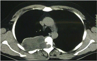
Figure 1
Pre- operative Computed Tomography Image.
Figure 2
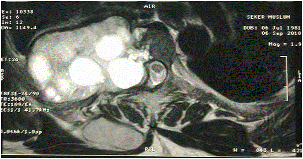
Figure 2
Thorax Magentic Resonance Imaging Image 1.
Figure3
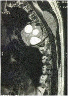
Figure 3
Thorax Magentic Resonance Imaging Image 2.
Figure 4
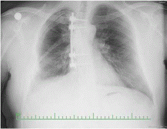
Figure 4
Post-operative Postero- Anterior Chest X-Ray Image
References
- Altintas N. Past to present: echinococcosis in Turkey. Acta Trop. 2003;85(2):105-12.
- Eroglu A, Kurkcuoglu C, Karaoglanoglu N. Bilateral pulmonary Echinococcosis. Thorax. 1991;46:599-600.
- Gouliamos AD, Kalovidouris A, Papailiou J, Vlahos L, Papavasiliou C. CT appearance of pulmonary hydatid disease. Chest. 1991;100(6):1578-81.
- Nhamoucha Y, Alaoui O, Doumbia A, Oukhoya M, Abdellaoui H, Tazi M, et al. [Bone hydatid cyst: a rare localization at the level of the hip bone]. Pan Afr Med J. 2016;24:226.