Research Article
Reconstruction Optıons of Lower Lip
Gamze Bektas and Ani Cinpolat*
Departemnt of Plastic Reconstructive and Aesthetic Surgery, Clinic Anka Private Practice, İstanbul, Turkey
*Corresponding author: Ani Cinpolat, Departemnt of Plastic Reconstructive and Aesthetic Surgery, Clinic Anka Private Practice, İstanbul, Turkey
Published: 03 Aug, 2017
Cite this article as: Bektas G, Cinpolat A. Reconstruction
Optıons of Lower Lip. Clin Surg. 2017;
2: 1629.
Abstract
Lip reconstruction can generate a considerable challenge to the plastic surgeon because of their role in aesthetic balance, facial expression, speech and deglutination. The goals of lip reconstruction are to provide adequate replacement of external skin, oral lining and vermilion while maintaining the aesthetic balance of the vermiliocutaneous junction and to maintain the competence of the orbicularis muscle sphincter, the adequate diameter for oral aperture and the adequate labial sulcus depth. This chapter presents techniques for vermillion, perioral cutaneous, and full-thickness lip reconstruction. Traditional techniques were summarized and some new techniques were broadly described to optimize the functional and cosmetic outcome.
Introduction
Lip reconstruction can generate a considerable challenge to the plastic surgeon in that the lips are the primary aesthetic and dynamic center of the lower third of the face. Their role in aesthetic balance, facial expression, speech, and deglutination is not replicated by any other tissue substitute. The goals of lip reconstruction are to provide adequate replacement of external skin, oral lining and vermilion while maintaining the aesthetic balance of the vermiliocutaneous junction and to maintain the competence of the orbicularis muscle sphincter, the adequate diameter for oral aperture and the adequate labial sulcus depth. Ideally, cutaneous sensation is preserved or reestablished to provide proprioceptive feedback for speech, animation, and management of secretions [1,2]. Lower lip reconstruction is more significant, because oral competence depends greatly on a functional lower lip having good muscular function, adequate height and sensation.
Anatomy and Etıology
The lips consist of four basic components: external skin, vermilion, the mucosa and the muscle. The vermilion separates the skin of the external lip and the mucosa of the inner lip. It is composed of keratinizing glabrous epithelium with numerous sebaceous glands. The primary muscles of the lip, the orbicularis oris muscles, maintain the oral competence by acting as a circumoral sphincter. They are paired, horizontally oriented muscles that link the modiolus and philtral columns producing a tightening of the lip. The orbicularis is acted upon by the surrounding elevating and depressing mimetic musculature. The levator labii superioris, levator anguli oris, and the zygomaticus major and minor elevate the upper lip, and the mentalis, depressor labii inferioris, and the depressor anguli oris draw the lip inferiorly [3]. The main arterial blood supply to the lips is from the facial artery with the inferior labial artery supplying the lower lip and the superior labial artery and branches from the angular artery supplying the upper lip [3,4]. Motor innervation of the lip is provided by the buccal and marginal mandibular branches of the facial nerve; sensory innervation of the upper lip is derived from (V2) the infraorbital nerve and the the lower lip receives its sensibility from (V3) the mental nerve [3,4]. The etiologies of acquired lip defects are mostly oncologic resection and trauma. Tumors are usually basal cell carcinoma in the upper lip and squamous cell carcinoma in the more sun-exposed lower lip. Melenoma is also quite common on the lip. Traumatic defects are usually seen in young, healty patients [5].
Figure 1
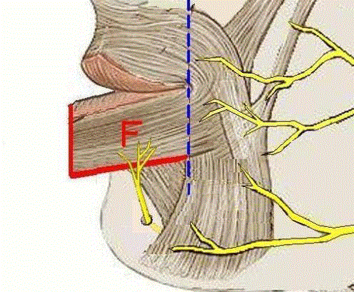
Figure 1
Schematic illustration of transverse lip advancement flap.
Figure 2
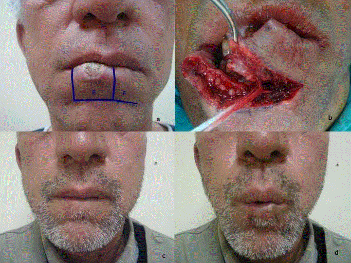
Figure 2
(a) Patient with lower lip squamous ca. (b) Excision, flap elevation
and mental nerve dissection. (c,d) Three months postoperative view.
Figure 3
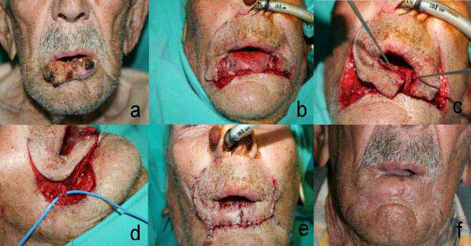
Figure 3
(a) 88 year-old patient with lower lip SCC. (b,c) Excision and
extended karapandzic flaps elevation. (d) Dissection of neurovascular
structures. (e) Immediate postoperative view. (f) Five months postoperative
view.
Figure 4
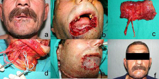
Figure 4
(a) 57 year-old patient with lower lip SCC. (b) Excision tumor. (c)
The gracilis muscle free flap. (d) Dissection of facial artery, vein, marjinal
mandibular nerve. (e) Immediate preoperative view after covering muscle
flap with the split-thickness skin graft. (f) Sixt months postoperative view.
Figure 5
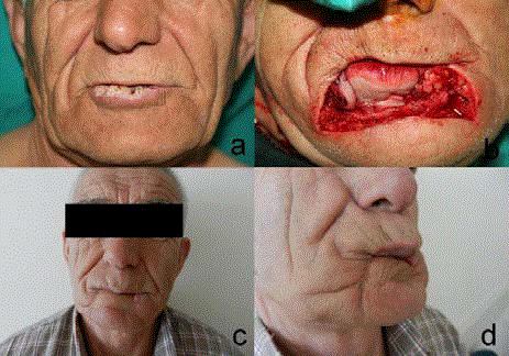
Figure 5
(a) The patient with lower lip SCC. (b) Excision of tumor. (c,d)
2 years postoperative view after reconstruction with functional free gracilis
muscle flap.
Vermılıon Defects
Small, superficial lateral vermilion defects can be left to heal by secondary intention. For the small central defects, primary closure is more suitable than healing by secondary intention in order to prevent depression or notching. For larger vermilion defects, flaps are indicated. These flaps are; V-Y advancement flaps from inside the vermilion, vermilion advancement flaps, vermilion switch flaps or nonlip flaps such as mucosal flaps and tongue flaps [6].
Cutaneous Defects
Partial-thickness lower lip defects are treated in same fashion as full thickness defects. The lower lip can tolerate wedge excision for the majority of defects. If the superficial defect is not amenable to the wedge excision, a skin graft or local transposition flaps from the chin and submandibular region are appropriate. Larger defects can be reconstructed with bilateral inferiorly based melolabial flaps [7].
Full-Thıckness Defects Less Than One Thırd Total Lıp Length
Defects of up to 30% of the transverse width of the lip in adults can typically be closed primarily without significant decrease in oral opening. Full-thickness defects are closed in three-layer of mucosa, muscle, and skin. The mucosal closure must provide a watertight mucosal seal. The deep and superficial fibers of the orbicularis muscle must be reapproximated to allow tension-free muscle coaptation and avoid attenuation. Alignment of the vermilion requires meticulous attention in order to to avoid a noticeable vermiliocutaneous mismatch or notch. Incisions that cross the white roll should be oriented perpendicularly to allow precise realignment of the while roll. Subtle misalignment of the lip can be aesthetically distracting, even in the absence of significant soft tissue loss. Some autors suggest that all of the deficiencies less than one-half the lip width can be repaired by primary closure. A recent publication by Soliman et al. [8] emphasized the superior functional and aesthetic results obtained with primary closure of full-thickness lower lip defects which comprise 50% of the lip and argued that local flaps are often overused in lip reconstruction. These size rule was especially applicable to elderly patients with greater tissue laxity.
Full-Thıckness Defects between One Thırd and Two Thırds Lıp Length
Central defects between 30% and 50% of the lower lip can be reconstructed with the Schuchardt procedure which facilitates primary closure with skin excision around labiomental fold. Recently, we described a new flap modification named transverse lip advancement flap for larger defects [9]. While designing the transvers lip advancement flap, a full thickness incision is made following the natural labiomental crease. The incision is ended at the level of commissure not to disturb the buccal and marginal mandibular branches of the facial nerve. The mental nerve was dissected and preserved to keep the sensorial innervation of the lower lip (Figure 1). Thus, the flap that includes the orbicularis oris with intact neurovascular supply is freed to provide the desired advancement (Figure 2). Utilizing the tremendous flexibility and expandability of the lip tissue, we tried to minimize the incisions and scar, and maximize the sensation and function with this flap. In planning a lip reconstruction, we think that transverse lip advancement flap should be considered as a second ladder after primary closure. Other two major techniques to reconstruct the defects between one third and two thirds lip length are lip-switching procedures, Abbe and Estlander flaps. Abbe flap is used for reconstruction if the commissure has not been resected and the Estlander flap is used if the commissure has been resected. One advantage of the cross-lip flap is the ability to replace a vertical segment of both vermilion and cutaneous lip tissue. The most significant disadvantage of these flaps is that they are denervated. Return of sensory function may begin after several months. Hypersensitivity of the flap may also occur but usually resolves after the first year. Fan-shaped orbicularis oris musculocutaneous flaps have been the mainstay of lip reconstruction, after Gillies introduced “fan flap” in 1920. This denervating reconstruction that gives form but no function was popularized and modifications were added periodically. In 1974, Karapandzic principle was introduced, modifiying the former into an innervated orbicularis oris myocutaneous flap. Reconstruction with Karapandzic flaps has the advantage of being a single-stage technique that replaces the missing lower lip with like tissue, maintains an intact oral sphincter, and preserves sensation and blood supply to the lips. However, use of Karapandzic flaps is usually restricted to reconstructing defects comprising two-thirds of the lip or less to avoid microstomia [10]. McGregor suggested a rectangular modification of the Gilles’ flap in 1983 to reconstruct the full thickness lip defects even up to entire lip [11]. Nakajima et al. [12] in 1984, proposed a modification for McGregor’s flap, based on the facial artery instead of labial artery. In addition to giving a more robust blood supply, the preservation of motor and sensory innervation to flap was superior.
Total Lıp Defects
For subtotal or total lip defects, non-lip techniques must be
employed. The Webster- Bernard technique is essentially an cheek
advancement flap to create a new lip [13]. Excision of tissue at the
nazolabial fold allows for this advancement. Although these were
originally described as full-thickness excisions, these are generally
performed as cutaneous excisions only. The vermilion is reconstructed
with mucosal sliding flaps simultaneously. Reconstruction with this
technique is adynamic and results in drooling. Hanasano et al. [14]
recently reported a modification of the Karapandzic flap for near
total or total lip full-thickness lip defects. Instead of joining up the
circumoral incision to the border of the lip defect, Hanasano et al.
[14] described extending the circumoral incisions inferolaterally in
the cheek tissue adjacent to the labiomental grooves. This cheek tissue
is then used to reconstruct the central component of the lower lip. This
modification provides additional tissue recruitment compared with
the traditional Karapandzic method. We agree that this technique is
very useful because of being sensate and having good colour matching
with adjacent tissue. Especially, the old patients with high tissue laxity
and patients who are not appropriate to microvascular free-tissue
transfer operations with high morbidity are proper candidates for this
technique (Figure 3).
Free-flap reconstruction is often required for large-scale defects
with associated loss of mucosa, cheek, nasal, and chin skin that
exceed the availability of local soft tissue. This can be due to paucity of
available soft tissue, previous radiation therapy, or previous surgery.
While free-tissue transfer can provide an abundance of soft tissue,
care must be taken in selecting a donor site with an appropriate
match in color, texture, and pliability. The most commonly used free
flap to reconstruct total or near-total lip defects is the radial forearm
free flap [15-17]. To provide static support for oral competence
and to help prevent drooling, this flap is usually transferred with
the palmaris longus tendon. Additional static support can also be
obtained through the use of a fascia lata graft. This flap can also be
transferred as a sensate free flap by performing anastomosis of the
lateral antebrachial cutaneous nerve to the mental nerve. Another
composite flap for lip reconstruction is anterolateral thigh-fascia lata
flap [18]. However, these flaps do not have their own motility, and
the color and texture match is not perfect. To restore the dynamic
sphincter function of the lower lip, neighbouring muscles have been
included. Combinations with a depressor anguli oris [19] and with a
masseter muscle [20] have been described. However, their functions
were not synchronised. For more synchronous and stronger functions,
forearm flap was combined with a free gracilis muscle transfer and
achieved good sphincter function but still poor color matching is a
problem [21,22]. Ninkovic et al. [23] used functional gracilis muscle
transfer covered a graft and local mucosal flap without combined a
radial forearm flap to improve the functional/aesthetic result, with
minimal donor-site morbidity and reported good results of two
cases. We recently presented our experience in reconstruction of
total or subtotal lower lip defects due to SCC excision with functional
gracilis muscle flap covered STSG in our series of 8 cases [24]. After
tumor resection on the lower lip, the fasial arter, vein and marginal
mandibular nerve are prepared as recipient vessels and nerve. The free
gracilis muscle is harvested from the contralateral thigh. The pedicle
includes the branch of the obturator nerve to the muscle. The gracilis
muscle flap was divided and trimmed to the required width and
length. The flap pedicle is anastomosed to the fasial artery and vein,
then the marginal branch of the facial nerve is coapted to the motor
nerve of the gracilis muscle A split-thickness skin graft is used to
cover the whole inner and outer surface of the gracilis flap which will
form mucosal surface and skin surface. Skin graft is harvested from
thigh and sutured to the remaning mucosa for reconstruction of the
lining. In a second stage, vermillion reconstruction with a fasial arter
musculomucosal flap is performed (Figure 4 and 5). In our series, the
patients were happy with the functional and aesthetic results. There
were no problems with normal word articulation, mouth opening,
or eating. Oral continence was perfect, the mouth sphincter function
has been restored completely.The color match of the grafted skin gave
a pleasing cosmetic result. Presently, there is no universally accepted
technique for subtotal or total reconstruction of the lower lip.
According to our personal experience; extended karapandzic flap is a
good choice for old patient who has enough tissue laxity fort his flap
or who has comorbidities for long microvascular operations. Other
patients with subtotal or total lower lip defects may benefit from free
gracilis muscle flap transfer.
Conclusion
While planning a lip reconstruction, as far as possible, dynamic reconstruction should be attempted for restoration of facial expression, speech, and eating. Patient characteristics like local tissue laxitiy, age, comorbidities, previous treatment (surgery and/or irradiation) also must be paid attention before the final surgical plan. We think that planning a reconstruction regarding the patient characteristic and also considering new concepts, will allow the surgeon to reach the best results both aesthetically and functionally.
References
- Adams W, Beran S. Lip, cheek, and scalp reconstruction; hair replacement. Selected Readings in Plastic Surgery. 2001;15:2-16.
- Kroll S. Lip reconstruction. In: Kroll S, ed. Reconstructive Plastic Surgery for Cancer. St. Louis, MO: Mosby Year Book; 1996:201-9.
- Williams PL, Warwick R, Dyson M, et al. Gray’s Anatomy: fasciae and muscles of the head. 37th ed. Edinburgh: Churchill Livingstone; 1989.
- Whetzel TP, Mathes SJ. Arterial anatomy of the face: An analysis of vascular territories and perforating cutaneous vessels. Plast Reconstr Surg. 1992;89:591-603.
- Cruse CW, Radocha RF. Squamous cell carcinoma of the lip. Plast Reconstr Surg. 1987;80(6):787-91.
- Galyon SW, Frodel JL. Lip and perioral defects. Otolaryngol Clin North Am. 2001;34(3):647-66.
- Renner G. Reconstruction of the lip. In: Local Flaps in Facial Reconstruction Edited by Baker SR, Swanson NA. St Louis: CV Mosby, 1995:345-96.
- Soliman S, Hatef DA, Hollier LH Jr, Thornton JF. The rationale for direct linear closure of facial Mohs' defects. Plast Reconstr Surg. 2011;127(1):142-9.
- Bektas G, Cinpolat A, Biçici P, Seyhan T, Coskunfirat OK. Reconstruction of lateral lower lip defects with transverse lip advancement flap. J Craniofac Surg. 2013;24(3):984-6.
- Karapandzic M. Reconstruction of lip defects by local arterial flaps. Br J Plast Surg. 1974;27(1):93-7.
- McGregor IA. Reconstruction of the lower lip. Br J Plast Surg. 1983;36(1):40-7.
- Nakajima T, Yoshimura Y, Kami T. Reconstruction of the lower lip with a fan-shaped flap based on the facial artery. Br J Plast Surg. 1984;37(1):52-4.
- Webster RC, Coffey RJ, Kelleher RE. Total and partial reconstruction of the lower lip with innervated musclebearing flaps. Plast Reconstr Surg Transplant Bull. 1960;25:360-71.
- Hanasono MM, Langstein HN. Extended Karapandzic flaps for near-total and total lower lip defects. Plast Reconstr Surg. 2011;127(3):1199-205.
- Sadove RC, Luce EA, McGrath PC. Reconstruction of the lower lip and chin with the composite radial forearm-palmaris longus free flap. Plast Reconstr Surg. 1991;88(2):209-14.
- Furuta S, Sakaguchi Y, Iwasawa M, Kurita H, Minemura T. Reconstruction of the lips, oral commissure, and full-thickness cheek with a composite radial forearm palmaris longus free flap. Ann Plast Surg. 1994;33(5):544-7.
- Jeng SF, Kuo YR, Wei FC, Su CY, Chien CY. Total lower lip reconstruction with a composite radial forearm-palmaris longus tendon flap: a clinical series. Plast Reconstr Surg. 2004;113(1):19-23.
- Yildirim S, Gideroglu K, Aydogdu E, Avci G, Akan M, Akoz T. Composite anterolateral thigh-fascia lata flap: a good alternative to radial forearm-palmaris longus flap for total lower lip reconstruction. Plast Reconstr Surg. 2006;117(6):2033-41.
- Kushima H, Iwasawa M, Kiyono M, Ohtsuka Y, Hataya Y. Functional reconstruction of total lower lip defects with a radial forearm free flap combined with a depressor anguli oris muscle transfer. Ann Plast Surg. 1997;39(2):182-5.
- Shinohara H, Iwasawa M, Kitazawa T, Kushima H. Functional lip reconstruction with a radial forearm free flap combined with a masseter muscle transfer after wide total excision of the chin. Ann Plast Surg. 2000;45:71.
- Ueda K, Oba S, Ohtani K, Amano N, Fumiyama Y. Functional lower lip reconstruction with a forearm flap combined with a free gracilis muscle transfer. J Plast Reconstr Aesthet Surg. 2006;59(8):867-70.
- Ueda K, Oba S, Nakai K, Okada M, Kurokawa N, Nuri T. Functional reconstruction of the upper and lower lips and commissure with a forearm flap combined with a free gracilis muscle transfer. J Plast Reconstr Aesthet Surg. 2009;62(10):e337-40.
- Ninkovic M, Spilimbergo SS, Ninkovic M. Lower lip reconstruction:introduction of a new procedure using a functioning gracilis muscle free flap. Plast Reconstr Surg. 2007;119(5):1472e80.
- Bektas G, Coskunfirat OK, Cinpolat A, Unal K. Experiencies with Functional Gracilis Muscle Flap in Lower Lip Reconstruction. 14-18 September 2011, Turkish Society of Plastic Reconstructive and Aesthetic Surgeons 33. National Congress.