Research Article
The Experience Outcomes of the Cosmetic and Functional Surgery in Congenital Aural Atresia
Nermin Başerer*
Department of Otolaryngology, Istanbul University, Istanbul, Turkey
*Corresponding author: Nermin Başerer, Department of Otolaryngology, Istanbul University, Valikonağı Caddesi, 149/11, 34365, Şişli-Istanbul, Turkey
Published: 18 Sep, 2017
Cite this article as: Başerer N. The Experience Outcomes
of the Cosmetic and Functional Surgery
in Congenital Aural Atresia. Clin Surg.
2017; 2: 1616.
Abstract
In this article we presented the results obtained in functional and cosmetic surgery for congenital
aural atresia (CAA) along with literature data and discussed the factors important in the success of
the surgical treatment. A total of 544 cases (599 ears) of CAA including 489 unilateral and 60 bilateral
atresia were treated in the last 30 years. The ratio among males and females was 2/1 with frequency
in males. The age of patient treated ranged from 5 to 38 years old. All the patients had a clinic,
audiologic and radiologic evaluation before the surgery. The technique of the surgery applied to the
patients was transatresic approach. Our cases 30% of were syndromic (181 hemifacial microsomia, 3
Treacher Collins, 4 Goldenhaar) and the other 70% was nonsyndromic. The preoperative audiologic
evaluation of the air-bone gap was found to be 50-60 dB. In the functional outcomes increasing airbon
gap were: 40 dB in 10%, 30 dB in 40% and 20 dB in 40% of patients. Remaining 10% patients
did not have gain. There were no labirynt injury in any patients and no need of hearing aids. Modest
hearing gain could be achieved. Transient facial paralysis was developed in 5 patients and none
became permanent. The lateralization of the tympanic membrane was the most common late
complication. The most important problem in cosmetic surgery was keloid and scar formation. Free
skin graft and enlargement of the canal 1.5-2 times from that required were used to prevent the restenosis
of the new canal.
Keywords: Congenital aural atresia; Transatresic approach; Branchial arches malformations;
Congenital malformations of head and neck; Auricular reconstruction
Introduction
CAA: Congenital Aural Atresia; Db: Decibell; ABR: Auditory Brainstem Reflex; HZ: Hertz; CA: Computerized Tomography
Introduction
Congenital Aural Atresia (CAA) is the malformation of the middle and external ear resulting from the anomaly of the first and second branchial arches which develop as hypoplasia or aplasia of the tympanic bone. As this malformation might be isolated, it could also be a part of various syndromes. It occurs in 1 in 10.000-20.000 births. It affects males more often than females at a ratio of 2/1 [1,2]. This malformation appears to have a right-ear preponderance [1-3]. The external and middle ear begin to form at the fourth (fifth) week of gestation from the first and second branchial arches (mesoderm), first pharyngeal cleft (ectoderm), and the first branchial pouch (endoderm) and continue to develop until the thirtieth week [2-5]. The inner ear developes of otic placode at the third week of gestation and completes its development at the fifth month of gestation [2]. The inner ear involvement is rare at a ratio of 6-10% in patients with CAA [6]. The inner ear involvement is more frequent in patients with CAA who have associated syndromes [7]. The embryopathogenesis developing after the fourth month of gestation causes the agenesis or hypoplasia of the the tympanic bone. Also the auricula is originated from the first and second branchial arches as middle ear. Helix, the crus of helix and the tragus are derived from the first branchial arch. The antitragus, the inferior part of the helix, and the tragus are derived from the second branchial arch. About 85% of the auricula is originated from the second branchial arch [2,4,5]. The degree of the auricular malformation is a sign of malformation of the external and the middle ear. The auricula completes its development before the middle ear. There is an association between the malformation of the auricula and the middle ear. However, at the late appearing embryological developmental disorders, although the auricula is normal, the minor malformation of the middle ear is still seen [5]. The dislocation or dysplasia of the mastoid bone or tympanic cavity, the anomaly of the facial nerve (in diameter and course), and the dislocation of the temporomandibular joint (posterior location)arise as a result of the agenesis or hypoplasia of the tympanic bone in the CAA. Type 1 (mild dysplasia), type 2 (moderate dysplasia), and type 3. (severe dysplasia in 2/3 of the cases) microtia and anotia arise depending on the developmental disorders of the auricula [2,5-7]. Surgical difficulty due to some reasons can be stated as the associated complex pathologies of the CAA patients, experience requirement of the surgeon for prevention of the possible complications of surgery, and the postoperative tardive problems about retrecissemen of reconstructed new canal due to osteogenesis and scatris formation during the healing period. Our aim in this study was to present functional and cosmetic results in CAA surgery applied in our clinic along with literature data, and discuss the factors important in the success of the surgical treatment.
Figure 1
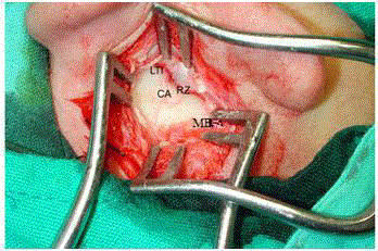
Figure 1
Transatresic approach (anterior approach) and leading points (MB:
mastoid bone, RZ: zygomatic root, LTI: linea temporalis inferior), CA: mastoid
bone cribriform area.
Figure 2
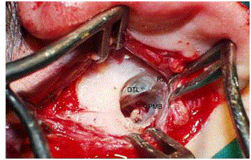
Figure 2
Transatresic approach (anterior approach) and boundaries DTL:
Dura of temporal lobe, I: Incus, PMB: Periost of mastoid bone, RZ: Zygomatic
root.
Figure 3
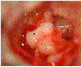
Figure 3
Exposition of the malformed incus malleus block in attic.
Patients and Methods
We conducted a retrospective review of 544 patients with CAA (599
ears) who underwent functional surgery (atresiaplasty-meatoplastycanalplasty
and tympanoplasty) and cosmetic reconstructive surgery
(auriculaplasty) in the last 30 years at the Department of Ear Nose
and Throat of Medical Faculty of İstanbul University. In total of 544
patients operated, 484 patients had unilateral and 60 patients had
bilateral CAA. The gender distrubition of 366 patients were male and
178 patients were female with male predominance in 2/1 ratio. Of 489
patients with unilateral involvement, 326 patients had right and 163
patients had left CAA. In the same way the right ear atresia was twice
as many as in the left. Conductive type of hearing loss was detected in
5% of CAA cases in the normal-looking ear. The age of the patients
ranged from 5 to 38 years and the main age of the patients opered for
unilateral CAA was 10 years. Whereas the main age of patients with
bilateral CAA was 6 years. In our case series, there are 187 syndromic
cases. The majority of these cases were hemifacial microsomia with
181 cases, there were only 7 cases remaining of which 4 Goldenhar,
3 Treacher Collin’s syndrome. These latter two groups of cases are
not suitable for functional surgery. All the patients were evaluated
clinically, audiologically and radiologically in the preoperative period.
In the clinical observation, mastoid apex (sign of pneumotization),
development of mandible, the state of facial nerve (the presence of
paralysis and/or paresis), the degree of auricula malformation have
been investigated. Other additional malformations due to branchial
arch anomaly (isolated or syndromic CAA) have also been under
investigation. In the audilogical evaluation, ABR (auditory brainstem
reflex) needs to be implemented for the new borns with bilateral
congenital aural atresia after the first month. On the otherhand, the
behavioural audiometry were performed for the unilateral CAA after
a month, and pure tone audiometry over 5 years old.
In the radiological evaluation, high resolution temporal bone
computed tomography in the axial and coronal plan with 1.5 mm
sections were performed for the children over 4-5 years (after
pneumotization of the mastoid bone). The thickness of the atresial
plaque, the ossicles, the diameter of the tympanic cavity, stapes, the
oval and round windows, the course of the facial nerve, dural level of
the middle cranial fossa, the state of the temporomandibular joint,
inner ear structures and especially the presence of narrow internal
acoustic canal and wide vestibular aquaduct to check the geyser risk
were carefully examined. Surgery for the congenital aural atresia
was performed with the aim of function (atresioplasty-meatoplastycanalplasty
and tympanoplasty), or cosmetical reconstruction
(auriculoplasty), or both function and cosmetically. The indications of
the functional surgery for the CAA were established by the evaluation
of the results of clinical, radiological and the audiometric studies.
In the presence of intact inner ear and adequate surgical anatomy is
necessary for the middle ear access. Developed and aerated mastoid
bone, not too narrow tympanic cavity, ossicular chain, the round
and oval windows should exist. The age of the patient >5-6 years
was accepted as a lower limit for the development of the mastoid
bone and costal cartilage. These surgical indications were noted
during this evaluation. Conntraindications of the functional CAA
surgery include undeveloped mastoid and middle ear cavity in CAA
cases and the low dura (prolapsed dura), the presence of significant
craniofascial malformation, the involvement of inner ear (the absence
of cochlear reserve), patients under the age of 5 years. The timing of
the surgery for the bilateral cases with CAA were performed earlier
than it is for the unilateral cases with respect to early prevention of
the patients’ psychological and learning problems. Usually 5-6 years
old children were preferred for the surgery because of adequate
development of mastoid bone and costal cartilage which is made for the auricular framework grafting and the permission of the children
in the dressing. Early use of bone conduction hearing aids and speech
education were planned as an important practice for the cases with
bilateral CAA until the surgical intervention. In cases with unilateral
CAA we conducted more electively; however we preferred to apply
the surgery early in these cases (before the primary school) to deal
with psychological problems related to microtia. Three different
approaches posterior, anterior, and modified anterior are being
performed in the functional surgery of CAA. Posterior approach is
an open cavity tympanomastoidectomy technique, and is performed
more than the transatresic anterior approach which can not be applied
in the cases with poorly developed mastoid bone. Current approach
for the modern CAA surgery is the transatresic-anterior technique
which was developed by Jahrsdoerfer. The anterior approach is
prefered in most of our cases. The modified anterior approach
technique is only performed in the cases with CAA who have poorly
developed mastoid bone as intact canal mastoidectomy.
We consider some leading points and boundaries to prevent the
surgical complications (related to labyrinth and facial nerve) in the
anterior transatresic approach (Figure 1). These leading points are
linea temporalis, root of the zygomatic bone, fossa glenoidalis, condyle
of the mandible (temporamandibular joint), and the cribriform area
in the mastoid cortex. The boundaries in the surgery are the dural
plate of the middle cranial fossa, the cortex or periosteum of the
fossa glenoidalis, mastoid air cell system, and the vertical segment
(mastoid) of the facial nerve (Figure 2). Functional surgery is generally
performed in the severe displasia after the first stage of auricular
reconstruction. Functional surgery starts with the opening of the
new outer canal, followed by complete removing of the atresic plaque
(Figure 3 and 4). The new canal must be as wide as possible, circular,
and smooth. Constitution of the sound conduction mechanism (Type
1-3 tympanoplasty), placement of the temporal muscle fascia graft is
centralized on the ossicular chain (Figure 5 and 6). In the formation
of a wide meatus grafting, a whole new canal with the thin free skin
flap is applied. Placement of canal skin graft should cover temporal
fascia graft. Suturing the canal skin graft circularly to the skin at the
level of the concha-tragus is performed (Figure 7 and 8). At the end
of the operation, the placement of antibiotique and cortico steroid
including merosel tamponade into the new canal is given in (Figure
8 and 9A). View of complete epithelization of new wide canal and
timpanic membrane one month after the atresiaplasty-timponaplasty
can be seen in (Figure 9B). Surgery in type 3 severe displasia of
the auricula begins with the first session of its reconstruction. Full
thickness cartilage graft for framework of reconstructed auricula that
is harvested from the contralateral sixth, seventh, and eighth costal
cartilage of the atresic ear costal cartilage graft is placed under the intact skin (Figure 10 and 11). In the tirth period of CAA surgery
with reconstruction of auricula, a free thin skin graft is placed behind
the ear. Temporo auricular angle is provided with the detacment of
the reconstructed auricula. Thin skin graft that is taken between the
umblicus and the pubis are used for recouvrement of the back of the
auricula (Figure 12 and 13).
Figure 4
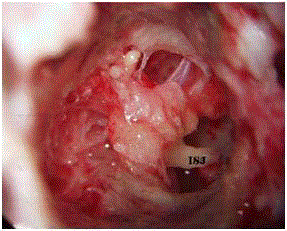
Figure 4
The widening of the middle ear cavity, ISJ: Incido stapedial joint.
Figure 5

Figure 5
The placement of the temporal fascia on the ossicular chain, FTM:
fascia of temporal muscle.
Figure 6
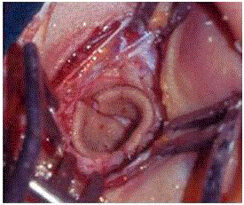
Figure 6
The covering of newly opened canal with the thin skin graft.
Figure 7
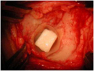
Figure 7
The placement of tamponade with merocell stent into the newly
opened canal.
Figure 8
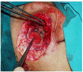
Figure 8
The suturing of the free skin greft to the skin of the meatus using
merocell stent.
Figure 9
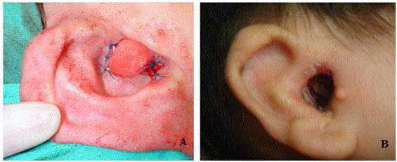
Figure 9
(A) The early postoperative appearance of wide external ear canal,
(B) tympanic membrane in a patient whose external ear canal has been
opened (one month after surgery).
Figure 10
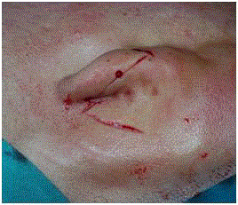
Figure 10
The appearance of the left ear type 3 displasia from the left lateral
view.
Figure 11
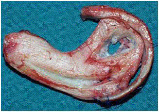
Figure 11
The auricular framework reconstructed from the autogenous
costal cartilages.
Figure 12
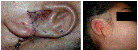
Figure 12
The appearance of the auricular framework placed under the skin.
Figure 13
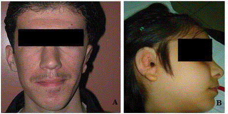
Figure 13
The postoperative appearance of patients who had auriculoplasty.
(A) CAA associated with hemifacial macrosomia, (B) nonsendromic CAA.
Results
In the clinical evaluation of our cases, the most common additional sign was hemifascial microsomia as a result of the developmental disorder of the mandibular arch (Figure 13). In the preoperative audiologic evaluation of the patients, the air-bone gap was detected as 50-60 dB. In the surgery, only 4 cases had temporary facial paralysis as an early surgical complication. Complete cure was obtained in 2 cases by performing facial decompression at the same session, and in the other 2 cases with anti-edema treatment. In 10% of cases, mild hearing loss (partial labyrinthization) was observed at the 6000-8000 Hz depending on the labyrinth trauma. Total hearing loss did not develop in any of the patients. The most common late complication was the lateralization of the tympanicmembrane. In the revision surgery, we usually detected that the reasons of the late hearing loss were the lateralization of the tympanic membrane and the new bone formation. In terms of re-stenosis of the meatus, no significant re-stenosis problem was encountered. Complication related to the auriculaplasty, scar, and keloid tissue were seen more than the clear skinned ones. Graft rejection and resorption were encountered while we were applying alloplastic (silicone, medpore) and heterogenous (costal cartilage of the calf ) auricular framework materials. In reconstruction, only autogenous costal cartilage is used in the last 25 years. Hearing improvement was better in cases who had type 1 tympanoplasty than the cases with type 3 tympanoplasty after the functional surgery. Evaluation of functional surgery showed that, in 10% of them 40 dB, in 40% 30 dB, and in 40% 20 dB increase in air bone gap was achieved. The air-bone gap remained the same in 10% of the patients.
Discussion
Aural atresi constitutes the most common congenital malformation in our speciality. Surgeon must have sufficiant experience in otolojic microsurgery and plastic reconstructive surgery to prevent serious risk of complications. In functional surgery; facial paralysis, labyrinthization, the re-stenosis of the newly opened canal, the lateralization of the tympanic membrane, the risk of neoosteogenesis, and the concern of inability to provide enough funcional gain make this surgery out of routin. In view of the circumstances, the supporters of the CAA surgery have not increased very much. When reviewing the literature, articles by the same surgeons prove that this issue is common. The experiences of the pioneering otologists in atresiaplasty, the determination of the criteria to choose the suitable candidates is a very important issue for the surgery. The modifications in the approach and grafting techniques were encouraging and guiding for the surgeons who perform this surgery. The experiences we have obtained for over 30 years in the surgery of 599 CAA have showed us that the effort in this surgery was too much but also satisfactory. Approximately 50% of all CAA cases are suitable candidates for functional surgery [8-10]. In 10% of unilateral CAA cases conductive or sensorineural type hearing loss can also be seen in the contralateral ear [6,11]. In our series, there were 5 patients who had conductive type hearing loss in the contralateral normal looking ear. In a normally developed auricula, fixation of the footplate of the stapes, anomaly of the facial nerve or fixation of the ossicular chain can be encountered as minor malformations [12]. In our series, fixation of the footplate has not been seen. In CAA, clinical and audiological evaluation should be performed in earlier ages of the patients (from the first month on). Early bone-conduction hearing aid and education of the patient are useful in the hearing and speech development of children with bilateral CAA. Despite bilateral CAA, it is not obligatory to employ functional surgery and hearig aids in the earlier phase of unilateral CAA cases. However, elective surgery is indicated to provide stereophonic natural hearing and to correct the malformation of auricula after 5 years of age. There is no concensus on the exact timing of the surgery. We support that the surgery should be performed when the patients are older than 5 years old. The development of the middle ear and the ossicles are completed in the seventh month. The development of the mastoid bone continues after the birth and throughout the early ages of the adulthood. A developed mastoid bone and surgical anatomy are needed for the external ear canal which will be newly opened. To evaluate the temporal bone and the inner ear anatomy with CT for surgical indication, it has to be waited until the age of 4. Narrow angled course of the facial nerve, little tympanic cavity, and low temporal lobe (undeveloped mastoid bone) are the anatomic contraindications of the functional surgery. In CAA, facial paralysis is the most important and distressing complication of the canalplasty and tympanoplasty. In our series temporary facial paralysis developed in 4 cases. Decompression surgery and the other two cases with steroid theraphy recovered after one month without sequelae. The facial nerve develops from the mesenchyme of the second branchial arch in the seventeenth week of the fetal life. At this period the facial nerve is in a position which is antero lateral to its normal course. Tympanic bone hypoplasia and aplasia which causes the malformations in the CAA prevent the posterioinferior development of the facial nerve. Therefore, the facial nerve follows a 60° narrow angled anterolateral course instead of a wide angle of 120°. The dislocation of the facial nerve is usually encountered in the vertical mastoid segment. In the horizontal tympanic segment mostly dehiscence of the Fallopian canal and the prolapsus of the facial nerve over the oval window can be seen. We adopted the transatresic (anterior) approach which Jahrsdoerfer descripted. In this approach, epitympanum is the first middle ear portion which is reached after the temporal lobe guidance, and the incus-malleus complex is the first structure that is seen. In the tympanic position, the facial nerve is always medial to the ossicles. There is no risk of trauma in this position. While the middle ear cavity is made wider through the mastoid bone in the posterior, the facial nerve can be especially opened or cut during the dissection of the atresia plaque which is performed in the posteroinferior segment. The use of facial nerve moniterization would be very helpful to the surgeons who have just beginned this surgery. The facial nerve travels superficially in the extratemporal parotis region in the children. During meatoplasty the dissection of the soft tissues must be careful. As an early complication, trauma to the inner ear usually results from the conduction of the vibration of the ossicles to the inner ear during the dissection of the atresic plaque with the high turnover burr. In the prevention of this complication the use of the low turnover diamond burr or in case of necessity the luxation of the incudo-stapedial joint is an important measure. There is no death labyrinth complication of any case. In the functional surgery, the lateralization of the tympanic membrane has been the major complication which decreased our success rate. While no improvement was noted in 10% of the cases, little improvement such as 20 dB was noted in 40% of the cases. So, we have succeeded functionally in 50% of the cases with 30-40 dB gain in air bone gap. However, although all precautions are taken, the extent of the structural deformity and neoosteogenesis can cause lateralization. The canal re-stenosis which was creating a problem in the first years has prevented with enlarging the newly opened canal 1.5 times bigger than the requested one in diameter (in the width that a finger can fit into). Grafting the canal with the thin free skin instead of pediculated regional skin flap, suturing this skin graft to the opening of the meatus in a 360° manner (circularly), and using the merocel stent on the post operative follow-up should be performed. In the severe displasia, we begin the surgery with the technique of Brent by placing the autogenous costal cartilage frame to the periauricular region, which has a good blood supply as the first session of the auriculoplasty. Patients having CAA, except the ones with multiple malformations and those with syndromes involving inner ear should undergo either cosmetic or cosmetic and functional surgery. The experience in this surgery and the patient satisfaction have made this difficult surgery very pleasant.
References
- ALTMANN F. Congenital atresia of the ear in man and animals. Ann Otol Rhinol Laryngol. 1955;64(3):824-58.
- Crabtree JA, Harker LA: Developmental anomalies of the ear. In: Cummings CW, Frederickson JM, Harker LA, Krausse CJ, Schüller DE, eds. Otolayrngology Head and Neck Surgery 2nd ed. St Louis Mosby. 1993;2746-55.
- Baserer N. Dogumsal aural atrezi. Çelik O. Kulak Burun Bogaz Hastaliklari Bas Boyun Cerrahisi. Turgut Yayincilik Istanbul. 2002:99-107.
- Mandell DL. Head and neck anomalies related to the branchial apparatus. Otolaryngol Clin North Am. 2000;33(6):1309-32.
- Lambert PR, Dodson EE. Congenital malformations of the external auditory canal. Otolaryngol Clin North Am. 1996;29(5):741-60.
- Cressman WR, Pensak ML. Surgical aspects of congenital aural atresia. Otolaryngol Clin North Am. 1994;27(3):621-33.
- Jahrsdoerfer RA, Aguliar EA. Surgical management of congenital ear malformations. In: Jahrdoerfer RA, Helms J. Eds. Head and neck surgery. Ear Stuttgart Georfthieme Verlag. 1996;35-66.
- De La Cruz A, Linthicum F, Luxford WM. Congenital atresia of external auditory canal. Laryngoscope 1985;95(4):421-31.
- Hashisaki GT, Lambert PR. Congenital aural atresia In. Gates GA ed. Current Therapy in Otolaryngology Head and Neck Surgery. St Louis Mosby. 1998;4-8.
- Lesinski SG. Congenital aural atresia. Current Therapy in Otolaryngology Head and Neck Surgery. Mosby Comp. London. 1984-1985;38-42.
- Bellucci RJ. Congenital aural malformations: diagnosis and treatment. Otolaryngol Clin North Am. 1981;14(1):95-124.
- Brent B. Technical advances in ear reconstruction with autogenous rib cartilage grafts: personal experience with 1200 cases. Plast Reconstr Surg. 1999;104(2):319-34.