Short Communication
Surgical Guidance in Foot Surgery by Means of 3D Impression and Minimally Invasive Surgery
Javier Ferrer-Torregrosa1*, Nadia Fernández-Ehrling2, Sergio García Vicente3 and Carlos
Barrios4
1Department of Podiatry, Physiotherapy and Podiatry School, Valencia Catholic University, Valencia, Spain
2Doctorate School, Valencia Catholic University, Valencia, Spain
3Private Clinic, Valencia, Spain
4Institute for Research on Musculoskeletal Disorders, Valencia Catholic University, Valencia, Spain
*Corresponding author: Javier Ferrer-Torregrosa, Department of Podiatry, Physiotherapy and Podiatry School, Valencia Catholic University, Valencia, C/Ramiro de Maeztu 14, Torrente-Valencia 963637412, Spain
Published: 07 Aug, 2017
Cite this article as: Ferrer-Torregrosa J, Fernández-Ehrling
N, Vicente SG, Barrios C. Surgical
Guidance in Foot Surgery by Means of
3D Impression and Minimally Invasive
Surgery. Clin Surg. 2017; 2: 1582.
Abstract
Background: Surgical accuracy in minimal incision foot surgery is an unexplored field. Optimal
gateways and more precise surgical processes to the patients' biomechanical problems must be
determined.
The purpose of this study is to compare the deviation between the position of the osteotomies
virtually planned and the position of the osteotomies made by means of the 3d printed surgical
guides for a human cadaver foot.
Methods: A left cadaver foot was scanned by means of a CAT scan and, by means of software; a
virtual surgery was carried out by positioning the cutting guides.
The virtual preoperative tomography was compared to the postoperative tomography and
measurements were made during the CAT scans. The data were analyzed with a t test (alfa=0,01).
Results:The average differences of measurement between the osteotomies planned by computer
and the osteotomies of the surgery were 0.0613 (99% CI: -0,42 -0,42).
Conclusion: The results showed that statistically there is no significant difference between virtual
and post-surgical osteotomies.
Keywords: Surgical guide; Minimal-incision surgery; Percutaneous foot surgery
Clinical Relevance (if basic Science Study)
The 3D-printed surgical glove could facilitate a fast, simple, and minimally invasive surgical technique. The use of this customized surgical glove should be considered to allow surgeons to preserve patient safety when performing bone osteotomies with precise correction of forefoot deformities.
Introduction
The minimally invasive surgery of Ihe foot is a method that allows carrying out surgery interventions through small incisions with a minimum anatomical damage [1]. Continuous radiation controls by means of intraoperative fluoroscopy are necessary since, as they are small incision, we do not visualize the surgical field, which creates further complications. The lack of sight or the lack of experience in the techniques themselves could injure important anatomical structures [2]. The new technologies as well as the progresses in medical image, result in the improvement of the traditional foot surgery techniques, introducing new forms of guidance to carry out such interventions. Nowadays, it is relatively simple to obtain a 3D reconstruction by means of DICOM images obtained through CAT scans/NMR and to rebuild them with specific software, obtaining a perfect image that we will be able to manipulate on the computer. Similar technology already exists in other areas like the dentists, where they place implants that have been planned and guided by computer [3], which avoids damaging significant structures surrounding the surgery. So far, presurgical planning was made by means of 2D X-rays and the convenient goniometry to quantify where and how much must the osteotomy be carried out. With three-dimensional planning, software like Mayan, 3D Max Sudio or Mimics, 3D medical segmentation software [4], a virtual planning can be carried out to subsequently send the surgical insole or the glove in this case to the manufacturing plant by means of rapid prototyping [5].
Figure 1
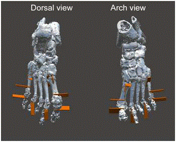
Figure 1
Osteotomy design.
Figure 2
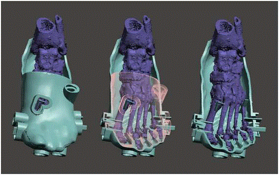
Figure 2
The surgical glove creation.
Figure 3
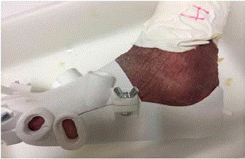
Figure 3
Glove printed in Polyamide 12.
Figure 4

Figure 4
Example of measurements.
Table 1

Table 1
Measurements made and differences between them.
Table 2
Table 2
Statistics of related samples.
Table 3
Table 3
Correlation of related samples.
Material and Methods
The first thing to do is to get a Computerized Axial Tomography
scan (CAT) of the foot that needs to be operated; in this case it was
a left foot. A reconstruction is carried out in a software of their own
created by the company’s Socinser (Asturias, Spain) and TEQUIR
(Valencia, Spain), it is a software allowing the surgeon to work
with medical image in 2D (from X-rays) and 3D (from CAT scans),
to visualize in detail the osseous structure and to carry out virtual
measurements, planning, simulations, of a surgery. Working on Web
collaborative environment, making possible communication online
in real time with one or more of the program users, is a process
that allows a comprehensive treatment of complex cases, making
possible the design and accomplishment of osteotomies along with
the corresponding surgical guides, resulting from all the process
followed and guaranteed by the surgeon, as a solution fully adapted
to the specific case.
The surgery was planned with the software in the following
osteotomies:
1. AKIN perpendicular to the axis of the phalanx of the 1st
incomplete.
2. Reverdin-isham, is an osteotomy that preserves the lateral
cortical.
3. Underlying osteotomy of the 1st.
4. Isham bunionette of the 5th.
5. Underlying osteotomy of the phalanx of the 2nd, 4th and 5th,
all of them are incomplete.
6. Once the intervention was planned and the osteotomies
were designed, the printing of the glove of the foot started with
the osteotomies incorporated in the cover itself, using for this
purpose the technology known as stereolithography of the AIMME
Technological Institute which provides an excellent dimensional and
superficial quality. Material such as PA 12 (polyamide 12) was used.
This material supports perfectly autoclave, its melt point EN ISO
11357-1 is around 172°C – 180°C.
7. Once the glove was printed, the surgery of all the
osteotomies established in the planning was carried out.
Results and Discussion
To assess the results both CAT scans were synchronised, through
Osirix Software, the pre-surgical one with the guides and the post- surgical one with the osteotomies carried out [6]. The 8 measurements
below were made:
• From the most proximal part to the dorsal of the Akin
osteotomy to the most proximal part of the Reverdin osteotomy.
• From the 1st metatarsal-cuneiform joint to the Reverdin
osteotomy at the arch level.
• From the most proximal joint to the beginning of the guide
at the pre-surgical and the osteotomy at the post-surgical.
• From the fat edge of the arch of the 5th finger to the
beginning of the osteotomy at the dorsal level.
• From the styloid apophysis to the osteotomy of the 5th at
the dorsal level.
• From the dorsal part of the proximal osteotomy of the 5th
finger to the edge of the distal osteotomy of the 5th finger at the dorsal
level and another one at the arch level.
• From the arch part of the distal osteotomy of 5th to the
osteotomy of the 5th finger at the arch level.
• Two osseous measurements were also made to verify the
existing deviation in the synchronisation of pre- and post-surgical,
the deviation among them was less than 0.02.
• Dorsal cleft of the first cuneiform bone to make sure we are
on the same plane.
• Measurement of navicular to make sure we are on the same
plane.
All the measurements were made by another investigator different
from the surgical one.
We can appreciate that on chart 1 there is almost no variation
between the two measurements, statistically there is no difference
between the sizes. If we analyze all the measurements we shall observe
how the maximum deviation is less than < 0,42 mm, something fully
reasonable in this kind of surgeries [7,8]. We made a T-student for twin samples observing that there is a correlation very close to 1 and
that statistically there are no significant differences with p-value =
-0.513. On the chart, we can see how only three measurements, the
4, 5, 8 do deviate, while the rest is almost equal between both CAT
scans.
Table 4
Table 4
Test of related samples.
Figure 5
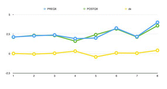
Figure 5
Comparison of measurements between the groups and deviation
among them.
Conclusion
The results of this study clearly show that the surgical glove provides accuracy in the accomplishment of the osteotomies. The cutting guides are accurate and helpful tools for the podiatrist during minimally invasive surgery of the foot, as it was already demonstrated by Hetherington in 2008 with other guides for exposed surgery. It has also has been proved in surgeries such as knee arthroplasty where more accurate cuttings were carried out. Nevertheless, there is not much information in literature about the use of osteotomy guides in podiatric surgery. The guides are very important for the osteotomies, especially for first metatarsal surgery. They must be considered for the treatment of deformities such as Hallux valgus and/or Tailor's bunion, to obtain the correct alignment of cutting and the angle, to recover the functional normality of the foot and to avoid possible side effects of the misalignment of the parts of the osteotomy. Further research is necessary on this subject and more types of surgical guides should be incorporated to establish a connection with the pre-surgical pressures emerged in consultation by means of simulation and trigonometry.
References
- Nieto-García E, Ferrer-Torregrosa J, Nieto-González E. Principios de la cirugía mínimamente invasiva. In: Cirugía Mínimamente Invasiva Del Pie. Valencia: Ediciones Glosa. 2017;297.
- De Prado M. Complications in minimally invasive foot surgery. Fuß Sprunggelenk. 2013;11(2):83-94.
- Pettersson A, Kero T, Gillot L, Cannas B, Fäldt J, Söderberg R, et al. Accuracy of CAD/CAM-guided surgical template implant surgery on human cadavers: Part I. J Prosthet Dent. 2010;103(6):334-42.
- Zeng Y, Zhang G, Lei T, Wu X, Tan Y. [Clinical application of mimics software in three dimensional CT images for treatment of zygomatic complex fracture]. Hua Xi Kou Qiang Yi Xue Za Zhi. 2012;30(2):123-7.
- Deshmukh TR, Kuthe AM, Vaibhav B. Preplanning and simulation of surgery using rapid modelling. J Med Eng Technol. 2010;34(4):291-4.
- Hetherington VJ, Kawalec-Carroll JS, Melillo-Kroleski J, Jones T, Melillo M, McFarland N, et al. Evaluation of surgical experience and the use of an osteotomy guide on the apical angle of an Austin osteotomy. Foot (Edinb). 2008;18(3):159-64.
- Teter KE, Bregman D, Colwell CW. Accuracy of intramedullary versus extramedullary tibial alignment cutting systems in total knee arthroplasty. Clin Orthop Relat Res. 1995;(321):106-10.
- Teter KE, Bregman D, Colwell CW. The efficacy of intramedullary femoral alignment in total knee replacement. Clin Orthop Relat Res. 1995;(321):117-21.