Case Report
Posterior Sterno Clavicular Joint Dislocation: A Case Report of a Surgical Stabilization Technique with PDSTM Cord
Castellani GC, Cerbasi S*, Massetti D, Maresca A, Fantasia R, Sangiovanni P and Pascarella R
Departemnt of Orthopedic and Traumatology, Ospedali Riuniti, Ancona, Italy
*Corresponding author: Simone Cerbasi, Departemnt of Orthopedic and Traumatology, Ospedali Riuniti, Ancona, Italy
Published: 07 Jul 2017
Cite this article as: Castellani GC, Cerbasi S, Massetti D,
Maresca A, Fantasia R, Sangiovanni P,
et al. Posterior Sterno Clavicular Joint
Dislocation: A Case Report of a Surgical
Stabilization Technique with PDSTM
Cord. Clin Surg. 2017; 2: 1546.
Abstract
Dislocation of sternoclavicular joint is an uncommon injury of shoulder girdle. It generally follows a high energy collision or a sport-related trauma. There is no unanimity on what the most adequate treatment management should be for such lesions in medical literature. Conservative treatment seems to be the choice of action in case of anterior sternoclavicular dislocations. Surgical procedure is to be reserved for posterior dislocations, due to possible complications which could arise given anatomical position. We here by present a case report on a posterior sternoclavicular dislocation following a sports trauma in a 15-year old boy surgically treated with a stabilizing technique using PDSTM Cord. The functional recovery after a 12-month follow up is extremely promising.
Introduction
Sterno Clavicular (SC) joint dislocation is an uncommon injury of shoulder girdle and it
represents 2-3% of all upper limb lesions [1-3]. Anterior dislocations are more common than
posterior dislocations with a 9 to 1 ratio [4]. In a review of 1600 SC joint dislocations, only one
subject was diagnosed with a posterior dislocation [5]. In 30% of cases, immediate complications
following posterior dislocations occur such as dyspnoea, dysphagia or vascular and neurological
damage, with a 3-4% mortality rate [6]. Such a lesion is potentially lethal due to proximity of
mediastinal structures (aortic arch, subclavian and carotid artery, esophagus, trachea, lungs and brachial plexus).
Two main mechanisms resulting in posterior sternoclavicular dislocation have been described.
On the one hand, injury can be caused by high energy traumas with a postero-lateral compressive
force to the shoulder. Second mechanism implies a force vector directed anteromedial to the clavicle,
thus causing posterior dislocation of clavicle [7]. Sternoclavicular joint is a diarthrosis saddle type
synovial joint. The joint is formed by two bone extremities covered with cartilage, a cavity limited
by synovial membrane, a joint capsule and ligaments. Joint cavity is composed of two portions
separated by a round articular disc (meniscus) connected to sternoclavicular anterior and posterior
ligaments and to the joint capsule. Both bones present alternated concave and convex areas which
fit together forming a saddle type joint which allows movements along anteroposterior and vertical
planes, besides a certain degree of rotation around the major axis of clavicle. As less than 50% of
the medial end of clavicle articulates with manubrium of sternum, it is not a stable joint. Its stability
is therefore derived from intrinsic and extrinsic ligaments [8]. It has been demonstrated that the
posterior capsule of the joint is more resistant than the anterior one, thus anterior sternoclavicular
dislocations are 9 times more frequent than posterior ones [9].
Case Presentation
A 15-year old boy was referred to our attention in February 2016 following a sporting accident
which took place during a rugby match. Patient presented functional limitation of left shoulder,
swelling and pain in sternoclavicular region with reduced mobility. Clinical history of the patient
was unremarkable and neurovascular examination of upper limb appeared normal. There were
no associated skeletal injuries. A standard chest x-ray and x-ray of shoulder were performed and
showed anomalies in the symmetry of the clavicles (Figure 1). Sternoclavicular joints, bones and
soft tissue as well as mediastinal structures such as heart, major blood vessels, esophagus and
trachea were more easily viewed on CT Angiography. Scans confirmed a full posterior dislocation
of proximal part of left clavicle from manubrium of sternum (Figure 2a) which was causing a compression on brachiocephalic vein (Figure 2b). As a reduction
maneuver was deemed not possible and given possible complications,
a multidisciplinary team (Orthopaedic surgeon and Cardio thoracic
surgeon) decided to treat surgically the lesion with an open reduction
and fixation. Patient was stable for following three days.
Surgical technique
Young athlete in supine position on operating table underwent
general anesthesia. Cardiothoracic surgeon was present in or in case
of damage to those vessels running posterior to SC joint occurred. An
arcuate incision of approximately 10 cm was made from manubrium
of sternum to proximal third of clavicle. Sternoclavicular joint
dislocation was identified through careful dissection. Meniscus was
repaired. Subsequently, through two 2.5 drill holes in medial end of
clavicle and sternum, manual reduction of dislocation was performed
and it was fixed using a figure-of-eight suture with PDSTm Cord
(Ethicon Johnson & Johnson International) (Figure 3a). An additional
suture anchor between clavicle and sternum was performed to have
more stability (Figure 3b). Repair and reinforcement of capsule was
then obtained. At the end of procedure joint was absolutely stable
with no mobility.
Follow up
In early post-op period and for the following 4 weeks a Gilchrist
shoulder brace was positioned. A careful passive joint mobilization
was initiated with pendulum exercises and gradual functional
recovery was observed together with an increased muscle tone and
tropism thus obtaining a complete ROM in 10-12 weeks. Patient
was clinically assessed during follow up at two weeks, one month,
three months and one year. Pain, ability to work and satisfaction with
treatment received were also evaluated, using an analog centesimal
scale (VAS). DASH questionnaire [10] (disability of the arm, shoulder
and hand), a validated tool for measurement of functional disabilities
with a range of values between 0 and 100, latter being worst outcome,
was used to perform clinical evaluation of patient. During last check
up, patient did not report any pain or disability, he was satisfied about
treatment that he had received and presented a full range of motion
(Figure 4). Six months after trauma, the young athlete went back to
playing rugby. As a secondary outcome, CT scans at three months
and one year were performed, in order to evaluate the joint and
recognize possible failures in result. No clinical aspects of a recurrent
dislocation were seen and CT confirmed joint stability (Figure 5).
Figure 1
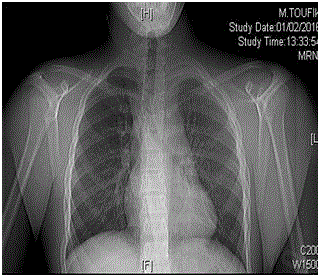
Figure 1
Radiography shows sternoclavicular dislocation to the left side.
Figure 2a
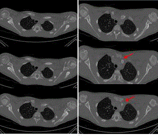
Figure 2a
Axial views of CT scan. Images show posterior dislocation of
clavicle.
Figure 2b
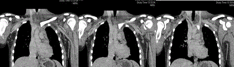
Figure 2b
Coronal views of CT scan. Compression on brachiocephalic vein.
Figure 3a
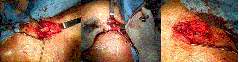
Figure 3a
Intra-operative pictures.
Figure 3b
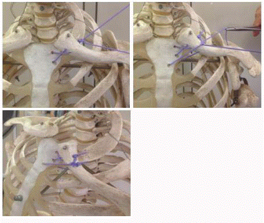
Figure 3b
Demonstration of technique using drill holes in clavicle and
sternum with PDSTM Cord.
Discussion
Sternoclavicular dislocations are rare and they usually follow a
high energy trauma. Due to their low frequency, there are no precise
guidelines regarding either conservative or surgical treatment.
Recent reviews have been published, which allow us to deduct
possible management guidelines [11-13]. In anterior dislocations a
closed reduction can be obtained in acute phase by applying traction to the arm and direct pressure over medial clavicle; shoulder is
then stabilized with a figure-of-eight bandage for a 6-week period.
Recurrent anterior dislocations are usually asymptomatic and require
no treatment, however, in presence of pain and functional limitation,
surgery is indicated. High disability rates have been reported in nontreated
anterior dislocations (90% of patients presented recurrent
dislocations and 28% of these referred to ongoing pain) [14]. For
the invalidating outcomes of sternoclavicular joint instability, Rotini
[15] and Abiddin [16] propose a stabilizing technique which implies
use of bony anchors and suture cut off plates, with excellent results
after a 2-year and 4-year follow up respectively. Others suggest a
reparative arthroplasty (with resection of the medial 1.5 cm of the
clavicle) [17,18] or other types of surgical reconstructions using
soft tissues (such as subclavian tendon tenodesis, fascia lata graft
or sternocleidomastoid muscle) [19-21], all associated with positive
clinical results or a very low rate of dissatisfaction.
In acute posterior dislocations a closed reduction following
sedation should be attempted. Rockwood described a reduction
technique with use of a sterile towel clip percutaneously to dislodge
the medial end of clavicle in its anterior position [22]. However,
posterior dislocations should always be treated with stabilizing
techniques in order to avoid compression on retrosternal structures
which could be life threatening both in immediate or long term
future [23-26]. Different techniques to stabilize sternoclavicular joint
in acute phase have been described such as use of figure-of-eight
technique [27-28], fixation with a locking plate and monocortical
screws [29] or sternoclavicular joint repair with reconstruction of
costoclavicular ligaments [30]. All methods had a long term positive
outcome, apart from few cases of dissatisfaction. Although certain
authors have previously recommended percutaneous Kirscher wire
fixation after closed reduction, it is not recommended now a days
due to possibility of wire migration, breakage and penetration into
major vessels [31]. No long term studies with a large number of
patients have been conducted, therefore there is no demostration
of one surgical technique being better than other. Our technique
proved to be safe and valid, with excellent functional results in long
term period. It does not imply accessory manoeuvers and the low
cost of materials used represents another advantage. Furthermore,
we underline importance of CT axial images to evaluate joint during
follow up. We suggest this technique as an alternative to those more
articulate ones present in medical literature. However, due to lack of
a large number of patients with a long follow up, only future research
will help us decide which is the best approach in order to obtain best
functional results or which is the best technique to be used as surgical
management of a sternoclavicular dislocation.
Figure 4a
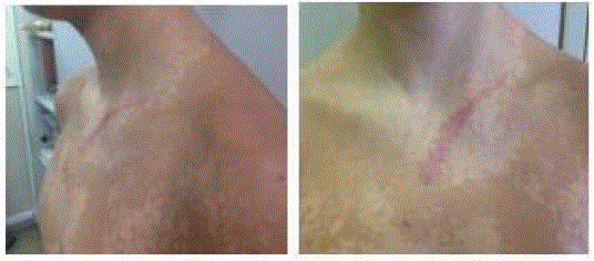
Figure 4a
Clinical outcome 12 months after surgical procedure.
Figure 4b
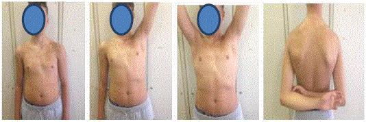
Figure 4b
Functional outcome 12 months after surgical procedure.
Figure 5a
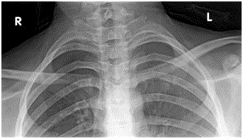
Figure 5a
Radiograph 12 months after surgical reduction.
Figure 5b
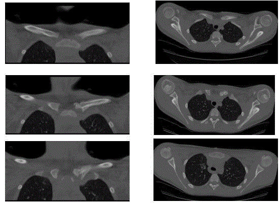
Figure 5b
CT scan showing correct position of sternoclavicular joint.
References
- Kocher MS, Waters PM, Micheli LJ. Upper extremityinjuries in the paediatric athlete. Sports Med. 2000;30(2):117-35.
- Wirth MA, Rockwood CA. Acute and Chronic TraumaticInjuries of the Sternoclavicular Joint. J Am Acad Orthop Surg. 1996;4(5):268-78.
- Nettles JL, Linscheid RL. Sternoclaviculardislocations. J Trauma. 1968;8(2):158-64.
- Lim KS, Lingaraj K, Das De S. Traumaticretrosternaldislocation of the sternoclavicular joint of a young adultwithgeneralized ligamentous laxity. Injury Extra. 2008;39(9):302-4.
- Cope R. Dislocations of the sternoclavicular joint.Skeletal Radiology. 1993;22(4):233-38.
- Daniel J Morell, David S Thyagarajan. Sternoclavicularjoint dislocation and its management: A review of the literature. World JOrthop. 2016;7(4):244-50.
- Bulstrode CKJ, editor. Oxfordtextbook of orthopedics and trauma. 10th ed. Oxford: OxfordUniversity Press. 2001;693:2053-5.
- Sewell MD, Al-Hadithy N, Le Leu A, Lambert SM.Instability of the sternoclavicular joint: current concepts in classification,treatment and outcomes. Bone Joint J. 2013;95(6):721-31.
- Spencer EE, Kuhn JE, Huston LJ, Carpenter JE, HughesRE. Ligamentous restraints to anterior and posterior translation of thesternoclavicular joint. J Shoulder Elbow Surg. 2002;11(1):43-7.
- S Bot, C Terwee, D A W M van der Windt, L Bouter, JDekker, H C W de Vet. Clinimetric evaluation of shoulder disabilityquestionnaires: a systematic review of the literature. Ann Rheum Dis. 2004;63(4):335-41.
- Bicos J, Nicholson GP. Treatment and results ofsternoclavicular joint injuries. Clin Sports Med. 2003;22(2):359-70.
- Glass ER, Thompson JD, Cole PA, Gause TM 2nd,Altman GT. Treatment of sternoclavicular joint dislocations: a systematicreview of 251 dislocations in 24 case series. J Trauma. 2011;70(5):1294-8.
- Thut D, Hergan D, Dukas A, Day M, Sherman OH.Sternoclavicular Joint Reconstruction A Systematic Review. Bull NYU Hosp JtDis. 2011;69(2):128-35.
- Rockwood CA, Odor JM. Spontaneous atraumatic anteriorsubluxation of the sternoclavicular joint. J Bone Joint Surg Am.1989;71(9):1280-8.
- Rotini R, Guerra E, Bettelli G, Marinelli A, Frisoni T.Sterno clavicular joint dislocation: a case report of a surgical stabilizationtechnique. Musculoskelet Surg. 2010;94:91-4.
- Abiddin Z, Sinopidis C, Grocock CJ, Yin Q, FrostickSP. Suture anchors for treatment of sternoclavicular joint instability. JShoulder Elbow Surg. 2006;15(3):315-8.
- Rockwood CA, Groh GI, Wirth MA, Grassi FA. Resectionarthroplasty of the sternoclavicular joint. J Bone Joint Surg Am.1997;79(3):387-93.
- Bae DS, Kocher MS, Walter PM, Micheli LM, Griffey M,Dichtel L. Crhonic Recurrent anterior sternoclavicular joint instability: resultsof surgical management. J Pediatr Orthop. 2006; 26(1):71-4.
- Armstrong AL, Dias JJ. Reconstruction for instabilityof the sternoclavicular joint using the tendon of the sternocleidomastoidmuscle. J Bone Joint Surg Br. 2008;90(5):610-3.
- Burrows HJ. Tendodesis of subclavius in the treatmentof recurrent dislocation of the sterno-clavicular joint. J Bone Jt Surg Br.1951;33(2):240-3.
- Castropil W, Ramadan LB, Bitar AC, Schor B, deOliveira D'Elia C. Sternoclavicular dislocation--reconstruction withsemitendinosus tendon autograft: a case report. Knee Surg Sports TraumatolArthrosc. 2008;16(9):865-8.
- Rockwood CA. Dislocations of thesternoclavicular joint. In: Evans E, editor. American academy of orthopaedicsurgeons instructional course lectures: Volume XXIV. st. Louis: CV Mosby. 1975:144-59.
- Ono K, Inagawa H, Kiyota K, Terada T, Suzuki S,Maekawa K. Posterior dislocation of the sternoclavicular joint with obstructionof the innominate vein: case report. J Trauma. 1998;44(2):381-3.
- Jougon JB, Lepront DJ, Dromer CE. Posteriordislocation of the sternoclavicular joint leading to mediastinal compression.Ann Thorac Surg. 1996;61(2):711-3.
- Cheng J. A rare cause of pediatric dysphagia:posterior dislocation of the sternoclavicular joint. Int J PediatrOtorhinolaryngol. 2014;78(1):152-3.
- Nakayama E, Tanaka T, Noguchi T, Yasuda J, Terada Y.Tracheal stenosis caused by retrosternal dislocation of the right clavicle. AnnThorac Surg. 2007;83(2):685-7.
- Adamcik S, Ahler M, Gioutsos K, Schmid RA, Kocher GJ.Repair of sternoclavicular joint dislocations with Fiber Wire®. ArchOrthop Trauma Surg. 2017;137(3):341-5.
- Aydin E, Dülgeroglu TC, Ates A, Metineren H. Repair ofUnstable Posterior Sternoclavicular Dislocation Using Nonabsorbable Tape Sutureand Tension Band Technique: A Case Report with Good Results. Case Reports inOrthopedics. 2015;2015:750898.
- Shuler FD, Pappas N. Treatment of posteriorsternoclavicular dislocation with locking plate osteosynthesis. Orthopedics.2008;31(3):273.
- Groh GI, Wirth MA, Rockwood CA. Treatment of traumaticposterior sternoclavicular dislocations. J Shoulder Elbow Surg. 2011;20(1):107-13.
- Smolle-Juettner FM, Hofer PH, Pinter H, Friehs G,Szyskowitz R. Intracardiac malpositioning of a sternoclavicular fixation wire.J Orthop Trauma. 1992;6(1):102-5.