Research Article
Guide Surgery Corticotomy System (GSCS) a New Device in Orthodontic Treatment
Giuseppe Salvato1, Carlo Chiavenna2* and Maria Costanza Meazzini3
1Department of Maxillofacial Surgery, ISI Istituto Stomatologico Italiano, University of Milano, Milan, Italy
2Department of Orthodontics, San Paolo Hospital, University of Milan, Italy
3Department of Maxillofacial Surgery, and orthodontic consultant, Cleft Lip and Palate Center, University of Milan,
Milan, Italy/p>
*Corresponding author: Pejman Janbaz, Department of Oral and Maxillofacial Surgery, Hamedan University of Medical Sciences, Shahid Fahmideh Street, Postal Code: 654178- 38741, Hamedan, Iran
Published: 12 Jun, 2017
Cite this article as: Yaripour S, Dehghan A, Janbaz P.
Diffuse Large B-Cell Lymphoma
Involving of Face - A Case Report and a
Brief Review. Clin Surg. 2017; 2: 1505.
Abstract
Introduction: This article proposes an innovative diagnostic and therapeutic protocol for performing
corticotomies in office under local anaesthesia with piezoelectric surgery using a surgical acrylic
guide produced through software-based planning.
Methods: The method was applied in late adolescent or adult patients that request short treatment
time, patients with mild dentoskeletal discrepancies and patients with poor periodontal support.
Performing a preoperative CT with a special splint, optical scanning of the models and subsequent
planning with software has enabled us to produce a model with rapid prototyping with the design
of the corticotomies on which the surgical guide was shaped. The use of the guide associated with
piezoelectric surgery, allowed to perform surgery under local anaesthesia, with minimal invasiveness
and high accuracy.
Results: Immediate movement of teeth after corticotomies allows a reduction of the treatment times
up to one year, with preservation of the roots of teeth involved. Corticotomies allow the orthodontist
to achieve with accuracy the objectives required by the treatment plan.
Conclusion: GSCS is a new method, which, utilizing 3D optical scanning images of models,
software and piezoelectric surgery, allows to perform dental movements which may be difficult or
dangerous for the periodontal support, with orthodontics only. It dramatically reduces total surgicalorthodontic
treatment time, with obvious great patient satisfaction.
Keywords: Corticotomy system; Orthodontic Treatment; Anaesthesia
Introduction
Orthodontic treatment of late adolescent or adult patients can be challenging; often these patients request short treatments. If growth modification is no longer possible, surgical procedures might be necessary to attain treatment goals [1,2]. With an osteotomy, both the cortical and trabecular bone is cut, followed by repositioning of the segments by the surgeon. Damage to the nerves and blood supply is a possible complication [3,4]. For patients with the need of minor facial reshaping, orthognathic surgery might not be a good choice and that is the reason why other surgical techniques, like low-level laser therapy, pulsed electromagnetic fields, electrical currents, mechanical vibration and corticotomy, have been proposed [5]. With a corticotomy, shallow perforations or cuts are made on the cortical alveolar bone only; the trabecular bone is left intact, in contrast to an osteotomy. Orthodontic force is applied shortly after surgery to produce the desired dental movement and optimal bone remodeling. It has been claimed that orthodontic treatment proceeds faster, and that the results are more stable after a corticotomy, with minimal risk of complications [6]. Corticotomy facilitated orthodontics has been indicated for non-extraction treatment of crowding, shortening treatment duration, borderline orthognathic surgery patients, extrusion of ankylosed teeth, intrusion of posterior teeth to close anterior open bites, faster canine retraction in extraction patients, and impacted canines. The use of rotary instruments can be potentially damaging to the teeth and to the periodontium [7-8] and reduces the compliance of patients under local anesthesia. Piezoelectric surgery facilitates dentoalveolar osteotomy under local anesthesia, as it is a more conservative approach on the soft tissues and is less taxing for patients [9]. The methods commonly used in guided implant surgery have been applied to the managing of post-traumatic deformities [10] and in the planning and execution of corticotomies to allow these minimally invasive, more accurate and more conservative outpatients’ procedures.
Figure 1
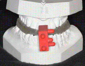
Figure 1
Extra oral volume transfer element (3DMarker - 3DIEMME, Italy).
Figure 2
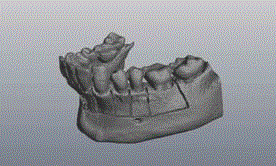
Figure 2
Optical scanning of the inferior plaster model.
Figure 3
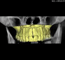
Figure 3
Virtual anatomical model with the corticotomies lines.
Figure 4
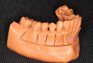
Figure 4
Anatomical model.
Figure 5
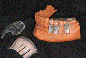
Figure 5
Anatomical model with the surgical guide for the osteotomy.
Materials and Method
AThis study follows a protocol in compliance with the World
Medical Association Declaration of Helsinki on medical research
protocols and ethics.
The method involves performing a CT scan (spiral or cone
beam) with a custom made radiological guide, consisting of a splint
incentric occlusion with the addition of an extra oral volume transfer
element (3DMarker e 3DIEMME, Italy) (Figure 1). Using this device
the optical scanning of the plaster models may be inserted into the
reconstructed volume by CT, in a precise and operator independent
manner. Corticotomies may be planned on a virtual model comprising
the full volume of the bone, the anatomy of the teeth and the gingival
thickness. The superimposition with the 3DMarker model is based on
the software recognition of the volumetric element, which is present
in both the CT volume and in the optical scanning. It is also facilitated
by the pre-alignment of volumes through best-fit algorithms that
analyze the clouds of points of the 3DMarker, overlap points in
common (from 5000 to 25,000 points for elaboration) minimizing the
distance. Upon completion of the pre-alignment on the 3DMarker,
the software (3Diagnosys 4.0 e 3DIEMME, Italy) verifies surfaces in
relation to anatomical profiles and further refines the alignment of
volumes returning a complete virtual anatomical model, replacing
the teeth present in the CT image with the surfaces obtained with
the optical scanning of the model (virtually eliminating artefacts that
may be present in the CT) and reconstructing the soft tissue thickness
(directly and not by difference of radiopaque radiographic guides).
The optical scanning of the models on which the traditional set-up was
carried out, allows confirmation that what was programmed virtually
is correct (Figure 2, 3). The virtual treatment plan is transferred on
to an anatomical model (RealMODEL_, 3DIEMME, Italy), using
CAD/CAM or rapid prototyping, in which the design of the planned
osteotomy is reproduced. On this model the laboratory constructs a
surgical guide. The corticotomy path is 0.75 mm as the cutters of the
piezoelectric system have a thickness of 0.6 mm (Figure 4, 5). The
corticotomies are performed with Piezotome (tip BS1 long; Satelec,
Acteon Group, Merignac, France).We extrapolate the measurements
of the alveolar thicknesses from the CT examination software so that,
during surgery, given the presence of the surgical template, we have
an additional 3D guide for millimetric drills (Figure 6, 7); therefore,
allowing observation of the soft tissues and the adjacent roots, even in
cases of reduced inter-radicular spaces.
We present a case of a Class II Division II in which we performed
corticotomies on the mandibular and maxillary arch (Figures 7-12).
After infiltration of all the superior vestibular fornix with local
anesthetic with vasoconstrictor, full thickness flaps were raised on
lingual surfaces to expose the alveolus surrounding the first molar
from side to side. Vertical releasing incisions are performed on the
second molar and however at least one tooth away from the bone
activation.
To allow the correct positioning of the surgical mask it is necessary
to check the correct fitting by exploration windows (the stamp of the
cuspid on the surgical guide), as show on virtual planning and it must
be absolutely stable. These tips are marked to check the depth and the
surgeon has just to follow the line inside the guide.
start the alignment (Figure 11) and the wires are changed weekly
for the first 2 months. Similarly, after one month, we executed the
corticotomies on the lower arch and the orthodontist may proceed
with the bonding of the lower arch too (Figures 11-13).
Figure 6
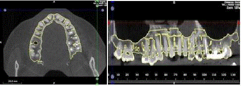
Figure 6
Template guide for the drilling.
Figure 7
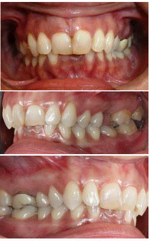
Figure 7
Before surgery.
Figure 8
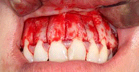
Figure 8
Corticotomy segments.
Figure 9
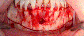
Figure 9
Corticotomy segments and excision of granuloma
Figure 10
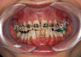
Figure 10
Bonding of the upper arch.
Figure 11-13
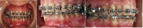
Figure 11-13
Four months after corticotomies.
Figure 14-18
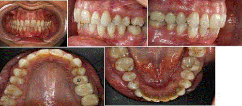
Figure 14-18
End of treatment after 12 months.
Result
The case was completed under local anesthesia without the use of sedation. The postoperative course, given the minimal dissection and the use of piezoelectric surgery, was straight forward and painless. The teeth involved remained viable and periodontal support was preserved (Figures 14-18). The treatment has been performed in 12 months, forty per cent less than a conventional treatment and the follow up is now at two years and all teeth are still viable.
Discussion
In a time when more and more patients seek minimally invasive,
rapid and predictable treatment, the GSCS method achieves these
goals in full. It can dramatically reduce the time of conventional
orthodontic treatment by, reducing treatment time, there is twice as
much tooth movement with than without corticotomies. However,
this window of opportunity used to accelerate tooth movement is
limited to 2-3 months, in which 4-6 mm of tooth movement might
be expected. Due to their micrometric and selective cut, piezoelectric
devices have been claimed to produce safe and precise osteotomies
without osteonecrosis damage. Other advantages like poor bleeding
and no vibrations increase the patient’s compliance since the surgery
is under local anesthesia.
All this is possible because, by creating a multidisciplinary team
consisting of orthodontist, maxillofacial surgeon, dental technician
and engineer, the principles of corticotomy, guided implant surgery;
piezoelectric surgery and 3D software are combined.
The use of software and a surgical template allows a simpler
procedure in executions of the corticotomies respecting radicular
anatomies, especially in the posterior areas where it is more difficult
to locate the root apices.
This technique allows a reduction of treatment time of about forty
percent if compare with a traditional treatment without corticotomies
[11].
Conclusion
GSCS is an aid to orthodontic. In patients who do not wish to undergo long treatments which include surgery under general anaesthesia. GSCS may be a valid alternative. It is useful to speed up treatment and the outcomes may be better. Studying the outcomes of a larger number of cases will allow clinicians to modify or increase the clinical indications for this approach. This technique necessarily requires the use of instruments suitable for piezo surgery.
References
- Reyneke JP. Diagnosis and treatment planning. In: Reyneke JP, editor. Essentials of orthogenetic surgery. Hanover Park, 1L: Quintessence. 2002;69-110.
- Chung CJ, Jung S, Baik HS. Morphological characteristics of the symphyseal region in adult skeletal Class III crossbite and openbite malocclusions. Angle Orthod. 2008;78:38-43.
- Foushee DG, Moriarty JD, Simpson DM. Effects of mandibular orthognathic treatment on mucogingival tissues. J Periodontol. 1985;56(12):727-33.
- Graber T, Varnarsdall R, Vig K. Orthodontics: current principles and techniques. St Louis: Elsevier. 2005:901-36.
- Long H, Pyakurela U, Wangb Y, Liaoa L, Zhoua Y, Lai W. Interventions for accelerating orthodontic tooth movement. A systematic review. Angle Orthod. 2013;83:164–171.
- Hoogeveen EJ, Jansma J, Ren Y. Surgically facilitated orthodontic treatment: a systematic review. Am J Orthod Dentofacial Orthop. 2014;145:S51-64.
- Morgan TA, Fridrich KL. Effects of the multiple-piece maxillary osteotomy on the periodontium. Int J Adult Orthodon Orthognath Surg. 2001;16(4):255-65.
- Schultes G, Gaggl A, Kärcher H. Periodontal disease associated with interdental osteotomies after orthognathic surgery. J Oral Maxillofac Surg. 1998;56:414-7.
- Robiony M, Polini F, Costa F, Vercellotti T, Politi M. Piezoelectric bone cutting in multipiece maxillary osteotomies. J Oral Maxillofac Surg. 2004;62(6):759-61.
- Salvato G, Chiavenna C, Meazzini MC. Guide surgery osteotomy system (GSOS) a new device for treatment in orthognathic surgery. J Craniomaxillofac Surg. 2014;42(3):234-8.
- Caserta M, Giansanti M, Di Mambro A, Calasso S, Barbato E. Minimally invasive corticotomy in orthodontics using a three-dimensional printed CAD/CAM surgical guide. Int J Oral Maxillofac Surg. 2016;45:1059–64.