Case Report
Unilocular Cystic Lymphangioma of the Small Omentum in a Girl of 4 Years
Missoki Azanlédji Boume1*, Ayi Kossigan Adodossi Amavi2, Komlan Adabra2, Komlan Anani
Mihluedo-Agbolan1, Sessime Yawa Sanni1, Serge Codjo Metchihoungbe1, Gamedzi1 And
Komlatsè Akakpo-Numado1
1Department of Paediatric Surgery, Sylvanus Olympio Teaching Hospital, BP 57 Lomé, Togo
2Department of General Surgery, Sylvanus Olympio Teaching Hospital, BP 57 Lomé, Togo
*Corresponding author: Missoki Azanlédji Boume, S/C M Gameti K. Evenyo, 08 BP 80025 Lomé 8, Togo
Published: 26 May, 2017
Cite this article as: Boume MA, Amavi AKA, Adabra K,
Mihluedo-Agbolan KA, Sanni SY,
Metchihoungbe SC, et al. Unilocular
Cystic Lymphangioma of the Small
Omentum in a Girl of 4 Years. Clin
Surg. 2017; 2: 1487.
Abstract
Background: Cystic lymphangiomas (CL) are congenital benign malformations of lymphatic
vessels. Their usual locations (90%) are neck and axilla in subcutaneous tissue. Abdominal locations
are rare and represent 10-12%. The incidence of mesenteric and omental cysts is 1 in 20,000 among
children, and even lower among infants. Only 2.2% are omental cysts of these two types of cystic
mass. Of omental CL, unilocular forms are rarer but more common in boys than in girls. We aim to
present clinical, radiological and therapeutic management of a small omental unilocular CL, slowly
evolved in a girl.
Observation: A girl of 4-years suffered of chronic intermittent abdominal pain without vomiting
and fever, managed like parasitic infection during a year. One year after, appears an abdominal
swelling leading to pediatric surgery department. There was no prenatal diagnosis. The preoperative
radiological diagnosis by ultrasonography and scan was simple mesenteric cyst, especially because
of the unilocular presentation aspect. One week before scheduled surgery it was complicated by
intracystic hemorrhage. The lesion of 17 cm × 8 cm was implanted in the small omentum under
the liver between hepatic pedicle and the gastric artery. It had sero-hematic content and was totally
removed by laparotomy. Hystological examination concluded to CL. The outcome was favorable
after 13 months follow up.
Conclusion: CL has many forms, locations and cystic contents. Unilocular cystic lymphangiomas
located in the small omentum are very rare and can be complicated at any time. It is more often
confused with mesenteric cysts but they are distinct from one another by pathological examination
which gives the final proof of the diagnosis. Surgical resection should be as complete as possible,
putting the patient free from recurrences.
Keywords: Cystic lymphangioma; Abdomen; Omentum ; Children; Surgery
Introduction
Cystic lymphangiomas (CL) are congenital benign malformations of lymphatic vessels. Lack of lymphatic vascular connections secondary to an abnormal embryological development of the lymphatic system lead to a sequestration of the lymphatic tissue. It results in obstruction of the local lymphatic flow and the development of lymphagectasias. Their origin remains uncertain [1- 3]. Described for the first time by Koch one century ago, it is known that this anomaly occurres primarily in children and 60% of cases are diagnosed before the age of 5 years [4]. Usual locations (90%) of cystic lymphangioma (CL) are neck, extremities and axillary in subcutaneous tissue. Abdominal locations are rare and represent 10%-12% [5]. In abdomen, locations are also variable and involve mesentery, mesocolon, retroperitoneum, omentum, splenic loge, liver and pancreas [2- 4,6]. Abdominal locations cause diagnostic and therapeutic problems. We report a CL located in the gastro-hepatic ligament and focus by literature reviewer on its abdominal locations.
Case Presentation
A 4-year-old girl was admitted on a scheduled basis in pediatric surgery department for an
abdominal mass about 1 month before, associated to chronic abdominal pain. Second child of a
fourth’s siblings without pass history of abdominal mass in the family, she was born at term. There
was no previous antenatal diagnosis of abdominal mass. Her parents are alive and well.
In fact, she had abdominal intermittent pain since one year
without vomiting or fever. One abdominal ultrasound realized at
the beginning of the symptoms was normal. She had analgesics and
ant parasitic drug, four times in year duration. One month before
scheduled admission, her parents realized abdominal epigastric
swelling. That time it was not associated with pain. She had a preserved
transit and general condition. She had no fever. The examination
noticed an epigastric abdominal swelling (Figure 1) where a moving
painless mass was felt. There was no node clinically felt. She had a
good cutaneo-mucous coloration. The rest of the examination was
normal. Abdominal ultrasound showed a unicystic mass with a
regular wall and a homogeneous content. An abdominal scan (Figure
2) revealed a unicystic anterior abdominal tumor with homogeneous
content resulting to cystic mesenteric cyst.
The patient was scheduled for exploratory laparotomy and
tumor excision. One week before surgery, she had moderate acute
abdominal pain. She had maintained a normal hemodynamic state.
At laparotomy we discover a unicystic tumor implanted in the
small omentum (Figure 3). It was very limited measuring 17 cm
× 8 cm. It had cracked and bleeding with hematomas in the great
omentum. The content was sero-hematic, mixing yellowish fluid with
blood due to the intracystic hemorrhage. Figure 4 shows the postresection
operative site. The patient left hospital five days after. The
final histological diagnosis was cystic lymphangioma. The patient was
doing well 13 months postoperatively.
Figure 1
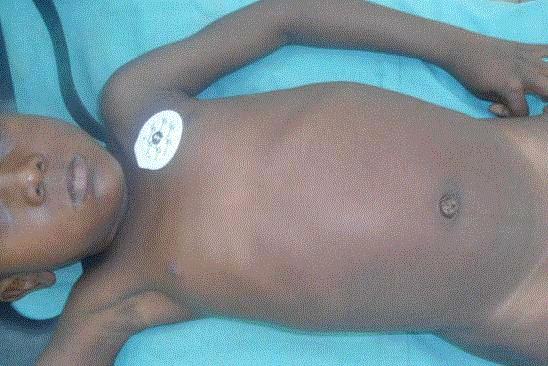
Figure 1
Abdominal located swelling in the girl; look upper part of the
abdomen.
Figure 2
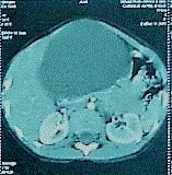
Figure 2
Abdominal CT scan (axial section over meso-colon) showing a
large intra-abdominal unicystic lesion with thin walls and homogenous
content.
Figure 3
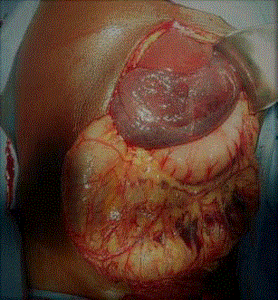
Figure 3
Intra operative appearance of the cyst implanted in the small
omentum (look between stomach and liver) with sero-hematic content.
Figure 4
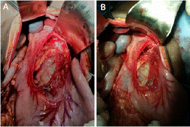
Figure 4
The cystic site after resection.
A. Look hepatic pedicle at the right and the gastric artery at the left.
B. The choledoc duct in the right of the tumor site.
Discussion
Cystic lymphangioma (CL) is conjunctival malformative vascular
tumours corresponding to a detention of lymphatic tissue due to an
abnormal embryonic development of the lymphatic system [7]. It is
a congenital benign malformation of lymphatic vessels. In fact, the
matter would be a lack of lymphatic vascular connections secondary
to an abnormal embryological development of the lymphatic
system which lead to a sequestration of the lymphatic tissue. These
arrested developments of lymphatico-venous connections during
the embryogenesis would cause absence of drainage of the primitive
lymphatic bags leading to the formation of a cystic lesion containing
some lymph. It results in obstruction of the local lymphatic flow and
the development of lymphagectasias. Their origin remains uncertain
[1-3,8]. Described for the first time by Koch one century ago, it is
known that this anomaly occurres primarily in children according
to its embryological development and 60% of cases are diagnosed
before the age of 5 years [4]. This congenital theory is strengthened
by observations of CL detected in the prenatal period. Or mostly in
children population however, the diagnosis can be made at any age
[8]. In our case there was no any prenatal discover of abdominal mass
but the cyst occurs at 4 years old. CL is usually benign but can be
locally invasive [9].
Usual locations (90%) of cystic lymphangioma (CL) are neck,
extremities and axillary in subcutaneous tissue [10,11]. But variety
of other sites have been described including the mediastinum,
pleura, pericardium, groin, bones and abdomen [12,13]. Abdominal
locations are rare and represent 10%-12% [5]. In abdomen, locations
are also variable and involve mesentery, mesocolon, retroperitoneum, omentum, splenic loge, liver and pancreas [2-4,6]; however mesentery
and retroperitoneum are the most common sites. We reported
omental location of CL. Omental and mesenteric cysts are both rare
pathologies in children. The incidence of mesenteric and omental
cysts is 1 in 20,000 among children and lower in infants. Of these two
types of cystic mass, 2.2% are omentalcysts [14-16]. Lymphangioma is
the most common cause of these cysts, which are generally restricted
to the lesser or greater omentum [15,16].
Clinical presentation can be variable and nonspecific. Acute
symptoms include acute abdomen, distension, vomiting, and fever.
Chronic symptoms include progressive abdominal distension and
pain. Patients admitted to the hospital may be classified into two main
groups: those with acute clinical symptoms and those with non-acute
clinical symptoms [17]. Although symptoms correlate to the location
and size of the cyst, non-acute clinical symptoms include pain less
abdominal mass, abdominal pain, abdominal distention, and possible
ascites [14,16,18,19]. Diagnosis of a cyst should be considered even
if the findings are non-specific and the patient exhibits symptoms
over a long period of time [20]. The presence of complicating factors,
including hemorrhage, torsion, and infection, rupture, or pressure
to other structures, is relevant with acute presentations that require
urgent surgery [17] like in our case. These complications had no
appropriated time and come occurs any time. At once evoked, even
if complications are absent the treatment must quickly accompanied
to avaoid complications which threaten life of the child. Choledochal
cysts, splenic cysts, multicystic dysplastic kidneys, intestinal
duplication cysts, and ovarian cysts are all cystic lesions that can be
included in the differential diagnosis of omental cysts [16, 21].
Ultrasonography has been reported as the initial diagnostic tool
in all cases. Sonographic findings frequently feature multiloculated,
fluid-filled, and predominantly cystic lesions [15,16,22].
Pathologically, they can be unilocular or multilocular [17], knowing
that unilocular form are more rare. Preoperative diagnoses are more
difficult in unilocular case, as in our case.
CL is more often confused with mesenteric cysts that arise from
mesothelial, not lymphatic tissue. This differentiation is important
because lymphangiomas often behave in an invasive and aggressive
manner, where as mesothelial cysts do not. Despite being difficult to
differentiate between imaging studies, they are histologically distinct
from one another. Lymphangiomas have an endothelial lining, foam
cells, and a wall that contains lymphatic spaces, lymphoid tissues, and
smooth muscles.
Clearly the case we described caused more preoperative diagnosis
difficulties probably because of its location (smallo mentum), form
(unilocular) and complication (intracystic hemorrhage).
The content of the cyst is sero-hematic in our case. The content of
CL can be, serous or sero-sanguineous. These different aspects can be
explained by different degrees of lymphaticstasis, a variable number
of connections with the lymphatic system and the protein content
of the cyst contained. The sero-hematiccyst appearance is secondary
to intra cystic hemorrhage. Rarely CL can be purulent by infection
[6,23].
The preferred treatment of omental cysts is complete excision,
whether laparoscopic or not. Resection of the bowel and recurrence
are rare. Malignant transformation of cystic lesions is also rare
[14,16,21]. Laparoscopic management has the advantages of lower
cost and decreased morbidity compared to open surgery [14,16].
Conclusion
Cystic lymphangioma has many forms, locations and cystic contents. Unilocular cystic lymphangiomas located in the small omentum are very rare and can be complicated at any time. It is more often confused with mesenteric cysts but they are distinct from one another by pathological examination which gives the final proof of the diagnosis.
References
- Colovic RB, Grubor NM, Micev MT, Atkinson HD, Rankovic VI, Jagodic MM. Cystic lymphangioma of the pancreas. World J Gastroenterol. 2008;14(44):6873-5.
- Mabrut JY, Grandjean JP, Henry L, Chappuis JP, Partensky C, Barth X, et al. [IMesenteric and mesocolic cystic lymphangiomas. Diagnostic and therapeutic management]. Ann Chir. 2002;127(5):343-9.
- Bezzola T, Buhler L, Chardot C, Morel P. Surgical treatment of abdominal cystic lymphangioma in adults and children. J Chir. 2008;145:238-43.
- Ghatak S, Ray S, Sanyal S, Sonar PK, Khamrui S, Basu K, et al. An unusual cause of acute abdomen in adults: giant cystic lymphangioma of the pancreatic head. A clinical case and literature review. JOP. 2011;12(3):266-70.
- Khmekhem R, Rahay H, Ghorbel S, Jlidi S, Douira W, Bellagha I. Abdominal Cystic Lymphangioma. About Seven Cases. SM J PediatrSurg. 2016;2(5):1029.
- Weeda VB, Booij KA, Aronson DC. Mesenteric cystic lymphangioma: a congenital and an acquired anomaly? Two cases and a review of the literature. J Pediatr Surg. 2008;43(6):1206-8.
- Guivarc'h M. [Tumors of the mesentery. Apropos of 102 cases]. Ann Chir. 1994;48(1):7-16.
- Solovei G, Alame A, Elchardus JF, Glavier F, Petit J, Cart P, et al. [Mesenteric cystic lymphangioma in children. Report of a case manifested by anemia]. Ann Pediatr (Paris). 1990;37(6):405-8.
- Sakhri J, Benali A, Ltaeif R, Dahmen Y, Derbel F. Cystic abdominal lymphangioma: diagnosis and treatment. Ann GastroentérolHépatol. 1997;33:113-6.
- Khandelwal M, Lichtenstein GR, Morris JB, Furth EE, Long WB. Abdominal lymphangioma masquerading as a pancreatic cystic neoplasm. J Clin Gastroenterol. 1995;20(2):142-4.
- Kullendorff CM, Malmgren N. Cystic abdominal lymphangioma in children. Case report. Eur J Surg. 1993;159(9):499-501.
- Goh BK, Tan YM, Ong HS, Chui CH, Ooi LL, Chow PK, et al. Intra-abdominal and retroperitoneal lymphangiomas in pediatric and adult patients. World J Surg. 2005;29(7):837-40.
- Leung TK, Lee CM, Shen LK, Chen YY. Differential diagnosis of cystic lymphangioma of the pancreas based on imaging features. J Formos Med Assoc. 2006;105(6):512-7.
- Pampal A, Yagmurlu A. Successful laparoscopic removal of mesenteric and omental cysts in toddlers: 3 cases with a literature review. J Pediatr Surg. 2012;47(8):e5-8.
- Adikibi BT, Wood R, Pillay K, Millar AJ. Omental cyst presenting with profound anaemia. Afr J Paediatr Surg. 2013;10(2):180-4.
- Motie MR, Asadi M. Large omental cyst: a case report and review of the literature. Acta Med Iran. 2011;49(10):690-3.
- Walker AR, Putnam TC. Omental, mesenteric, and retroperitoneal cysts: a clinical study of 33 new cases. Ann Surg. 1973;178(1):13-9.
- Joshi N, Yadav S, Singh B, Gupta A. Omental cyst presenting as tubercular ascites. J Infect Dev Ctries. 2010;4(3):183-6.
- Karhan AN, Soyer T, Gunes A, Talim B, Karnak I, Oguz B, et al. Giant Omental Cyst (Lymphangioma) Mimicking Ascites and Tuberculosis. Iran J Radiol. 2016;13(3):e31943.
- Kurtz RJ, Heimann TM, Holt J, Beck AR. Mesenteric and retroperitoneal cysts. Ann Surg. 1986;203(1):109-12.
- Moralioğlu S, Sönmez K, Türkyilmaz Z, Başaklar AC, Kale N. A child with a giant omental cyst. Acta Chir Belg. 2007;107(6):724-5.
- Kokhanovsky N, Nachtigal A, Reindorp N, Shinhar D, Zeina AR. Giant omental hemorrhagic cyst presenting as acute hemorrhagic anemia in a 21-month-old infant. Pediatr Emerg Care. 2014;30(3):188-90.
- Losanoff JE, Richman BW, El-Sherif A, Rider KD, Jones JW. Mesenteric cystic lymphangioma. J Am Coll Surg. 2003;196(4):598-603.