Case Report
Lately Recognized Left Main Bronchial Rupture and Fail Chest
Ming-Ho Wu* and Han-Yun Wu NP
Department of Surgery, Tainan Municipal Hospital, Taiwan
*Corresponding author: Ming-Ho Wu, Department of Surgery, Tainan Municipal Hospital, 670 Chung- Te Rd, Tainan, Taiwan
Published: 24 Apr, 2017
Cite this article as: Ming-Ho Wu, Han-Yun Wu NP. Lately
Recognized Left Main Bronchial
Rupture and Fail Chest. Clin Surg.
2017; 2: 1412.
Abstract
Blunt bronchial rupture and flail chest are usually early detected and managed. However, in
multiple trauma patients, bronchial rupture and flail chest could be lately diagnosed. Herein, we
present a 50-year-old woman who had multiple trauma caused by traffic accident. In this patient,
left main bronchial rupture was lately detected at the first aid hospital. Furthermore, the flail
chest was confirmed after bronchoplasty of left main bronchus at the referred hospital. Reasons
of delayed diagnosis of left main bronchial rupture are lack of significant pneumothorax and
pneumomediastinum. Reasons of delayed diagnosis of flail chest are the patient was initially on
ventilator support and the ribs fracture occurred at the costo-cartilages.
Keywords: Bronchial rupture; Flail chest; Bronchoplasty; Chest wall fixation
Case Presentation
A 50 year-old woman with diabetes mellitus suffered from traffic accident resulting in multiple injuries, including ribs fracture, hemopneumothorax, pulmonary contusion, left scapular fracture, left acetabular fracture, pars fracture of 5th lumbar spine, and right olecranon fracture. The chest film taken in the Emergency Department showed haziness of bilateral lower lung fields only (Figure 1). She was admitted to the intensive care unit for ventilator support because of respiratory distress at the first aid regional hospital. She underwent left tube thoracostomy for suspected hemothorax, total hip replacement, and open reduction of right olecranon fracture in the regional hospital. Bronchoscopy was performed to find out the cause of partial lung collapse in 3 weeks after traffic accident. She was referred to our hospital because of suspected left main bronchial rupture based on the bronchoscopic finding of bronchial stenosis. After referral, bronchoscopy revealed bilateral vocal cords paralysis, subglottic ulcerations and stenosis of left main bronchus at 3 cm away from the carina (Figure 2). She underwent bronchoplasty of left main bronchus via left posterolateral thoracotomy with left 4th rib cut on the next day after referral. Choking while eating was noted. To prevent aspiration in the situation of bilateral vocal cords paralysis, a gastrostomy was additionally performed on post-bronchoplasty day 3. In the following days, paradoxical respiration was noted. Physically, the left 2nd up to 6th costo-cartilage fractures were detected. To improve respiratory effect, chest wall fixation was performed using 5 bone plates (Figure 3) on post-bronchoplasty day 16. Her breaths got improved after operative fixation of left chest wall. She was discharged uneventfully on post-bronchoplasty day 21. The bone plates on left chest wall were removed 10 months later.
Comments
Tracheobronchial rupture caused by blunt chest injury may be overlooked at the initial management [1,2]. Early recognition and primary repair of these injuries clearly decrease morbidity and provide excellent anatomic and functional results in most cases. In patients with bronchial rupture all had ipsilateral pneumothorax and most had associated pneumomediastinum, the degree of each component relating to the site of rupture [1]. In the presenting case, the initial chest film did not lead to consider the airway injury. We reviewed the chest CT taken in Emergency Department of the regional hospital, the mediastinal window of showed bilateral mild hemothorax and deformity of left main bronchus (Figure 4), and lung window showed localized peribronchial emphysema and mild pneumothorax (Figure 5). Left main bronchial rupture is confirmed by the operative finding. Delayed diagnosis of bronchial rupture may result in healing with scar tissue and total obliteration of the bronchus by granulation tissue [3]. In this case, the bronchoplasty required careful dissection because the surrounding tissue got fibrotic change (Figure 6). The patient experienced choking while eating. A gastrostomy was performed instead of tracheostomy because the temporary non-movement of the vocal cords and subglottic ulcerations were due to prolonged endotracheal intubation. In our previous report, 4 of 8 patients with bronchial rupture occurred at left main bronchus. Two patients with left main baronial rupture were delayed diagnosis [4]. In patient with multiple ribs fragment fracture, the flail chest is usually presented with paradoxical respiration without use of ventilator. In the present case, flail chest was lately recognized following bronchoplasty. Chest CT performed on post-bronchoplasty day 10, revealed depression of left anterior chest wall and atelectasis of left lower lobe lung (Figure 7). The above conditions got improved after chest wall fixation. Surgical stabilization is associated with a shorter ICU time, less hospital cost, and recovery of pulmonary function in a select group of patients with flail chest [5]. In conclusion, reasons of late diagnosis of left main bronchial rupture are lack of significant pneumothorax and pneumomediastinum. Reasons of late diagnosis of flail chest are the patient care initially on ventilator support and the ribs fracture occurred at the costo-cartilages.
Figure 1
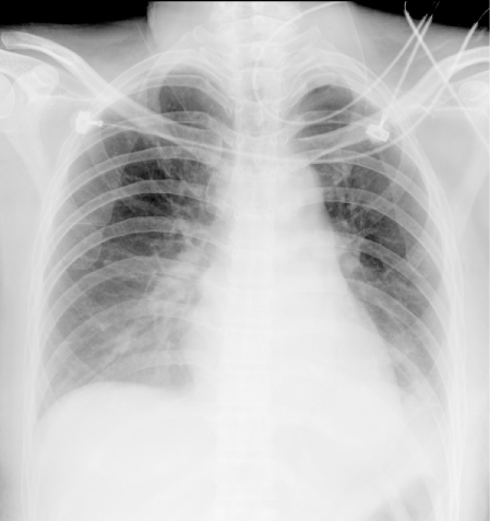
Figure 1
Chest film taken in Emergency Department showed no significant pneumothorax except haziness of bilateral lower lung fields.
Figure 2
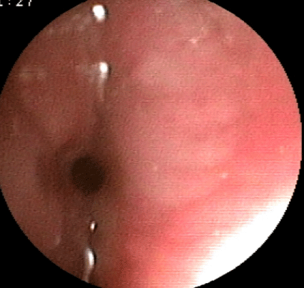
Figure 2
Bronchoscopy performed on post-trauma day 20 revealed stenosis
of left main bronchus at 3 cm away from the carina.
Figure 3
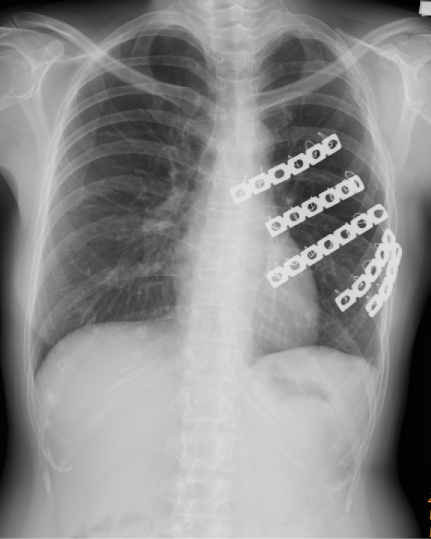
Figure 3
Chest wall fixation using bone plates was performed on postbroncoplasty
day 10.
Figure 4
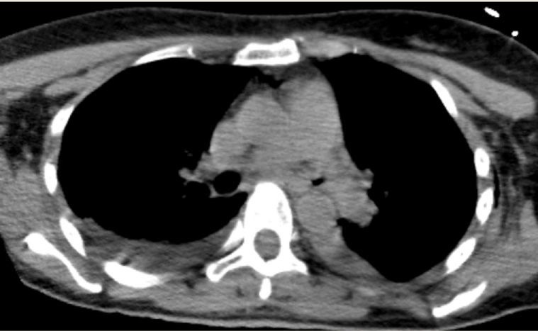
Figure 4
Mediastinal window of chest CT taken in Emergency Department
showed bilateral mild hemothorax and deformity of left main bronchus.
Figure 5
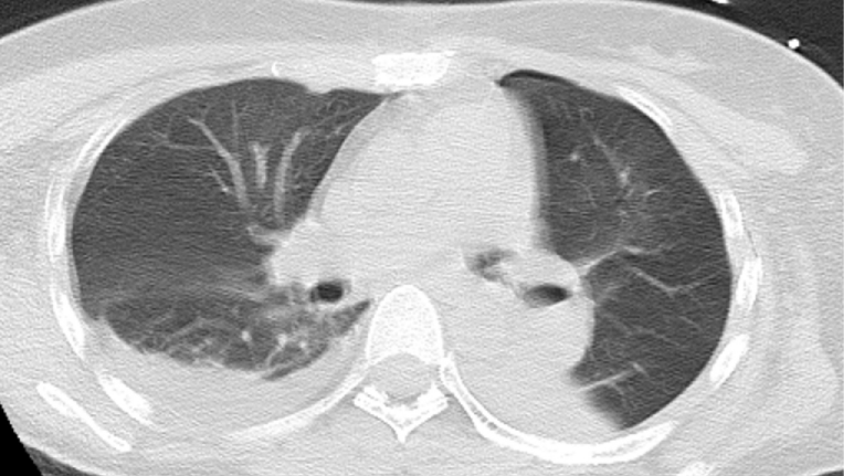
Figure 5
Lung window of chest CT taken in Emergency Department showed
localized peribronchial emphysema and mild pneumothorax.
Figure 6
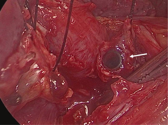
Figure 6
At surgery, the end of bronchial tube of double-lumen endotracheal
tube was visulized when proximal end of left main bronchus was mobilized
(arrow).
Figure 7
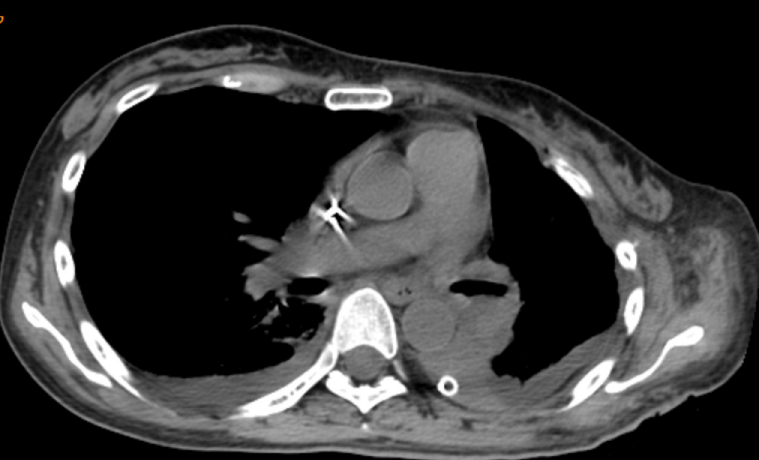
Figure 7
On post-bronchoplasty day 10, chest CT revealed depression of
left anterior chest wall and atelectasis of left lower lobe lung.
References
- Spencer JA, Rogers CE, Westaby S. Clinico-radiological correlates in rupture of the major airways. Clin. Radiol. 1991;43(6):371-6.
- Baumgartner F, Sheppard B, de Virgilio C, Esrig B, Harrier D, Nelson RJ, et al. Trachealand main bronchial disruptions after blunt chest trauma: presentation and management. Ann. Thorac Surg. 1990;50(4):569-74.
- Lin MY, Wu MH, Chan CS, Lai WW, Chou NS, Tseng YL. Bronchial rupture caused by blunt chest injury. Ann. Ernerg. Med. 1995;25(3):412-5.
- Wu MH, Tseng YL, Lin MY, Lai WW. Surgical results of 23 patients with tracheobronchial injuries. Respirology. 1997;2(2):127-30.
- Pettiford BL, Luketich JD, Landreneau RJ. The management of flail chest. Thorac Surg Clin. 2007;17(1):25-33.