Case Report
Refractory Odontogenic Infection Associated to Candida Albicans: A Case Report
Daya A Mikhail*, Mederos Heidi BS and McClure Shawn
Department of Oral and Maxillofacial Surgery, Nova Southeastern University /Broward Health Medical Center, USA
*Corresponding author: Daya A Mikhail, Department of Oral and Maxillofacial Surgery, Nova Southeastern University /Broward Health Medical Center, 3301 College Ave, Fort Lauderdale, FL 33314, USA
Published: 31 Mar, 2017
Cite this article as: Mikhail DA, Mederos Heidi BS, McClure
Shawn. Refractory Odontogenic
Infection Associated to Candida
Albicans: A Case Report. Clin Surg.
2017; 2: 1397.
Abstract
Background: Multifascial space infections from an odontogenic origin have been attributed to
a different number of microorganisms. These infections can be serious, involving multiple deep
spaces in the head and neck, and potentially compromising the airway. Fungal etiology including C.
albicans has been reported in the past as a rare agent involving multifascial space infections.
Case Description: We present an unusual case of a severe deep space infection associated with
carious teeth numbers 31 and 32. This specific infection proved to be resistant to multiple antibiotic
therapy and required incision and drainage on two different occasions. After the initial surgery,
the patient remained febrile with an elevated white blood cell count; thus, the patient was taken to
the operating room again for a re-drainage and new cultures. Cultures obtained during the second
surgery were positive for C. albicans. The patient was responsive to antifungal therapy, showing
quick improvement in his condition.
Conclusion: Although multiple factors could have contributed to this patient’s vulnerability
to odontogenic infection of fungal etiology including history of alcoholism and broad spectrum
antibiotic therapy, it remains an infrequent finding in the literature. This case illustrates the need
to consider a fungal cause in patients with odontogenic infections who are not responsive to broad
spectrum antibiotics and surgical drainage.
Keywords: Odontogenic infection; Candida albicans; Microorganisms
Introduction
Many microorganisms have been identified in multifascial space infections of the head and
neck region. These infections are most commonly caused by bacteria [1] and can be life-threatening
with potential loss of airway and drainage to the mediastinum requiring immediate attention and
treatment.
Fungal etiology has been reported in the past as a rare causative factor. Badiee “et al.” [2]
reported a case of mediastinitis caused by C. albicans in an immune competent patient occurring
after dental extractions. The most commonly occurring mycoses are caused by commensal fungi
of the human body [3]. In healthy individuals, Candida species can be found in the oral cavity,
and other areas of the human body such as the gastrointestinal tract, groin and vaginalcanal [3].
Most these fungi are opportunistic pathogens, causing infections in the event of a change of normal
microbiota following administration of broad-spectrum antibiotics, in immune compromised
individuals, and when protective mechanisms have been disrupted [3].
We present a rare case of severe multi space infection, which proved to be resistant to multiple
antibiotic therapy and surgical drainage. The patient was taken to the operating room for a redrainage
and new cultures. Cultures obtained in the second surgery were positive for C. albicans.
Antifungal therapy was subsequently implemented and a favorable response and improvement of
the patient’s overall condition ensued. This case illustrates the need to consider a fungal cause in
patients with odontogenic infections who are not responsive to broad spectrum antibiotics and
surgical drainage.
Case Presentation
A 45-year-old male with a history of uncontrolled hypertension and severe alcohol abuse
presented to the emergency department with a complaint of worsening right facial swelling. The
patient reported that he started to develop a tooth pain on the right lower quadrant one week prior
to admission. Two days before presenting to the hospital he was seen by his dentist who diagnosed him with facial cellulitis from an odontogenic origin likely associated
to tooth #32. At that time, the patient was prescribed amoxicillin and
Percocet. The morning of admission he awoke with increased right
facial swelling and pain, and sought treatment in the Emergency
Department.
At the time of admission vital signs were: Heart rate 93, blood
pressure 115/75, and O2 saturation 96% on room air. Laboratory
findings at time of admission showed White blood cell count
(WBC) of 29.4, hemoglobin of 16, hematocrit 46%, platelets 342,000.
Clinically the patient was alert, in no evident distress, and tolerating
his secretions. Extraoral exam revealed significant swelling of the
right temporal, buccal, masseter, submandibular and submental
spaces, generalized cellulitis with erythema and warmth to touch was
notable. Intraoral examination showed severe swelling on the right
buccal mucosa, trismus and no signs of active drainage. The floor of
mouth was soft and not elevated. The tongue had free range of motion,
mild edema in the posterior right oropharynx, the pterygomandibular
raphe was slightly swollen and tooth #32had severe caries most likely
the source of the infection (Figure 1).
Computerized tomography with contrast of the face and neck revealed extensive soft tissue infection extending in the right
sub-masseteric space and right parapharyngeal space, and severe
enlargement of right parotid gland with no fluid collection and slight
airway deviation (Figure 2).
The patient was admitted to ICU and was started on intravenous
(IV) 900 mg clindamycin. On hospitalization day 2, he was taken
to the operating room for incision and drainage of multifascial
space abscess. Extraoral and intraoral approaches were used to
drain right sub-masseteric space and right parapharyngeal space,
purulent discharge was noted in both spaces, Penrose drains were
left in place. Teeth #31 and #32 were extracted and, a significant
amount of purulence was evacuated from the extraction site of tooth
#32. During drainage, it was noted that there was frank purulence
coming through the patient’s right ear exiting his auditory canal
(Figure 3). The patient remained intubated post operatively due to
severe swelling and possible compromise of airway. Ciprofloxacin
and Vancomycin were added to the antibiotic regimen per Infectious
disease recommendations. ENT consultation was requested; upon
evaluation, it was deemed that the EAC discharge was an extension
of the facial abscess.
No significant improvement was noticed after two days
post operatively. Culture and sensitivity showed “rare growth
of Streptococcus viridans isolated group and no anaerobe
microorganisms”, however, the patient showed minimal clinical
response to treatment, had continuous spikes in fever and a
maintained leukocytosis. No fungal cultures where collected from
the first surgery since multiple studies in the past an associated
multifascial space infections to be bacterial in nature.
A repeat CT (Figure 4) of head and neck was done and showed
persistent phlegmonous cellulitis with abscess of the right face
associated with adenopathy. Swelling and edema did extend to and
involve the deeper soft tissues medial to the right mandibular ramus.
Fluid was seen around the ramus laterally and medially representing
abscess collection. When compared to the previous CT, there is
evidence of obstruction of the right EAC as well as well as a well
differentiated collection adjacent to the mandibular condyle; this
finding has significant relevance do to drainage noted from the right
ear explained before (Figure 5).
Post operative day number 4 decision was made to take the patient to the OR for re-drainage. Once the patient was on the operating
table, Stensen’s duct was milked and there was purulent drainage was
elicited from the duct, this finding added to the enlargement of the
parotid gland and the fact that the patient was worsening brought
to the differential diagnosis suppurative parotitis. However, this
was ruled out by making a Modified Blair incision to expose parotid
capsule and three different punctures were made with a curved Kelly
with no significant drainage. Previously existing approaches were
used to re-drain right submasseteric and right parapharyngeal spaces,
in addition intraoral incision made to approach infratemporal space.
Purulent content was evacuated in all spaces. New cultures for aerobic,
anaerobic and fungus were taken. Patient remained intubated post
operatively and was transferred back to ICU.
Intraoperative cultures from the second surgery were positive
for Candida albicans growth. Empirical therapy with intravenous
antifungals micafungin and fluconazole were started as part of the
antimicrobial therapy. There was progressive improvement in
swelling and WBC. The patient was extubated on postoperative day
four following the second surgery and discharged day eight from
admission.
Figure 1
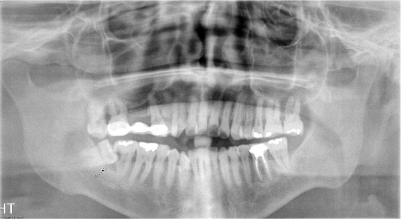
Figure 1
Panoramic X-ray showing malposition tooth #32 with gross carious #s 31 and 32.
Figure 2
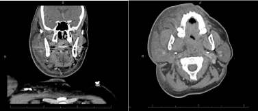
Figure 2
Coronal view of CT from initial presentation. Transverse view of
initial presentation demonstrating soft tissue swelling.
Figure 3
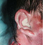
Figure 3
Frank purulence was evacuated from external auditory canal.
Figure 4
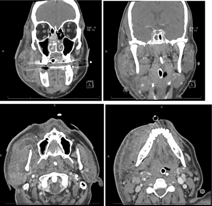
Figure 4
Repeat CT scan after persistent clinical signs of infections where
seen with minimal improvement.
Figure 5
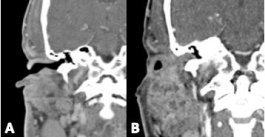
Figure 5
A. EAC clear and patent on initial CT. B.Repeat CT scan shows
obstruction of canal.
Discussion
Bacteria are the most frequent cause of odontogenic infections
[1]. The literature is replete with antimicrobial recommendations for
treatment of odontogenic infections attributed to bacterial etiology.
However, there have been rare case reports of odontogenic infections
caused by fungi, namely C. albicans. Farmahan “et al.” [1] showed
that C. albicans was seen in 3 cases (5%) of their patients. Candida
species can cause a broad range of infections in humans, ranging
from mild to dangerously wide spread diseases [3,1]. The most
commonly occurring mycoses are caused by commensal fungi of the
human body [3].
We present an unusual case of severe deep fascial spaces infection
of odontogenic origin with purulent discharge through the external
auditory canal. The infection was unresponsive to conventional
treatment with broad-spectrum antibiotics and surgical incision
and drainage. Fungal culture was requested at the time of the second
surgical incision and drainage showing growth of C. albicans.
Antifungal therapy was started with micafungin and fluconazole
following and the patient’s condition began to resolve.
The present case exemplifies the importance of considering both
bacterial and fungal etiology of odontogenic infections even when
patients are otherwise healthy. A review of the literature revealed
only one other case, by Badiee “et al.” [2], of widespread odontogenic
infection associated to C. albicans. The case report involves an
immune competent man who presented with Candida mediastinitis
due to a retropharyngeal abscess following a dental extraction [2].
The patient did not show improvement following administration of
broad-spectrum antibiotics and surgical drainage. The second culture
obtained indicated C. albicans growth and the patient’s condition
subsequently responded to antifungal treatment [2].
Odontogenic infections are very rarely associated to fungal
etiology, thus, fungi are not commonly included in the initial
differential diagnosis. However, due to the lack of response to initial
treatment, other possible etiologies should be included such as fungus.
Consequently, there has been significant research in attempt
to identify the bacterial population of infected root canals and
periapical lesions. Vigil “et al.” [4] analyzed the periradicular tissues
of 28 refractory endodontic cases and found the most common
isolates to be Propionibacterium acnes, Staphylococcus epidermidis,
Streptococcus intermedius, Wolinella recta, Fusobacterium species,
and Clostridium species. Though their sample size was small, one
culture was also positive for C. albicans [4]. Several other studies
have also reported that C. albicans is more frequently encountered
in these lesions that was previously expected [5,6,3]. C. albicans is
the most commonly isolated fungal species in the oral cavity and is
found in both healthy and immune compromised individuals [5,3].
However, infections arising from C. albicans usually occur in immune
suppressed individuals and are not commonly seen in otherwise
healthy patients.
Though uncommon, there are a few studies in the literature
reporting instances of C. albicans found in infected root canals.
Baumgartner “et al.” [5] identified C. albicans in 5 of 24 infected
root canal samples. Waltimo “et al.” [6] isolated 48 strains of fungi
out of 692 root canal samples positive for microorganisms (7%). The
fungi were mostly isolated together with bacteria (87%) but pure
cultures were also obtained (13%) [6]. C. albicans comprised 80% of
the identified yeasts [5]. Moreover, Portela “et al.” [7] demonstrated a
direct correlation between collagen degradation and C. albicans due
to type I collagen degradation in HIV-infected children. These reports
illustrate that C. albicans may be involved in root canal infections
more often than believed.
In healthy individuals, Candida species can be found in the oral
cavity, the gastrointestinal tract, anus, groin, vaginal canal, and vulva
[3]. The incidence of C. albicans has been reported to be 30% to 45%
in healthy adults and 95% in HIV patients [3,7]. The majority of
these fungi are opportunistic pathogens in nature, causing infections
in the event of change in normal microbiota as well as the event of
the administration of broad-spectrum antibiotics, or by immune
compromised hosts [3].
Our patient’s history of alcohol abuse could have played a role
in predisposing him to the C. albicans infection. Molina et al. [8]
reported that alcohol abuse impairs various aspects of the human
immune response giving way to an increased risk of infections.
Additionally, the C. albicans infection could have resulted from
the initial administration of broad-spectrum antibiotics, leading
to modification of the patient’s normal microbiota and increased
susceptibility to fungal colonization [9].
An important aspect to highlight in this case is the fact that at
the time of the first surgical drainage, frank purulence was noted to
be draining from the right external auditory canal. A review of the
literature revealed only two other reported cases of odontogenic infections involving drainage from the external auditory meatus,
though not associated to C. albicans. Pepato et al. [10] reported
a case of a mandibular third molar fascial abscess with purulent
secretion through the left external auditory meatus. They attributed
this phenomenon to two possible etiologies: multiple fissures in the
anterior wall of the cartilaginous portion of the external acoustic
meatus, and congenital defects sometimes present in the anteriorsuperior
aspect of the external acoustic meatus, known as the foramen
of Huschke [10]. The foramen allows communication between the
external acoustic meatus and the mandibular fossa. Biron “et al.” [11]
reported a similar occurrence of a deep neck abscess of dental origin
draining through the external ear canal.
Although multiple factors could have contributed to this patient’s
vulnerability to odontogenic infection of fungal etiology, it remains
an infrequent finding in the literature making this case a noteworthy
manifestation.
Conclusion
Candida albicans is an opportunistic pathogen, most frequently
causing disease in those individuals who are immune suppressed.
Although it is a rare etiology of odontogenic infections, it should be
suspected in individuals that present with this type of odontogenic
infections with possible immune compromised state who show no
improvement to broad-spectrum antibiotics and surgical incision
and drainage. This case illustrates the need to consider a fungal
cause in patients with odontogenic infections who do not respond to
conventional treatment.
Further studies are required to evaluate fungal cultures at the
initial presentation to establish a baseline treatment in immune
compromised individuals, since fungal super infection can also be
associated to broad spectrum antimicrobial treatment.
References
- Fatmahan S, Tuopar D, Ameerally P. The clinical relevance of microbiology specimens in head and neck space infections of odontogenic origin. Br J Oral Maxillofac Surg. 2014; 52:629-631.
- Badiee P, Alborzi A, Farhoudi F. A case of Candida mediastinitis after dental extraction. J Infect Dev Ctries. 2011; 5: 75-78.
- Siqueira JF, Sen BH. Fungi in endodontic infections. Oral Surg Oral Med Oral Pathol Oral Radiol Endod. 2004; 97: 632-641.
- Vigil GV, Wayman BE, Dazey SE, Fowler CB, Bradley DV. Identification and antibiotic sensitivity of bacteria isolated from periapical lesions. J Endod. 1997; 23: 110-114.
- Baumgartner JC, Watts CM, Xia T. Occurrence of Candida albicans in infections of endodontic origin. J Endod. 2000; 26: 695-698.
- Waltimo TMT, Siren EK, Torkko HLK, Olsen I, Haapasalo MPP. Fungi in therapy‐resistant apical periodontitis. Int Endod J. 1997; 30: 96-101.
- Portela MB, Souza IP, Costa EM, Alviano CS, Soares RM, Santos AL. Differential recovery of Candida species from sub gingival sites in human immunodeficiency virus-positive and healthy children from Rio de Janeiro, Brazil. J Clin Microbiol. 2004; 42: 5925-5927.
- Molina PE, Happel KI, Zhang P, Kolls JK, Nelson S. Focus on: alcohol and the immune system. Alcohol Res. 2010; 33: 97-108.
- OrlandiSardi JC, Fusco Almeida AM, Mendes Giannini MJ. New antimicrobial therapies used against fungi present in subgingival sites-A brief review. Arch Oral Biol. 2011; 56: 951-959.
- Pepato AO, Yamaji MAK, Sverzut CE, Trivellato AE. Lower third molar infection with purulent discharge through the external auditory meatus. Case report and review of literature. Int J Oral Maxillofac Surg. 2012; 41: 380-383.
- Biron A, Halperin D, Sichel JY, Eliashar R. Deep neck abscess of dental origin draining through the external ear canal. Otolaryngol Head Neck Surg. 2005; 133: 166-167.