Case Report
Chondrosarcoma of the Foot: A Rare Occurrence in the Distal Phalanx of the Fourth Digit
Mark J Mendeszoon*, Ewald R. Mendeszoon and Jason Snyder
Department of Foot and Ankle Orthopedic Surgery, Precision Orthopaedic Specialties Inc, OH 44024, USA
*Corresponding author: Mark J Mendeszoon, Department of Foot and Ankle Orthopedic Surgery, Precision Orthopaedic Specialties Inc, 150 Seventh Ave., Suite 200, Chardon, OH 44024, USA
Published: 30 Mar, 2017
Cite this article as: Mendeszoon MJ, Mendeszoon ER,
Snyder J. Chondrosarcoma of the
Foot: A Rare Occurrence in the Distal
Phalanx of the Fourth Digit. Clin Surg.
2017; 2: 1382.
Abstract
Chondrosarcomas are among the most common types of tumors seen in the body. However, these lesions are very rarely found in the foot as there are few documented cases reported in literature. In this article, we present a case study of a primary chondrosarcoma arising in the foot involving the distal phalanx of the right 4th toe in a 38-year-old female. Initial radiographs did not reveal a fracture; however slight abnormalities were seen in the bone that led to a high suspicion of a bone tumor. MRI confirmed the presence of neoplasm. Subsequently, a bone biopsy was performed with an intra-operative pathology consult. Pathology results revealed and intermediate-grade chondrosarcoma of the distal phalanx of the right fourth toe. The patient later went on to have an amputation of the right 4th toe at the level of MTPJ. Upon follow-up, a CT scan of the chest, abdomen, and pelvis was ordered to ensure that the spread of metastatic disease was prevented. The patient went on without progression of disease after 4 years.
Introduction
Chondrosarcomas are the third most common malignant tumors of bone, being less common
than only myelomas and osteosarcomas. Chondrosarcomas are a group of tumors with highly
diverse features and behavior patterns, ranging from slow-growing non-metastatic lesions to highly
aggressive metastasizing sarcomas [1]. Chondrosarcomas are malignant tumors of cartilaginous
origin in which tumor matrix formation is entirely chon-droid in nature. Chondrosarcomas can
be divided into several different subtypes depending on clinical, radiographic, and histologic
characteristics. The subtypes are: conventional chondrosarcoma, chondrosarcoma of small bones,
secondary chondrosarcoma, periosteal chondrosarcoma, clear cell chondrosarcoma, dedifferentiated
chondrosarcoma, and mesenchymal chondrosarcoma [2].
Chondrosarcomas are most frequently found in men between the ages of 30-60 years although
there is a slight female predominance for phalangeal chondrosarcomas. Chondrosarcomas are
typically slowly evolving neoplasms. The most common symptoms are mild pain and tenderness.
Other physical findings include localized swelling, well defined soft tissue masses, pathologic
fractures, and/or antalgic gait. The risk factors for chondrosarcoma include enchondromas, multiple
exostoses, Ollier’s disease and Maffucci’s syndrome. Metastasis of chondrosarcoma from the foot is
through a hematogenous route and is typically to the lungs. The overall prognosis is related to the
size of the lesion, its anatomic location, and its histologic grade. Patients with axial lesions have a
worse prognosis than those with lesions of the appendicular skeleton. The 5 year survival rate for
patients with grade 1 lesions is 90%; the rate decreases to 29% with grade 3 tumors. Grade 1 lesions
do not metastasize. Metastatic spread, typically pulmonary, is more frequently associated with grade
3 lesions than with other grades.
The radiographic appearance of these tumors varies, but conventional chondrosarcomas
are usually seen on radiographic examination as large radiolucent areas with thick walls. These
radiolucent areas have trabeculation with multilocular medullary bone destruction in the central
areas. Secondary chondrosarcomas may have features that indicate the transformation of a benign
osteochondroma into a malignant chondrosarcoma. These features usually consist of a bulky
cartilaginous cap (more than 2 cm thick), scattered calcifications in the cartilaginous part of the
tumor, focal areas of radiolucency, significant soft-tissue mass, pressure erosions or adjacent bone,
and rapid growth. The most significant radiographic findings that indicate whether a tumor is a low
grade or high-grade tumor are the pattern of calcification, the nature of the tumor margin, and the
size of the soft-tissue mass. The pattern of calcification of low-grade tumors is also widespread but
without soft-tissue extension. There is a small zone of transition between normal and abnormal appearing tissue. High-grade tumors also have large areas with no
calcification, as well as soft-tissue extension.
Low-grade chondrosarcomas may not be visible on radiographs,
or they may show few or no signs of malignancy. It is important,
therefore, to consider the use of other imaging modalities such as
magnetic resonance imaging (MRI) or computed tomography (CT).
MRI allows complete visualization of the intra-osseous and soft-tissue
extent of the chondrosarcomas. Cartilaginous areas of the tumor
have intermediate signal intensity relative to adjacent skeletal muscle
on T1-weighted images and high signal intensity on T2-weighted
images. Calcifications appear as areas devoid of signal [3-6].
Computed tomography and standard radiographs are the
best modalities for visualization of calcification. If only minimal
calcification is seen on standard radiographs or CT scans, it will not
be seen on MRI. MRI not only is the best modality for visualization
of the soft-tissue extent of the chondrosarcoma, but is also best for
observing endosteal scalloping and cortical erosion. MRI can be used
to characterize tumor grade. When MRI is enhanced with gadolinium,
it may be helpful in determining biopsy location by delineating areas
of necrosis within the lesion.
These physical and radiographic findings help in distinguishing
malignant and non-malignant tumors of the foot. The differential
diagnosis of chondrosarcoma in the foot includes benign lytic bone
tumors such as enchondroma or osteochondroma, malignant bone
tumors such as those that have metastasized from other areas or
osteosarcoma, and infection, such as mycetoma or tuberculosis.
Conventional chondrosarcomas are divided into four histological
grades based upon their appearance under a microscope. The grading
is based primarily on nuclear size of tumor cells, nuclear staining, and
cellularity. Grade I (low grade) tumors resemble normal cartilage, but may surround areas of lamellar bone, or show atypical cells
including bi-nucleate forms. Grade II (intermediate grade) is more
cellular with a greater degree of nuclear atypia, hyperchromasia
and nuclear size. Grade III (high grade) tumors have significant
areas of marked pleomorphism, large cells with more hyper
chromatic nuclei than grade II, occasional giant cells and abundant
necrosis. Mitoses are frequently detected. Mesenchymal and
dedifferentiated Chondrosarcomas are considered to be Grade IV
tumors. Dedifferentiated chondrosarcomas, along with mesenchymal
Chondrosarcomas, are highly malignant, particularly aggressive (i.e.
rapidly growing and disturbing surrounding tissues) and carry a poor
prognosis.
Secondary chondrosarcomas arise in the presence of a preexisting
condition. These may arise in exostoses, either single or
multiple, or in chondrodysplasias. Most Chondrosarcomas arising
in an osteochondroma are extremely well differentiated and, hence,
are difficult to diagnose histologically. The radiographic features are
important. Osteochondromas have a thin, regular cartilage cap. When
a chondrosarcoma supervenes, the cartilage cap becomes thicker and
irregular with fuzzy borders. Most osteochondromas have a smooth
cartilage cap that is usually less than 1 cm thick. A thick cartilage cap,
especially one showing myxoid change, suggests chondrosarcoma.
Most secondary chondrosarcomas are low grade. The prognosis
in secondary chondrosarcoma is generally good. Periosteal
chondrosarcomas occur on the surface of a bone. These tend to be large
lesions, usually more than 5 cm in greatest dimension. The radiographs
show a poorly defined mass with uneven calcification. Histologically,
the tumor tends to permeate surrounding soft tissues. Clear cell
chondrosarcoma is an unusual chondroid neoplasm. The lesion tends
to occur at the ends of long bones, similar to chondroblastomas and
giant cell tumors. The radiographic appearance may mimic that of a chondroblastoma in that the lesion is usually well circumscribed and
may even have a sclerotic border. Aneurysmal bone cyst-like changes
often are found in clear cell chondrosarcoma. The clinical behavior
of clear cell chondrosarcoma is that of a low-grade chondrosarcoma.
Dedifferentiated chondrosarcoma occurs in older adults. The imaging
studies show classic features of chondrosarcoma but, juxtaposed
to it, there is a more destructive-appearing area. Dedifferentiated
chondrosarcomas are of a low-grade chondrosarcoma. Juxtaposed
to it is a soft, fleshy sarcoma-like tumor. Microscopically, one sees a
low-grade chondrosarcoma juxtaposed to a high-grade spindle cell
sarcoma. The spindle cell malignancy is always high grade and may
have features of fibrosarcoma, osteosarcoma, or malignant fibrous
histiocytoma. A poor prognosis is associated with a dedifferentiated
chondrosarcoma.
Dedifferentiated chondrosarcoma has to be differentiated from
chondroblastic osteosarcoma. Chondroblastic osteosarcoma usually
involves adolescents, whereas dedifferentiated chondrosarcoma
involves older adults. In chondroblastic osteosarcoma, the cartilage
cells look malignant and merge into a spindle cell sarcoma. In
dedifferentiated chondrosarcoma, the cartilage is well juxtaposed to it
rather than merging into it. This distinction is important because the
prognosis in dedifferentiated chondrosarcoma is much worse than in
chondroblastic osteosarcoma.
Mesenchymal chondrosarcoma affects mainly adolescents and
young adults. About one-third of mesenchymal chondrosarcomas
occur in soft tissues or the meninges. Mesenchymal chondrosarcoma
tends to involve the jaw bones and the ribs. The radiographic features
are non-specific. The radiographs usually suggest a malignant tumor,
with or without mineral. Grossly, the lesion usually is pink and fleshy
but may show foci of calcification. The microscopic appearance of mesenchymal chondrosarcoma is typical. There is a combination
of well-differentiated cartilage and small cell malignancy. The longterm
prognosis of mesenchymal chondrosarcoma is poor. This article
reports a case of an intermediate grade primary chondrosarcoma in
the proximal phalanx of the right fourth toe.
Figure 1
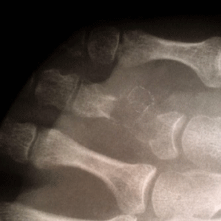
Figure 1
Pre-op AP X-ray with radiolucent lesion in 4th proximal phalanx.
Figure 2
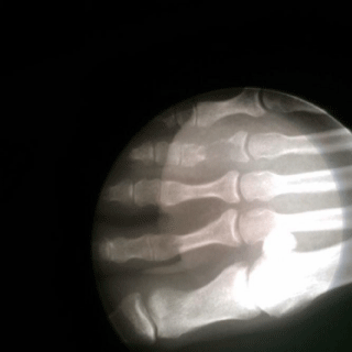
Figure 2
Pre-op MO X-ray.
Figure 3
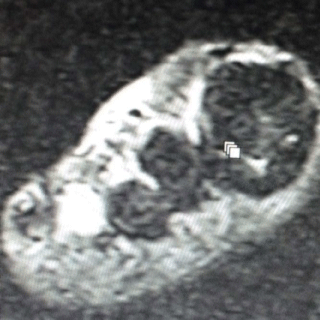
Figure 3
Pre-op T2 MRI showing enhancement at lesion
Figure 4
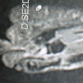
Figure 4
Pre-op T1 MRI with low signal at lesion.
Figure 5
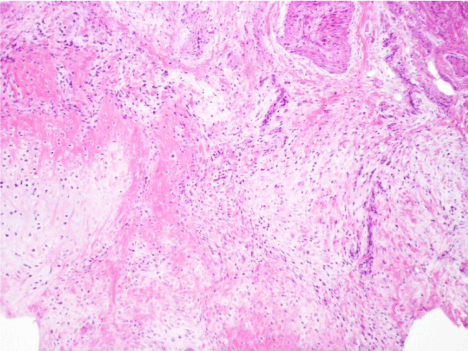
Figure 5
Soft tissue invasion: Neoplasm (concentrated in lower left) invading
soft tissue.
Figure 6
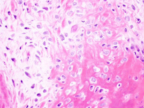
Figure 6
Nuclear atypia: Enlarged irregular nuclei with small nucleoli within
the tumor cells.
Case Presentation
A 38-year-old woman presented to the office for evaluation and
treatment of right foot pain for two weeks duration. She was doing
an activity where she was standing on her “tip toes” and she felt a
pop and had immediate pain and discomfort localized to the 4th toe.
She also complained of sharp, shooting pain with every step. Her
past medical history was unremarkable. Pain, swelling, ecchymosis
at the MTPJ of the fourth toe, and an antalgic gait were noted on
physical exam. X-rays did not demonstrate any fracture, but did show
an expanding osteolytic lesion with calcification in the distal phalanx
of the right fourth toe which was highly suspicious of a bone tumor.
Initial treatment consisted of a walking boot and an MRI to assess the
bone tumor (Figure 1).
Patient returned to office after getting an MRI. Findings on the
MRI showed a lesion in the fourth toe proximal phalanx with low
T1 and bright T2 signal. The lesion approximated 12 mm in length
(Figure 2). The medial and lateral cortices were grossly intact.
Subsequently, a bone biopsy was performed with an intra-operative
pathology consult approximately one week after receiving the MRI
results. Histological sections showed lobular lesions composed of a
combination of myxoid matrix and chondroid.
The cells were spindle or stellate in shape, some bi-nucleated
cells were also seen (Figure 3). The cellularity was much higher than
in normal cartilage. Chondroid cells were atypical. Final report
diagnosed the lesion as an intermediate grade chondrosarcoma. The
patient was referred to oncology once pathological results returned.
The patient was worked up clinically by oncology and noted not to
have any masses or ipsilateral inguinal masses. Blood work consisting
of BMP 21, complete blood count with differential, liver function tests
and thyroid panel all returned within normal limits (Figure 4).
The patient underwent diagnostic testing inclusive of CT scans
of thoracic, abdominal and pelvic areas post operatively, 6 months
and 1 year and 5 years after diagnosis which did not reveal metastasis
(Figure 5).
Two weeks after the biopsy, an amputation was performed at
the level of the 4th MPJ. The patient was followed up post operatively
weekly until the surgical site healed completely in 4 weeks. Sutures
were removed 2 weeks post operatively (Figure 6). The patient then
followed up with the surgeon every 3 months until one year post
op and then went to one year follow up appointments. The patient
was monitored every 6 months with oncology until one year post
operatively and then yearly (Figure 7). Five years post operatively
the patient remains to do well with no complications from surgery or
recurrence of cancer (Figure 8).
Figure 7
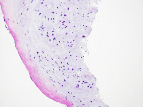
Figure 7
Myxoid matrix: Devoid of mitotic figures or signs of tumor necrosis.
Figure 8

Figure 8
Post-op X-ray s/p 4th digit amputation.
Discussion
Chondrosarcomas account for 10% to 15% of primary bone
tumors. The most common sites for these tumors are the pelvis,
shoulder, and the metaphysis of long tubular bones, particularly the
femur. The incidence of primary bone tumor in the foot is about 2%,
chondrosarcoma is the most common primary malignant bone tumor
of the foot. According to Harkless, 36% of the tumors occurred in the calcaneus, 41% in the metatarsals, and only 19% in the phalanges [1-
3]. Correct classification of the grade of chondrosarcoma is important
because the prognosis and treatment are different for different stages
and grades. Evans used their own grading system: low grade is a lowlevel
malignancy, intermediate grade-as in our case-is moderately
malignant, and high grade is highly malignant. Reported 10-year
survival rates for chondrosarcoma were 83% for low grade, 64% for
intermediate grade, and 29% for high grade. Local recurrence was
40% for low grade, 60% for intermediate grade, and 47% for high
grade. The incidence of metastatic disease is rare for low grade, 10%
for intermediate grade, and 71% for high grade [4-7].
Bovee reported that phalangeal chondrosarcoma recurs in 36%
of cases but does not metastasize [8-10]. Phalangeal chondrosarcoma
also has a much better prognosis than chondrosarcoma in other
locations. Mohammadianpanah described a case of a phalangeal
chondrosarcoma at the proximal phalanx of the third toe that went
on to an amputation at the MPJ [11,12]. Lo reported on a case of a
low-grade chondrosarcoma at the base of the fifth metatarsal, lateral
aspect of the cuboid and third cuneiform. The patient went on to have
a below knee amputation.
Conclusion
In this case, early diagnosis of chondrosarcoma before metastasis and treatment with definitive amputation led to a malignancy free patient at 5 years post operatively. The patient continues to do well and functions without difficulty. Proper collaboration between the surgeon, pathologist, oncologist, radiologists and other medical specialists led to, at this point and hopefully for her foreseeable future, a healthy patient.
References
- Lo EP, Pollak R, Harvey CK. Chondrosarcoma of the foot. J Am Podiatr Med Assoc. 2000;90(4):203-7.
- McGlamry ED, Banks AS, Downey MS. “Tumors”, in Comprehensive Textbook of Foot Surgery, 2nd edn. McGlamry ED, editor. Williams & Wilkins, Baltimore. 1992.
- Harkless L. Soft-tissue and bone tumors. Clinics in Podiatric Medicine & Surgery. 1993;10:587.
- Evans HL, Ayala AG, Romsdahl MM. Prognostic factors in chondrosarcoma of bone. A clinicopathologic analysis with emphasis on histological grading. Cancer. 1977;40(2):818-31.
- Bovee JVMG, Van der Heul RO, Taminiau SHM, Hogendoorn PCW. Chondrosarcoma of the phalanx: a locally aggressive lesion with minimal metastatic potential. A report of 35 cases and a review of the literature. Cancer. 1999;86(9):1724-32.
- Kinoshita G, Maruoka T, Matsumoto M, Futani H, Maruo S. Bone and Soft Tissue Tumors in the Foot. The J Bone Joint Surg Am. 2002;84:216.
- Mohammadianpanah M, Torabinejad S, Bagheri MH, Omidvari S, Mosalaei A, Ahmadloo N. Primary Chondrosarcoma of Phalanx. The Foot. 2004;14:159-63.
- Unni KK. Cartilaginous lesions of bone. J Orthop Sci. 2001;6(5):457-72.
- Ogose A, Unni KK, Swee RG, May GK, Rowland CM, Sim FH. Chondrosarcoma of small bones of the hands and feet. Cancer. 1997;80(1):50-9.
- Bashir SI, Gupta R, Khan HN, Ahmed R, Mohd A, Salaria AQ. Trans-articular chondrosarcoma grade 2 of proximal phalanx resulting in its fracture along with destruction of middle phalanx of 2nd toe right foot: a case report and review of the literature. Cases J. 2009;2:7488.
- Masuda T, Otuka T, Yonezawa M, Kamiyama F, Shibata Y, Tada T, et al. Chondrosarcoma of the distal phalanx of the second toe: a case report. J Foot Ankle Surg. 2004;43(2):110-2.
- Hatori M, Watanabe M, Kokubun S. Chondrosarcoma of the distal phalanx of the great toe. J Am Podiatr Med Assoc. 2007;97(2):156-9.