Case Report
An Unusual Presentation of Urachal Carcinoma Treated by Complete Laparoscopic Excision
Selman Uranues* and Yusuf Alper Kilic
Department of Surgery, Medical University of Graz, Austria
*Corresponding author: Selman Uranues, Department of Surgery, Medical University of Graz, Auen bruggerplatz 29, 8036 Graz, Austria
Published: 30 Mar, 2017
Cite this article as: Uranues S, Kilic YA. An Unusual
Presentation of Urachal Carcinoma
Treated by Complete Laparoscopic
Excision. Clin Surg. 2017; 2: 1379.
Abstract
Urachal carcinoma is an exceedingly rare tumor which may not present alarming clinical findings in the early stages and has a predilection for local invasion and recurrence. We present a case of urachal carcinoma managed by complete excision through laparoscopic approach. The patient two years later developed a pancreatic tumor and undergone a Whipple procedure.
Introduction
Urachal carcinoma constitute 0.34%–0.7% of all bladder tumors. The tumor may not cause dysplasia in the bladder mucosa and may not present with alarming clinical findings like hematuria. Besides the delays in diagnosis, predilection of the tumor for local invasion and recurrence leads to a poor prognosis, with 5-year survival rates of 6.5% to 55% [1]. Traditionally patients with respectable tumors are treated by en bloc cystoprostatectomy and wide excision of the urachus and umbilicus. But several reports have also shown reliability of extended partial cystectomy and excision of umblicus with comparable survival rates to those of radical cystectomy. Additionally a few groups have reported successful excision of urachal tumors by laparoscopic approach [2].
Case Presentation
On a routine postoperative follow up examination three years after a left heminephrectomy for
renal cell carcinoma, 64 years old male patient has been diagnosed to have a tumor at the anterior
abdominal wall (Figure 1). At the time of diagnosis he had no complaints, computed tomography
revealed a cystic mass of 9 cm in size, below the level of umblicus. There were no calcifications
in the direct abdominal graphies, and cystoscopy and intravenous urography examinations were
negative. The only positive finding was the presence of mucus in urine examination (Figure 2).
Despite the medical advice to respect the tumor, he delayed the operation for one year, and then
finally agreed to undergo laparoscopic surgery (Figure 3). At the time of operation patient was
symptom free and repeated laboratory examinations revealed no progression. The tumor has been
respected laparoscopically using the surgical technique described below. Since the intraoperative
frozen section examination was inconclusive and the tumor has been respected completely no
further attempts for a more radical resection were undertaken.
Surgical technique
The urachal mass was excised with safe tumor free margins through three trocars. The first trocar
was inserted in midline above the umblicus using the open technique, and the other two trocars
were inserted in both flanks. Dissection has been performed by using UltraCision, the urachal mass
was completely excised with its urachal attachment at the umblicus and a cuff of perivesicular tissue
which was macroscopically tumor free. No tumoral infiltration in the fascia transversalis, bladder
wall or other intra abdominal structure was observed during the dissection (Figure 4).
Histologically presence of a safe tumor free margin has been confirmed within the medial
umblical fold, but the frozen section examination was inconclusive related to the histologic type of
the tumor, so that a more radical resection including a partial cystectomy was not attempted (Figure
5).
Pathologic examination of the tumor has revealed a mucin-positive adenocarcinoma. The patient
was symptom free for two years, and follow up computed tomography examinations have revealed
no recurrence. After two years the patient has developed a pancreatic tumor (adenocarcinoma)
for which a Whipple procedure was performed. The fast-growing and highly invasive tumour led
bowel obstruction requiring two further surgeries. The patient deceased due to tumor progression of the pancreatic carcinoma 15 months after the Whipple procedure (Figure 6).
Figure 1
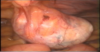
Figure 1
Macroscopic appearance intra operatively showing an isolated mass.
Figure 2
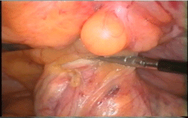
Figure 2
Dissection of the urachus with Ultra Cision.
Figure 3
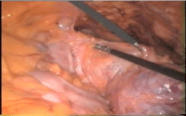
Figure 3
Dissection towards bladder dome.
Figure 4
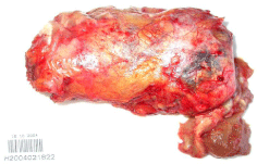
Figure 4
Macroscopic appearance of specimen.
Figure 5
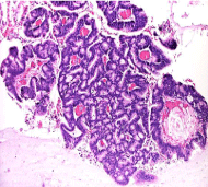
Figure 5
Microscopic view (H&E).
Figure 6
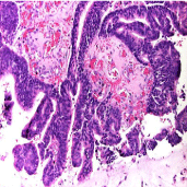
Figure 6
Microscopic view (H&E).
Discussion
Urachal carcinoma constitute 0.34–0.7% of all bladder tumors.
Histologically urachal tumours have been classified as mucin-positive
adenocarcinoma (69%), mucin-negative adenocarcinoma (15%),
sarcoma (8%), squamous cell cancer (3%), transitional cell carcinoma
(3%), and other mesenchymal neoplasmas like desmoid and leiomyoma, and myofibroblastic tumor arising from the wall of the
urachus (2%) [3]. Other urachal disorders that must be considered in
the differential diagnosis include Castelman`s disease of the urachus
(a lymphoid tissue disorder), urachal leiomyosarcoma and congenital
urachal anomalies [4].
Diagnosis of urachus carcinoma prior to surgery is difficult in
view of the nonspecific abdominal or urinary signs and symptoms.
The tumor may not cause dysplasia in the bladder mucosa and
may not present with alarming clinical findings. Hematuria or the
presence of a suprapubic mass is the most frequent features. Other
symptoms described include umbilical discharge, dysuria or the
finding of mucus in urine. In 50% to 70% of cases psammomatous
calcifications can be detected with computed tomography or
ultrasound, while plain abdominal films detect only about 5% of these
calcifications. A filling defect of the bladder dome may be detected
with intravenous urography. Ultrasound is very useful as it can also
differentiate the cystic components of an urachal carcinoma. Colour
Doppler ultrasonography can demonstrate neovascularisations [5].
Besides the delays in diagnosis, predilection of the tumor for
local invasion and recurrence leads to a poor prognosis, with 5-year
survival rates of 6.5% to 55% [6].
Distant metastases from urachal carcinoma are reported to be a late
event. Few patients had distant metastases without local recurrence
after excision of clinically localized tumor. Both dissemination during
the radical surgery and distant metastasis could be considered. The
prognosis for metastatic urachal cancer is generally very poor, and no
consensus has been reached on how to manage the disease best [7-9].
Complete surgical resection of the tumour appears to offer
the best chance for prolonged survival. Postoperative irradiation
and chemotherapy may be beneficial in some patients. Urachal
adenocarcinomas of the colonic type are well differentiated
histologically and have a good prognosis; they could be treated with
segmental rather than radical excision. Traditionally patients with
resectable tumors are treated by en bloc cystoprostatectomy and wide
excision of the urachus and umbilicus [10-12]. But several reports
have also shown reliability of extended partial cyctectomy and
excision of umblicus with comparable survival rates to those of radical
cystectomy. Additionally a few groups have reported successful
excision of urachal tumors by laparoscopic partial cystectomy with en
bloc resection of the urachus. Both transperitoneal and extraperitoneal
methods have been described. The laparoscopic approach allows for
precise tissue dissection and affords the well established benefits of
diminished blood loss, shorter hospital stay, and faster convalescence.
Conclusion
Complete surgical resection of the tumour as part of a multimodal strategy involving radiation and chemotherapy appears to offer the best chance for prolonged survival. The newly developed laparoscopic instrument and also improving surgical skills through newest learning methods are steps leading to safety and technically easily performance of laparoscopic urachal carcinoma resection. The method is safe and offers a great patient satisfaction.
References
- Porpiglia F, Cracco CM, Terrone C, Cossu M, Renard J, Billia M et al. Combined endoscopic and laparoscopic en bloc resection of the urachus and the bladder dome in a rare case of urachal carcinoma. Int J of Urol. 2007;14(4):362–64.
- Wadhwa P, Kolla SB, Hemal AK. Laparoscopic en bloc partial cystectomy with bilateral pelvic lymphadenectomy for urachal adenocarcinoma. Urology. 2006;67(4):837–43.
- Milhoua PM, Knoll A, Bleustein CB, Ghavamian R. Laparoscopic partial cystectomy for treatment of adenocarcinoma of the urachus. Urology.2006;423(2):15–17.
- Hong SH, Kim JC, Hwang TK. Laparoscopic partial cystectomy with en bloc resection of the urachus for urachal adeno carcinoma. Int J Urol. 2007;14(10):963–65.
- Morii A, Furuya Y, Fujiuchi Y, Akashi T, Ishizawa S, Fuse H. Urachal signet ring cell carcinoma. Int J Urol. 2007;14(4):360–61.
- Henly DR, Farrow GM, Sincke H. Urachal cancer: role of conservative surgery. Urology. 1993;42(6):635.
- Siefker-Radtke AO, Gee J, Shen Y, Wen S, Daliani D, Millikan RE, et al. Multi-modality management of urachal carcinoma: the M.D. Anderson Cancer Center experience. J Urol. 2003;169(4):1295–8.
- Cadeddu JA, Boyle KE, Fabrizio MD, Schulam PG, Kavoussi LR. Laparoscopic management of urachal cysts in adulthood. J Urol. 2000;164(5):1526–8.
- Sheldon CA, Clayman RV, Gonzalez R, Williams RD, Fraley EE. Malignant urachal lesions. J Urol.1984; 131(1):1–8.
- Kakizoe T, Matsumoto K, Andoh M, Nishio Y, Kishi K. Adenocarcinoma of the urachus: report of 7 cases and review of literature. Urology. 1983;21(4):360–6.
- Satpathy RS. Carcinoma of the urachus. J Indian Med Assoc. 1966;46:38–9.
- Hurwitz SP, Jacobson EB, Ottenstein HH. Mucoid adenocarcinoma of the urachus invading bladder. J Urol. 1951;65(1):87–92.