Research Article
Hallux Valgus Correction by Opening Wedge Lapidus Fusion and Intercuneiform Stabilization
Carl T Hasselman1*, Bradley Cambell2, Tejas Patel1, Chad M Ferguson2 and Alex J Kline2
1Department of Orthopedic Surgery, University of Pittsburgh Medical Center, USA
2Department of Mechanical Engineering, University of Pittsburgh, USA
*Corresponding author: Carl T Hasselman, Department of Orthopedic Surgery, University of Pittsburgh Medical Center, 200 Delafield Road, Suite 1040, Pittsburgh, PA 15215, USA
Published: 30 Mar, 2017
Cite this article as: Hasselman CT, Cambell B, Patel T,
Ferguson CM, Kline AJ. Hallux Valgus
Correction by Opening Wedge Lapidus
Fusion and Intercuneiform Stabilization.
Clin Surg. 2017; 2: 1374.
Abstract
Background: The Lapidus procedure is commonly utilized for severe hallux valgus correction,
but concerns exist regarding union rates, shortening of the first ray, transfer metatarsalgia and
recurrence of deformity. We hypothesized that a Lapidus fusion with an “opening wedge” technique
would yield excellent rates of fusion while minimizing shortening of the first ray. Furthermore, we
hypothesized that stabilization of the intercuneiform joints using a semi-rigid device would prevent
the intermetatarsal angle widening during the postoperative period, thereby causing a recurrence
of the hallux valgus.
Materials and Methods: A retrospective review of all hallux valgus corrections performed by a
single surgeon over a one year period using an “opening wedge technique” through the first
tarsometatarsal joint combined with stabilization of the intercuneiform joints using a suture button
device was performed. Specifically, we evaluated successful arthrodesis rates, time to weight bearing,
complications, and radiographic parameters including changes in the intermetatarsal angle and
hallux valgus angle. Additionally, the magnitude of length change between the first and second ray
was evaluated.Placement of a low profile plate over the first TMT joint to give final
fixation of the pre= reduced joint.
Results: Thirty four patients (42 feet) were followed for a minimum of 6 months postoperatively. The
decrease in the intermetatarsal (IM) angle averaged 8.3 degrees (mean preop IM angle 14.6 degrees
and mean post op IM angle 6.3 degrees). The average decrease in the hallux valgus (HV) angle was
21.2 degrees (mean preop HV angle 35.4 degrees and mean post op HV angle 14.2 degrees). The
length difference between the first and second rays revealed a minimal change of an increase in 0.89
mm length of the first ray. There were no hardware failures or non unions determined by clinical
exam or radiographs.
Conclusison: In our experience, the technique described in this paper for hallux valgus correction
yielded excellent correction with a low rate of complications and no significant changes in the length
of the first ray.
Keywords: Lapidus; Bunion; Hallux valgus; Metatarsalgia; Opening wedge
Introduction
A multitude of options are available for correcting severe hallux valgus deformities. One of the more popular choices for these deformities is the Lapidus procedure, a fusion of the first tarsometatarsal (TMT) joint combined with a distal soft tissue procedure and medial eminence resection. Since Lapidus described the technique in 1934 there have been several modifications of the technique but the main premise is to fuse the first TMT joint in appropriate alignment with reduction of the intermetatarsal angle between the first and second ray [1]. Benefits include the ability to correct severe deformities, the avoidance of fusing the metatarsophalangeal joint, and addressing any hypermobility or arthritic symptoms at the first TMT joint [2-4]. This procedure has been popular for addressing hypermobility in the first TMT joint, arthritis of the first TMT joint and as a secondary procedure when a primary procedure for hallux valgus has resulted in recurrence of the deformity [2-4]. In spite of all of the potential benefits, the Lapidus procedure is still not a popular procedure as a primary hallux valgus correction due to many potential issues. First, it can be a technically demanding procedure and if performed incorrectly, elevation of the first ray or persistent valgus may result in failure [5]. Other potential complications with the Lapidus procedure include nonunion, delayed union or malunion and the time to begin full weight bearing can be longer than other procedures to correct hallux valgus [6]. Shortening of the 1st ray is common with previously described Lapidus procedure variants and a relative contraindication to these techniques is a shortened first ray due to the higher risk of transfer metatarsalgia [7]. Finally, any coronal plane instability within the joint between the medial and middle intercuneiform bones could potentially allow medial drift of the first ray and a recurrence of the hallux valgus deformity could occur even with a technically well done Lapidus procedure [8,9]. Based on these complications associated with previous Lapidus procedures, we have developed an “opening wedge” Lapidus procedure. The premise of this technique was to create a procedure that would fuse the first TMT joint, allow for earlier weight bearing, avoid shortening of the first ray and minimize the risk of nonunion. The procedure was also designed to allow more accurate reduction of the intermetatarsal angle and stabilization of any intercuneiform instability that is not readily apparent preoperatively. The study was designed to determine whether such a procedure could minimize the degree of shortening of the first ray while still achieving an acceptable degree of correction and fusion rate.
Figure 1
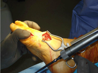
Figure 1
Debridement of the joint with only the medial two thirds of the joint
debrided of the cartilage and subchondral joint.
Figure 2
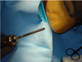
Figure 2
Calcaneal bone being harvested through a small incision at the
calcaneal tuberosity.
Figure 3
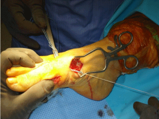
Figure 3
Placement of the mini tightrope from the second to first metatarsal
to reduce the IM angle.
Materials and Methods
A retrospective chart review of consecutive patients undergoing
an “opening wedge” Lapidus arthrodesis for correction of hallux
valgus in which the surgery was done between January and December
of 2010 was performed. Inclusion criteria included bunion correction
via the opening wedge technique, age over 18, and adequate followup
data to determine fusion and complication rates (minimum 6
months). Exclusion criteria included hallux valgus correction via
another technique, age under 18, and incomplete post-operative
follow-up. A total of 42 feet in 34 patients were found who met all
of the inclusion criteria. All 34 patients were women. Institutional
review board approval was obtained for this retrospective study that
required records review and follow up radiographs. The mean follow
up was 28 months. All cases were performed by a single orthopedic
surgeon. A complete review of the medical records was performed
to document the time to weight bearing, any intra-operative or postoperative
complications that were encountered, and any secondary
procedures that were performed.
Operative technique
All cases utilized regional anesthesia and sedation. A calf
tourniquet was utilized during the procedure for hemostasis. A
dorsal first web space incision was made to perform a lateral release
of the first metatarsophalangeal (MTP) joint. The lateral release was
performed by releasing the adductor tendon off the proximal phalanx
and fibular sesamoid and an L shaped release of the lateral capsule
was made between the fibular sesamoid, metatarsal and proximal
phalanx. Subsequently, a medial incision over the first MTP joint was
made and the capsule of the MTP joint longitudinally split. The joint
was inspected, and a medial eminence resection was undertaken. The
1st tarsometatarsal joint was exposed via a medial approach with care
to protect the tibialis anterior tendon. The cartilage and subchondral
bone of the first TMT joint was removed of only the medial two thirds
of the joint to avoid damaging the lateral capsule (Figure 1). Calcaneal
bone graft was then harvested with a core reamer device through
a small incision over the lateral wall of the calcaneal tuberosity,
posterior and inferior to the peroneal tendons and sural nerve (Figure
2). A small incision was subsequently made over the lateral side of
the second metatarsal approximately 2 cm distal to the second TMT
joint. A 2.7 mm mini-tightrope device (Arthrex Inc, Naples, FL) was
placed from the second metatarsal base to the first metatarsal base
and then tightened to reduce the intermetatarsal angle to anatomic
alignment, essentially hinging on the lateral side of the joint (Figure
3). The position of correction was maintained during the procedure
by the self locking mechanism of the mini-tightrope. The calcaneal
bone graft was subsequently packed into the medial aspect of the
1st TMT joint. A low-profile locking plate was then placed over
the medial aspect of the joint with 2 screws placed in to the medial
cuneiform and 2 screws into the proximal metatarsal (Figure 4). The
alignment of the first ray, placement of the hardware and reduction
of the sesamoids were checked radiographically at this point with
simulated weight bearing intraoperative fluoroscopy. Reduction was
verified prior to medial first MTP capsule closure. Once verified, the
excess medial capsule was excised, and the capsule was closed with a non-absorbable suture. All wounds were then copiously irrigated, and a layered skin closure was performed. A bunion dressing was
placed, and the patient was placed into a hard soled post-operative
shoe. Heel weight bearing with crutches was allowed immediately
postoperatively. Radiographs were obtained at three and six weeks
and three months to assess fusion of the TMT joint. Full weight
bearing was allowed at six weeks in all cases. A complete radiographic
review was performed. All patients had pre and post-operative weight
bearing views including AP, lateral, and oblique. All radiographs
were examined by two fellowship trained orthopedic surgeons. Pre
and postoperative measurements including hallux valgus angle, the
1-2 intermetatarsal angles, and the difference between the first and
second ray lengths were measured. Additionally, the post-operative
films were reviewed for the rates of fusion, hardware complications
and recurrence of deformity. Patients were defined to have a union if
they were shown to have successful arthrodesis on plain radiographic
evaluation and were without symptoms of pain or swelling at the
arthrodesis site. Only patients suspected of delayed or nonunion had
the addition of diagnostic CT confirmation of arthrodesis.
Figure 4
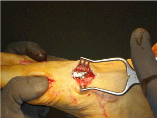
Figure 4
Placement of a low profile plate over the first TMT joint to give final
fixation of the pre= reduced joint.
Figure 5A and D
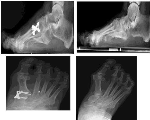
Figure 5A and D
Placement of the mini tightrope from the second to first metatarsal
to reduce the IM angle.
Results
Mean post operative follow up was 28 months (range 6-42 months). The mean preoperative IM angle was 14.6 degrees (range 11-20 degrees). The mean post operative IM angle was 6.3 (range 3-9 degrees). The mean preoperative hallux valgus angle was 35.4 degrees (range 23-52). The mean postoperative hallux valgus angle was 14.2 degrees (range 2-24 degrees). The preoperative distance between the first and second metatarsal heads averaged 1.92 mm (range -4.1-6.3). The postoperative distance between the first and second metatarsal heads averaged 1.03 (range -4.1-4.1). Complications included one delayed union that required a bone stimulator and avoidance of impact activities for an additional 2 month period. Hardware removal due to tibialis anterior tendon irritation was required in 4 patients. There was one postoperative infection that required local wound debridement and local antibiotics, which resolved without sequelae. There were no non-unions or loss of reduction in the patients studied during the follow up period. One case of mild hallux varus developed postoperatively; however the patient did not feel the symptoms were significant enough to warrant surgical intervention. There were no complications of nonunion, transfer metatarsalgia or recurrence of the hallux valgus deformity during the study period. No stress fractures of the second metatarsal were found during the follow up period. No secondary surgeries to address the lesser MTP joints were performed during the follow up period and no complaints of lesser MTP overload were noted.
Discussion
Correction of hallux valgus deformities that are challenging (IM angle greater than 15 degrees, HV angle over 50 degrees, hypermobility of the first TMT joint, arthrosis of the first TMT joint and obesity) have been typically corrected by the use of a Lapidus procedure to prevent recurrence [2-4]. However, even this procedure has shown a high complication rate and recurrence rate. Most of the complications are from malposition, shortening or nonunion [8,9]. Even in a wellpositioned Lapidus procedure, the shortening of the first ray has the potential to cause problems with the adjacent metatarsophalangeal joints. These can vary from metatarsalgia or the development of painful calluses all the way up to a dislocated 2nd metatarsophalangeal joint [6,10,11]. Because of these potential complications, it is imperative to minimize the degree of first ray shortening while still achieving the goals of a solid fusion and correction of the underlying deformity. The technique of the opening wedge Lapidus procedure described in this article has the potential to prevent many of these complications. One potential concern with a minimal joint resection and an opening wedge technique is that the rate of nonunion may be increased; however, our results do not support this conclusion. Technical considerations of the opening wedge Lapidus reduce the necessary joint resection may aid in fusion by preventing aggressive joint preparation and disruption of the blood supply necessary for fusion. The authors propose that any Lapidus procedure previously described required complete debridement of the joint which would require devitalization of the lateral collateral ligament complex. The dorsalis pedis artery sends communicating vessels to the first TMT joint through the first interspace of the foot. A saw or osteotome could easily damage this area and result in devitalization of the first TMT joint. By avoiding further dissection of the lateral joint with a medial based fusion, we feel that the lateral collateral circulation to the joint is preserved and one possible explanation for our low nonunion rate in this study. Most recent reports in the literature report nonunion rates ranging from 0 to 10% with the modified Lapidus Sangeorzan et al. [12] reported an overall nonunion rate of four out of forty patients (10%), three of whom ultimately required revision surgeries. Kopp et al. [7] reported an overall nonunion rate of 5% in their series of thirty-four feet. Coetzee et al. [5] reported seven non-unions in 105 feet (6.7%). All seven patients ultimately required revision surgeries. Catanzariti et al. [9] reported a nonunion rate of three out of fortyseven (6.4%) and an additional delayed union rate of two out of forty-seven (4.2%). We believe that our rate of one delayed union in forty two feet (2.3%) is due to a less invasive debridement technique thereby preserving the lateral first TMT blood flow. Recurrence of the deformity is another situation that is seen even after fusion of the first ray. The authors feel that this may be due to increased coronal plane intercuneiform hypermobility of the intertarsal joints as previously described by Hansen. Although first TMT hypermobility is seen and can result in hallux valgus, it is possible that hypermobility may be a more global foot condition with mobility within the mid foot region. The suture fixation device augmentation of this surgical construct provides coronal plane stabilization while allowing sagittal plane mobility due to the intermetatarsal device orientation. Other described techniques have proposed screw fixation of the intermetatarsal angle, but screw fixation has several pitfalls. First, Intermetatarsal screw fixation will not only stop coronal plane motion, but will stop sagittal plane motion which stiffens the sagittal plane movement of the medial column and if not placed anatomically, can result in lesser metatarsal overload and subsequent transfer metatarsalgia. Additionally, screw fixation places a completely rigid metallic implant between the first and second metatarsal, which continue to have micro motion in the sagittal plane. Rigid fixation in a micro motion environment leads to hardware failure (screw breakage) similar to screw fixation of the ankle syndesmosis. The opening wedge Lapidus technique proposed utilizing intermetatarsal suture fixation allows for motion in the sagittal plane and can allow for more anatomic motion and lessen the risk for hard ward failure. In addition, the suture device allows for more anatomic reduction of the intermetatarsal angle. The suture device allows for intermetatarsal reduction , but due to its soft design (unlike a screw) the coronalplane will reduce and prevent malalignment of the first and second metatarsalin the coronal plane but a moderate mobility will be allowed within the sagittal planeto allow the metatarsals to fit to their anatomic sagittal plane position. This technique has powerful correction of not only the forefoot, but can cause realignment of the hind foot as previously described by other authors (Figure 5A and D). The complication of symptomatic hardware was initially seen in our study. The tibialis anterior tendon seemed to be irritated by the plate or the plate was too stiff for this region. We initially used a stainless steel plate (Synthes Inc, Paoli, PA) This complication has not been seen since changing from a more prominent locking X plate (Synthes, Paoli, PA) to a more low profile titanium plate (Arthrex Inc, Naples, Fl). The precise reason for the improvement may be related to plate morphologic, stiffness, or metallurgy; however, the technical fixation change has resulted in no further hardware removal in the latter half of the study period. Weaknesses of our study include its retrospective nature, a relatively small number of patients, and the lack of preoperative and postoperative clinical scores. Additionally, union in this study was defined by plain radiography showing union in a symptomatic patient and without the utilization of CT scans. In spite of its shortcomings, we do feel that this significantly adds to the body of literature by highlighting a new technique that seeks to minimize potential complications inherent to the modified Lapidus, namely nonunion, recurrence, and shortening of the first ray with the development of transfer metatarsalgia. The treatment of hallux valgus must be tailored to the individual patient. Factors including radiographic parameters, clinical instability, arthritic symptoms, adjacent joint symptoms, and patient desires all play a role in determining the optimal procedure. For severe deformities with the absence of significant metatarsophalangeal joint arthritis, our study surgeons prefer the “opening wedge” Lapidus. This procedure addresses potential concerns for metatarsal shortening while also minimizing the risk of nonunion based on the results of this study. Since the study period, the senior author has performed over 200 cases with results similar as to report in this article. Further studies are needed to assess the long term function of this procedure and recurrence rates.
References
- Lapidus PW. Operative correction of the metatarsus primus varus in hallux valgus. Surg Gynecol Obstet. 1934;58:16.
- Clark HR, Veith RG, Hansen ST Jr. Adolescent bunions treated by the modified Lapidus procedure. Bull Hosp Jt Dis Orthop Inst. 1987;47:109- 22.
- Hansen ST Jr. Hallux valgus surgery. Morton and Lapidus were right! Clin Podiatr Med Surg. 1996;13:347-54.
- Coetzee JC, Resig SG, Kuskowski M, Saleh KJ. The Lapidus procedure as salvage after failed surgical treatment of hallux valgus. Surgical Technique. J Bone Joint Surg. 2004;86:30-6.
- Coetzee JC, Wickum D. The Lapidus procedure: a prospectice chohort outcome study. Foot Ankle Int. 2004;25:526-31.
- Thompson IM, Bohay DR, Anderson JG. Fusion rate of first tarsometatarsal arthrodesis in the modified Lapidus procedure and flatfoot reconstruction. Foot Ankle Int. 2005;26:698-703.
- Kopp FJ, Patel MM, Levine DS, Deland JT. The modified Lapidus procedure for hallux valgus: a clinical and radiographic analysis. Foot Ankle Int. 2005;26:913-7.
- Bednarz PA, Manoli A 2nd. Modified Lapidus procedure for the treatment of hypermobile hallux valgus. Foot Ankle Int. 2000;21:816-21.
- Catanzariti AR, Mendicino RW, Lee MS, Gallina MR. The modified Lapidus arthrodesis: a retrospective analysis. J Foot Ankle Surg. 1999;38:322-32.
- Myerson M. Arthrodesis for treatment of hallux valgus and metatarsus primus varus. Orthopedics. 1990;13:1025-31.
- Schmid T, Krause F. The modified Lapidus fusion. Foot Ankle Clin N Am. 2014;19:223-33.
- Sangeorzan BJ, Hansen ST Jr. Modified Lapidus procedure for hallux valgus. Foot Ankle Int. 1989;9:262-6.