Research Article
Study of the Biological Effect of Bufalin on Anti-Proliferation, Adhesion and Invasion in Liver Cancer Cells
Sheng X1, Zhu PF2 and Qin JM2*
1Department of Pathology, Second Military Medical University, China
2Department of General Surgery, Second Military Medical University, China
*Corresponding author: JIanmin Qin, Department of General Surgery, Affiliated to the Third Hospital, Second Military Medical University, North Moyu Road 700, Jiangding District, 201805, Shanghai, China
Published: 22 Mar, 2017
Cite this article as: Sheng X, Zhu PF, Qin JM. Study of
the Biological Effect of Bufalin on Anti-
Proliferation, Adhesion and Invasion in
Liver Cancer Cells. Clin Surg. 2017; 2:
1363.
Abstract
Background: Recurrence and metastasis are the main factors which seriously influence the
therapeutic effect and long-term survival for hepatocellular carcinoma (HCC). Bufalin is a
kind of topology isomerase II inhibitors. The pharmacological effects of bufalin are anti-tumor,
anticoagulant, analgesia, enhanced systole and immunity. Therefore, in the present study, we
explore the migration, invasion and adhesion influence of bufalin on the liver cancer cells, and
provide better theoretical basis for bufalin on preventing and treating the recurrence and metastasis
of HCC.
Methods: The human high metastasis potential LM3 hepatoma cells (HCC-LM3) were cultured in
vitro. The influence of bufalin on HCC-LM3 cell proliferation was detected by the CCK-8 living cells
staining technique, and the migration, invasion and adhesion influence of bufalin on the HCC-LM3
cells were detected by the Trans well Chambers technique.
Results and Discussion: The results showed that bufalin can inhibit the growth of liver cancer
cells with time and concentration of dual dependence. With the increase of dose concentration, the
inhibitive effect of bufalin on liver cancer cell adhesion, migration and invasion ability significantly
enhanced. Bufalin is a kind of agent being potential application value to inhibit the recurrence and
metastasis of HCC.
Keywords: Hepatocellular carcinoma; Bufalin; Proliferation; Metastasis
Introduction
The occurrence of malignant tumors is the result of a in vivo metabolic imbalance. At this time, the regulatory and controlled mechanisms in vivo for cell division and proliferation occur abnormally, which is the key to the development of excessive proliferation in tumor cells. Recurrence and metastasis are the main factors which seriously influence the therapeutic effect and long-term survival for hepatocellular carcinoma (HCC). The postoperative recurrence and metastasis rate of 5 years is as high as 60%-70% even if the radical resection is performed for liver cancer [1]. How to effectively inhibit the proliferation, adhesion and adhesion of liver cancer cells, is the key to reduce recurrence and metastasis, to improve therapeutic effect and long-term prognosis of HCC. Bufalin is the strongest toxicity legends that is extracted from Chinese toad venom, and is a kind of topology isomerase II inhibitors. The pharmacological effects of bufalin are anti-tumor, anticoagulant, analgesia, enhanced systole and immunity. The anti-tumor mechanism of bufalin mainly lies in inhibiting tumor cell proliferation and angiogenesis, promoting tumor cell differentiation, inducing tumor cell apoptosis, reversing drug-resistant, regulating the body's immune system and the gene expression of tumor cells [2]. This study will explore the inhibitive effect of bufalin on the human high metastasis potential LM3 hepatoma cells (HCC-LM3) in vitro by the CCK - 8 living cells staining technique, and detecting the migration, invasion and adhesion influence of bufalin on the HCC-LM3 cells by the Transwell Chambers technique. The study can provide better theoretical basis for bufalin on preventing and treating the recurrence and metastasis of HCC.
Material and Methods
Reagents
Bufalin, purchased from Sigma Chemical Co. (St. Louis, MO, U.S.A.), was dissolved in anhydrous
alcohol at a concentration of 10-1 mol/L and stored at 4°C. High glucose Dulbecco’s modified Eage’s
medium (DMEM) and fetal bovine serum (FBS) were purchased from Gibco (Gaithersburg, MD, U.S.A.). Matrigel glue was purchased from American BD Company.
Transwell Chambers was purchased from American Corning
Company. Crystal violet (0.1%) dyeing liquid was purchased from
Jiangsu Blue skies Biotechnology Company.
Cell lines
HCC-LM3 cells were obtained from Liver Cancer Institute
of Zhongshan Hospital affiliated to Fudan University. The cells
were cultured in high glucose DMEM supplemented with 10%
FBS, 100U/ml of penicillin and 100 μg/ml of streptomycin in a
humidified atmosphere with 5% CO2 in air at 37°C. Then the cells
in the logarithmic growth phase were collected for the following
experiments.
Grouping
HCC-LM3 cells were cultured in vitro; they were divided into
DMEM group and Bufalin group according to the experimental
design.
Drug concentration
Bufalin concentration was designed according to our previous
study [11], drug concentration gradient were respectively 0.02 μg/ml,
0.04 μg/ml, 0.08 μg/ml, 0.16 μg/ml, 0.32 μg/ml, 0.64 μg/ml, 1.28 μg/
ml and 2.56 μg/ml.
Cell proliferation assay
The Cell Counting Kit-8 (CCK-8, Dojindo, and Tokyo, Japan)
assay was used to evaluate cell proliferation. Briefly, cells in the
logarithmic growth phase were plated at a density of 10 × 104 cells/
ml, then 100 μl/well in 96-well plates. Culture mediums were changed
after cells were adherent growth. Various concentration of bufalin
was diluted with DMEM with 10% FBS. 200 μl bufalin was injected
into culture plate per well in bufalin group, 200 μl DMEM with FBS
was only injected into culture plate per well in DMEM group, four
wells were set in each drug concentration. The cells in each group
were respectively cultured at 24 h, 48 h and 72 h in a humidified
atmosphere with 5% CO2 in air at 37°C, then culture medium were
changed. 100 μl CCK-8 (dilution 9:1) was injected into culture plate
per well and cultured for 1.5 h in dark place. Optical density (OD)
value in per well was detected at 450 nm wavelength in automatic
enzyme mark instrument. Each experiment was performed in
triplicate. The cell inhibition ratio was calculated by the following
formula:
Cell inhibition ratio (%)=1-(average OD value of treated groupaverage
OD value of blank group)/(average OD value of control
group-average OD value of blank group) × 100%.
30 percent inhibitory concentration (IC30), half maximal inhibitory
concentration (IC50), and 70 percent inhibitory concentration (IC70)
at 24 h and 48 h are respectively calculated with SPSS 18.0 software.
Cell migration assay
Cells in the logarithmic growth phase were plated at a density of 10
× 104 cells/ml, 5 ml cell suspension liquid was cultured in a humidified
atmosphere with 5% CO2 in air at 37°C, and culture medium was
changed after cells were adherent growth. 5 ml bufalin of IC30, IC50,
IC70 at 24 h were respectively injected into culture flask in bufalin
group, and 5 ml DMEM with FBS was only injected into culture
flask in DMEM group. After the cells were cultured in a humidified
atmosphere with 5% CO2 in air at 37°C for twenty hours, the cells in
various groups were respectively harvested and centrifuged at 2000
rpm for 5 min. Then supernatant was removed, sediment was rinsed
with 0.01 MPBS solution. The cells in various groups were suspended
in serum-free DMEM at a density of 1 × 10⁶ cells/ml. Cell suspension
(100 μl) were seeded into the upper chamber of Transwell chamber,
and 600 μl DMEM with 10% FBS culture medium simultaneously
added to the lower chamber, the same cell suspension was set in three
wells per each group. After the cells were cultured for 48 h, culture
medium in the upper and lower chamber of Transwell chamber were
removed, and the upper and lower chamber were rinsed with 0.01
MPBS solution. 600 l crystal violet staining fluid was added into the
upper chamber of Transwell chamber, the cells were stained for 10
min, the cells on the upper surface of the filter were removed with
cotton swabs, and the number of migrated cells was counted in five
fields of each triplicate filter with an inverted microscope. The cell
migration ratio was calculated by the following formula:
Cell migration ratio (%)=the number of cell migration in the
treated group / the number of cell migration in the control group ×
100%.
Cell adhesion assay
The 96-well flat-bottomed plates were percolated with 50 μl/
well of 1:4 DMEM-diluted Matrigel at 4°C overnight. Cells in the
logarithmic growth phase were plated at a density of 10 × 104 cells/ml,
5 ml cell suspension liquid was cultured in a humidified atmosphere
with 5% CO2 in air at 37°C, and culture medium was changed after
cells were adherent growth. 5 ml bufalin of IC30, IC50, and IC70 at 24
h were respectively injected into culture flask in bufalin group, and 5
ml DMEM with FBS was only injected into culture flask in DMEM
group. After the cells were cultured in a humidified atmosphere with
5% CO2 in air at 37°C for twenty hours, the cells in various groups
were respectively harvested and centrifuged at 2000 rpm for 5 min.
Then supernatant was removed, sediment was rinsed with 0.01 MPBS
solution. The cells in various groups were suspended in serum-free
DMEM at a density of 1 × 10⁶ cells/ml. 100 μl cell suspension per
well were injected into 96 well plates in bufalin group, 100 μl culture
medium was only injected into 96 well plates in DMEM group. After
the cells in various groups were cultured for 2 hours in a humidified
atmosphere with 5% CO2 in air at 37°C. 100 μl CCK-8 (dilution 9:1)
was injected into culture plate per well and cultured for 1.5 h in dark
place. Optical density (OD) value in per well was detected at 450 nm
wavelength in automatic enzyme mark instrument. Each experiment
was performed in triplicate. The cell adhesion ratio was calculated by
the following formula:
Cell adhesion ratio (%)=average OD value of treated group/
average OD value of control group × 100%.
Cell invasion assay
Cells in the logarithmic growth phase were plated at a density
of 10 × 104 cells/ml, 5 ml cell suspension liquid was cultured in a
humidified atmosphere with 5% CO2 in air at 37°C, and culture
medium was changed after cells were adherent growth. 5 ml bufalin
of IC30, IC50, and IC70 at 24 h were respectively injected into culture
flask in bufalin group, and 5 ml DMEM with FBS was only injected
into culture flask in DMEM group. After the cells were cultured in a
humidified atmosphere with 5% CO2 in air at 37°C for twenty hours,
the cells in various groups were respectively harvested and centrifuged
at 2000 rpm for 5 min. Then supernatant was removed, sediment was
rinsed with 0.01 MPBS solution. The cells in various groups were
suspended in serum-free DMEM at a density of 1 × 10⁶ cells/ml. 100 μl cell suspension was added into the upper chamber with a filter coated
with 100 μl matrigel at 1:8 dilutions in serum-free medium. 100 μl
culture medium was added into the lower chamber of the Transwell
chamber. And the experiment was performed in triplicate. After the
cells were incubated at 37°C with 5% CO2 for 72 h, the cells that did
not invade the membrane were gently removed. Cells that invaded
the membrane were stained with 0.1% crystal violet. Six fields were
randomly selected and observed under an inverted microscope for
counting the cell number of stained cells and image collection. The
cell invasion ratio was calculated by the following formula:
Cell invasion ratio (%)=the number of cell invasion in the treated
group/the number of cell invasion in the control group × 100%.
Statistical analysis
Data were analyzed using analysis of variance (SPSS18.0 and
Graphpad Prism 5; Cary, NC, USA). Data are expressed as the
mean values ± standard of the mean. P values of less than 0.05 were
considered statistically significant.
Table 1

Table 1
The growth inhibition rate of HCC-LM3 cells after bufalin was used with different time and concentration (`x±s%,n=3).
Table 2
Table 2
IC30, IC50 and IC70 of bufalin on HCC-LM3 cells at 24h and 48h (x±s μg/ml, n=3).
Figure 1
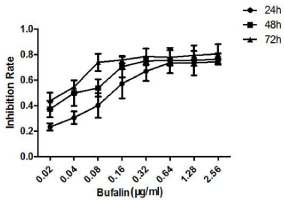
Figure 1
The growth inhibition rate of HCC-LM3 cells with different time and
concentration of buflin. It showed that growth inhibition rate of HCC-LM3 cells
increased with the extension of time and high drug concentration of bufalin.
Figure 2
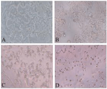
Figure 2
Morphologic changes of HCC-LM3 cells after different concentration
of bufalin were used for 24 hours (magnification×200).
A: the cells were dense and stronger adherent ability in DMEM group; B:
some cells became round and shrinkage in the IC30 of bufalin group; C: most
cells became round and shrinkage in the IC50 of bufalin group; D: the cells
became round and lower vigour in the IC70 of bufalin group.
Results
Inhibitive effect of bufalin on proliferation of HCC-LM3 cells
After different concentrations of bufalin were used to inhibit the
proliferation of HCC-LM3 cell at 24 h, 48 h and 72 h, growth inhibition
rate of bufalin on HCC-LM3 cells increased with the increase of drug concentration at the same time point(P<0.05). Growth inhibition rate
of bufalin on HCC-LM3 cells increased with the extension of time at
the same drug concentration (P <0.05). It indicated that bufalin could
inhibit the growth of HCC-LM3 cells with time and concentration of
dual dependence. (Table 1 and Figure 1).
IC30, IC50 and IC70 of bufalin on HCC-LM3 cells at 24 h and
48 h
IC30, IC50 and IC70 of bufalin on HCC-LM3 cells at 24 h were
higher than that at 48 h (P <0.05). There was statistical difference
between IC30 and IC70 at 24 h (P <0.05), and among IC30, IC50 and IC70
at 48 h (P <0.05). It indicated that the inhibitive effect of bufalin on the
proliferation of HCC-LM3 cells enhanced with the extension of time
and higher drug concentration Table 2.
Morphologic changes of HCC-LM3 cells
IC30, IC50 and IC70 of bufalin on HCC-LM3 cells at 24 h were
respectively used to treat the HCC-LM3 cells for 24 h. The results
showed that the HCC-LM3 cells were loss of polarity, and adherent
ability weakened. The cells became round, shrinkage and sparse. The
living cells gradually reduced, and the dead cells increased with the
concentration increase of bufalin (Figure 2).
Bufalin inhibits migration of HCC-LM3 cells
IC30, IC50 and IC70 of bufalin on HCC-LM3 cells at 24 h were
respectively used to treat the HCC-LM3 cells for 24 hours. The
results showed that the number of migrated cells in the bufalin
group was lower than that of the DMEM group (F=45.73, P<0.05),
and the number of migrating cells reduced with increase of bufalin
concentration. The migrating rate of the HCC-LM3 cells in the
bufalin group was lower than that of the DMEM group (F=48.98,
P<0.05), and the migrating rate decreased gradually with increase of
bufalin concentration. It indicated that the inhibitive effect of bufalin on the HCC-LM3 cells migration occurred in a dose-dependent manner (Figure 3 and Table 3).
Bufalin inhibits adhesion of HCC-LM3 cells
IC30, IC50 and IC70 of bufalin on HCC-LM3 cells at 24 h were
respectively used to treat the HCC-LM3 cells for 24 hours. The
results showed that the adhesive rates of the HCC-LM3 cells were
respectively 95.41 ± 3.17%, 83.76 ± 6.07%, 40.16 ± 3.42% in the IC30
of bufalin group, the IC50 of bufalin group and the IC70 of bufalin
group. The adhesive rate of the HCC-LM3 cells in the IC70 of bufalin
group was lower than that of the IC30 of the bufalin group and the
IC50 of bufalin group (40.16 ± 3.42% vs. 95.41 ± 3.17%, 83.76 ± 6.07%,
F=17.41, P<0.05). It indicated that bufalin could significantly inhibit
adhesion of HCC-LM3 cells, and the inhibitive effect of bufalin
enhanced with increase of drug concentration and occurred in a
dose-dependent manner.
Bufalin inhibits invasion of HCC-LM3 cells
IC30, IC50 and IC70 of bufalin on HCC-LM3 cells at 24h were
respectively used to treat the HCC-LM3 cells for 24 hours. The results
showed that the number of invaded cells in the bufalin group was
less than that of the DMEM group (F=35.85, P<0.05). The number
of invading cells reduced significantly with increase of bufalin
concentration. The invading rate of the HCC-LM3 cells in the bufalin
group was lower than that of the DMEM group (F=46.38, P<0.05),
and the invading rate decreased gradually with increase of bufalin
concentration. It indicated that the inhibitive effect of bufalin on
the HCC-LM3 cells invasion occurred in a dose-dependent manner
(Figure 4 and Table 4).
Table 3
Table 3
The migrating rate of the HCC-LM3 cells after bufalin was used with different concentration for 24 hours (`x±s%,n=3).
Table 4
Table 4
The invading rate of the HCC-LM3 cells after bufalin was used with different concentration for 24 hours (`x±s%, n=3).
Figure 3
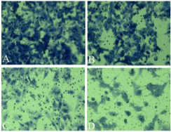
Figure 3
The migration of the HCC-LM3 cells after different concentration of
bufalin were used for 24 hours (magnification×200).
A: the cells migrated in a dense manner in the DMEM group; B: some cells
migrated in a scattered manner in the IC30 of bufalin group; C: a small number
of cells migrated sparsely in the IC50 of bufalin group; D: fewer cells migrated
dispersedly in the IC70 of bufalin group.
Figure 4
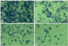
Figure 4
The invasion of the HCC-LM3 cells after different concentration of
bufalin were used for 24 hours (magnification×200).
A: most cells invaded in a dense manner in the DMEM group; B: some
cells invaded sparsely in the IC30 of bufalin group; C: fewer cells invaded
scatteredly in the IC50 of bufalin group; D: several cells invaded dispersedly
in the IC70 of bufalin group.
Discussion
Bufalin is one of the bufotoxin legends extracted from the white serous fluid of retro auricular gland of toad. Recent study indicated that anti-tumor mechanisms of bufalin involved in tumor cell proliferation, apoptosis, autophagy, related genes, signaling pathways, multi-drug resistance, etc. The main mechanisms lie in inhibiting DNA and RNA biosynthesis of cancer cells, destroying the rough surface endoplasmic reticulum of mitochondria of cancer cells, Inducing differentiation and apoptosis of cancer cells, and Increasing the intracellular cAMP and the cAMP/cGMP, changing the content of cyclic nucleotide in tumor tissue or cancer cells, promoting the cell differentiation and inhibiting the cell proliferation, regulating the intracellular metabolism of nucleic acid and protein, increasing the serum level of IgG and white blood cell count, enhancing the macrophage phagocytosis, stimulating the body's anti-tumor cytokine release. Through the above- mentioned roles, bufalin can promote the tumor cell differentiation, induce the tumor cell apoptosis, inhibit the tumor angiogenesis, improve the radiotherapy sensitization of tumor tissues, reverse the multiple drug resistance, regulate the body's immune system, change the related gene expressions in tumor cells, regulate signal transduction pathways of tumor cells, inhibit the cell proliferative cycle, topoisomerase and membrane Na + / K +-ATPase activity, enhance the mitogen activated protease activity etc. Bufalin has the stronger pharmacological effects on anti-tumor, anti-inflammatory, positive immunomodulation [3- 5]. Zhang et al. [6] found that bufalin could inhibit the proliferation and PI3K/AKT signal pathway of HepG2 cells. Meng et al. [7] found that bufalin could induce the apoptosis of Bel-7402 liver cancer cells with drug resistance in dose-dependent manner. The apoptotic rate of Bel-7402 liver cancer cells significantly increased with increase of bufalin concentration from 0.01mol/L to 1.00 mol/L. Bufalin could reverse the drug resistance to 5-fluorouracil in Bel-7402 liver cancer cells. Tumors can get rich nutrition from host through tumor blood vessels, and also output a large number of tumor cells through the tumor blood vessels, then result in tumor constant growth and metastasis. Lee et al. [8] cultured the endothelial cells of bovine artery in vitro, 5, 10, 20 nMol/L of bufalin were respectively added into cell culture medium to culture the endothelial cells for 5 days, the result showed that the growth inhibitive rates of the endothelial cells were respectively 45.3%, 62.8%, 75.6%, bufalin could reduce the endothelial cells of G0/G1, S phase and block the cell proliferation in G2/M phase. Cell proliferation includes the G1, S, G2 and M phase, the G1/S phase and G2/M phase of cell cycle are two key control points. The cell cycles are out of control once the two key control points occur dysfunction, and it will result in cell carcinogenesis. Abnormal cell proliferation and regulation defects are the most step of malignant tumor [9,10]. Our previous study indicated that bufalin could block the HCC-LM3 cells in S and G2 phase, and the cell apoptotic rate increased with the increase of bufalin concentration and time extension, it was one of the important mechanisms that bufalin inhibited the proliferation of liver cancer cells [11]. The adhesion between cells and extracellular matrix occur abnormal changes, because of genetic traits changes, it results in the microenvironment changes in vivo and cancer cell metastasis [12,13]. Recurrence and metastasis of tumor involves in multiple factors and links, including the related oncogenes, tumor suppressor genes, adhesion molecules, matrix protease, cytokines, and the related signal transduction pathways etc., and it result in cancer cells migration from the primary tumor lesion to other site to form a new tumor lesion [14]. In the process of recurrence, invasion and metastasis of HCC, tumor cell adhesion is an important step of tumor metastasis. When tumor cells break away from the primary lesion and adhere to extracellular matrix, blood vessels and lymphangion are vulnerable to invasion of tumor cells, and result in tumor recurrence and metastasis. When liver cancer cells occur invasion and metastasis, extracellular matrixes are degradation, the dynamic balance of the extracellular matrix is broken, it causes the cancer cells to diffuse and metastatize through the basement membrane [15]. Our present study showed that bufalin can inhibit the growth of liver cancer cells with time and concentration of dual dependence. The cancer cells are loss of polarity, and adherent ability weakened. The cells become round, shrinkage and sparse. The living cells gradually reduce, and the dead cells increase with the concentration increase of bufalin. Different concentrations of bufalin are used to treat the liver cancer cells for 24 h, the results showed that with the increase of drug concentration, the inhibitive effect of bufalin are significantly stronger on the liver cancer cell proliferation, and the number of adhesion, migration and invasion of liver cancer cells significantly reduce. It indicate that with the increase of dose concentration, the inhibitive effect of bufalin on liver cancer cell adhesion, migration and invasion ability significantly enhanced, and the metastatic potential of liver cancer cells significant weaken. In summary, there is a positive correlation between the inhibitive effect of bufalin and the action time and dose concentration. Bufalin is a kind of agent being potential application value to inhibit the recurrence and metastasis of HCC.
References
- Shimada K, Sakamoto Y, Esaki M, Kosuge T, Morizane C, Ikeda M, et al. Analysis of prognostic factors affecting survival after initial recurrence and treatment efficacy for recurrence in patients undergoing potentially curative hepatectomy for hepatocellular carcinoma. Ann Surg Oncol. 2007; 14: 2337-2347.
- Gao YR, Zhang L, Zhang L, Hu WL, Qi G. Progress on pharmacology and mechanism of Chan Su with the active components. Acta Academiae Medicinae CPAPF. 2003; 12: 406-408.
- Jiang CL, Zhu YQ. Research progress on Chansu anti-tumor. Natural Product Research & Development. 1999; 12: 67-72.
- Han JT, Chen XY, Xu RC. Research progress on pharmacological activity of bufalin. Chinese Remedies & Clinics, 2002; 2: 120-122.
- Chen XY, Hu WL, Xu RC, Chen L, Qian J. Effect of bufalin on cytotoxicity and growth related gene expression of human hepatoma cell line SMMC 7721. Chinese J Pharmacology & Toxicology. 2001; 15: 293-296.
- Zhang N, Wang C, Gu W, Ling CQ. Effect of apoptosis induced by bufalin on hunman hepatocarcinoma cell line HepG2 in vitro. Chinese J Integrated Traditonal & Western Medicine on Liver Diseases. 2009; 19: 343-345.
- Meng XY, Fang FF, Gu W. Influence of bufalin on proliferation activity of human liver multi-resistant BEL - 7402/5 - FU cells. Shandong Medical J. 2009; 49: 37-39.
- Lee DY, Yasuda M, Yamamoto T, Yoshida T, Kuroiwa Y. Bufalin inhibits endothelial cell proliferation and angiogenesis in vitro. Life Sci. 1997; 60: 127-134.
- Hartwell LH, Kastan MB. Cell cycle control and cancer. Science. 1994; 266: 1821-1828.
- Tsai SC, Yang JS, Peng SF, Lu CC, Chiang JH, Chung JG, et al. Bufalin increases sensitivity to AKT/mTOR-induced autophagic cell death in SK-HEP-1 human hepatocellular carcinoma cells. Inl J Oncol. 2012; 41: 1431-1442.
- Chang J, Sun K, Sheng X, Qin JM. Experimental study of bufalin on inhibiting cell proliferation and apoptosis in liver cancer cells with high metastatic potential. Chinese J Experimental Surgery. 2015; 32: 2388-2391.
- Tang ZY, Qin LX, Sun HC. Studies on postoperative recurrence and metastasis of hepatocellular carcinoma. Chinese J General Surgery. 2000; 15: 517-520.
- Otto C, Heuschen U, Hofmann WJ, Krumm G, Hinz U, Herfarth C. Survival and recurrence after liver transplantation versus liver resection for hepatocellular carcinoma: a retrospective analysis. Ann Surg. 1998; 227: 424-432.
- Shi H, Feng YM. Research progress on liver cancer metastasis. Modern Oncology. 2007; 15: 1850-1853.
- Gngionl WF, Garbisa S, Errieo AD, Baccarini P, Stetler-Stevenson WG, Liotta LA, et a1. Evaluation of hepatocellular carcinoma aggressiveness by a panel of extracellular matrix antigens. Am J Pathol. 1991; 138: 647-654.