Case Report
Surgical Intervention for Recurrent Catamenial Haemopneumothorax is Inadequate
Salim Tayeh1, Amer Harky2, Mohamad Bashir2 and Wael Awad2*
1Department of Cardiothoracic Surgery, London Chest Hospital, UK
2Department of Cardiothoracic Surgery, St. Bartholomew’s Hospital - Barts Heart Centre, UK
*Corresponding author: Wael Awad, Department of Cardiothoracic Surgery, Barts Heart Centre,West Smithfield, London, EC1A 7BE, UK
Published: 07 Mar, 2017
Cite this article as: Tayeh S, Harky A, Bashir M, Awad W.
Surgical Intervention for Recurrent
Catamenial Haemopneumothorax is
Inadequate. Clin Surg. 2017; 2: 1353.
Abstract
Thoracic endometriosis (EMS) syndrome is a rare which includes four recognized clinical entities,
namely catamenial pneumothorax, catamenial haemothorax, catamenial haemoptysis and lung
nodules. Catamenial haemothorax may present in association with catamenial pneumothorax,
but this occurrence is uncommon. We report a case of recurrent catamenial haemopneumothorax
(CHP) in a young patient who underwent two surgical procedures without complete recovery and
review literature for optimum medical and surgical management of recurrent CHP for best outcome.
Keywords: Thoracic endometriosis; Catamenial haemopneumothorax
Case Presentation
A 33 year old African woman presented with right sided chest pains and shortness of breath with every menstrual cycle she had for 5 consecutive years. In between the menstrual cycles she was completely asymptomatic apart from periods of dysmenorrhea. Following numerous admissions to her local hospital and computerized tomography (CT) scans which confirmed right sided haemopneumothorax, she had repeated aspiration of at least 500 mls of blood each month. She underwent two laparoscopic procedures for acute and severe abdominal pains which showed severe pelvic EMS. The diaphragm was reported as normal during the laparoscopic procedures. During her last admission, a repeated CT scan of thorax confirmed a right sided haemopneumothorax and a 1 x 1 cm nodule in the right upper lobe of the lung, which was thought to represent ectopic endometrial tissue. A diagnosis of thoracic EMS was made and the patient was offered medical and surgical therapy, however she was reluctant to have hormone manipulation therapy as she had been trying to conceive for over 7 years, and was keen to pursue a surgical option. She underwent mini thoracotomy and drainage of 800ml of dark blood. At the time of surgery the parietal pleura was found to be thickened and a parietal pleurectomy was undertaken with excision of the nodule in the upper lobe of lung. This nodule was proud from the lung parenchyma, and was the likely source of the haemothorax as it contained fresh blood. The diaphragm was inspected and no abnormality was found at that time. Histological examination of the excised specimens confirmed ectopic endometrial tissue in both lung and parietal pleural specimens. Immunohistochemistry was positive for CD10. Her immediate post-operative course was uneventful and she was discharged home. The patient was well for three months and then had recurrent episodes of haemopneumothorax. She still refused hormone manipulation therapy but elected to undergo a second thoracotomy two years later. On this occasion, she was found to have multiple diaphragmatic fenestrations (Figure 1). The fenestrations were covered with a bovine pericardial patch (Figure 2). The patient continues to develop chest pain with menses and is left with a chronic pneumothorax. However, the second procedure completely abolished associated haemothorax.
Figure 1
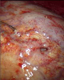
Figure 1
Diaphragmatic fenestrations.
Figure 2
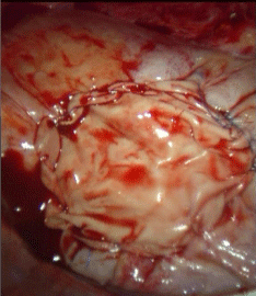
Figure 2
Bovine pericardial patch covering diaphragm.
Discussion
EMS is defined as the presence of ectopic endometrial tissue outside its normal location (the uterine cavity); thoracic EMS syndrome refers to the varying clinical and radiological manifestations associated with the growth of endometrial tissues in the lungs or on the pleural surface. It is a rare condition that includes four recognized clinical entities (lung nodules, catamenial pneumothorax (CP), haemothorax (CH) and haemoptysis) [1]. Thoracic EMS was first described by Maurer et al. [2]. Clinical presentation of CH thought to be a consequence of sloughing of thoracic endometrial tissue [1]. Pleural EMS is seen in approximately 30%, and diaphragmatic fenestrations in 40%, of patients. These findings appear to be more prevalent at the time of, or soon after, menses and may explain the absence of diaphragmatic fenestrations at the two laparoscopies and the first thoracotomy in our patient. Definitive management of the underlying EMS should be considered if patients become severely troubled. Several therapeutic methods have been proposed to tone down oestrogen receptors or block pituitary gonadotropin [3]. These include oral contraceptives, alkyl derivatives of testosterone (danazol) and gonadotropin–releasing hormone agonists. Ravindran P et al. [3] reported that a remarkable response can be obtained with hormonal therapy to CH and haemopneumothorax. Surgical management includes pleurodesis, pleurectomy and/or pleural abrasion, resection of ectopic intrathoracic tissue and repair of diaphragmatic abnormalities by partial resection, plication or covering the diaphragm by a mesh. Covering the diaphragm would seem a better option than plication or limited resection of the diaphragm, as fenestrations may be absent or difficult to visualize, depending on the relationship to menses, as in our case. We used bovine pericardium to cover the diaphragmatic fenestrations in our case, as we felt this would be a more resistant barrier to air and fluid entering from the peritoneal cavity. Recurrence rates are lower when surgery is combined with hormone manipulation therapy. In our case, medical treatment was not an option since the patient was trying to conceive.
Conclusion
Recurrent CHP is a rare condition of multifactorial aetiology. Hormonal therapy can have a statisfactory results, however surgery plays an important role in the management of patients with this condition, but is rarely definitive. Covering the diaphragm with a mesh or patch should be considered in all patients, irrespective of whether fenestarions are visualised at surgery. Hormonal treatment in combination with surgery is likely to offer the best outcome.
References
- Alifano M, Trisolini R, Cancellieri A, Regnard JF. Thoracic Endometriosis: Current Knowledge. Ann ThoracSurg. 2006; 81:761-769.
- Maurer ER, Schaal JA Mendez FL. Chronic recurring pneumothorax spontaneus due to endometriosis of the diaphragm. J Am Med Assoc. 1958; 168: 2013-2014.
- Ravindran P, Raj RJ, Parameswaran K. Concurrent catamenial haemothorax and haemopneumothorax. Chest 1993; 103: 646-648.