Clinical Image
Not Deep Vein Thrombosis, But Delayed Enhancement with Acute Limb Malperfusion
Motohiko Goda*, Naoto Yabu, Norihisa Tominaga, Daisuke Machida, Yukihisa Isomatsu, Shinichi Suzuki and Munetaka Masuda
Department of Cardiovascular Surgery, Yokohama City University Hospital, Japan
*Corresponding author: Motohiko Goda, Department of Cardiovascular Surgery, Yokohama City University Hospital, Fukuura 3-9, Kanazawa-ku, Yokohama, 236-0004, Japan
Published: 06 Mar, 2017
Cite this article as: Goda M, Yabu N, Tominaga N,
Machida D, Isomatsu Y, Suzuki S, et
al. Not Deep Vein Thrombosis, But
Delayed Enhancement with Acute Limb
Malperfusion. Clin Surg. 2017; 2: 1338.
Clinical Image
A 73-year old woman was transferred with acute back pain. Contrast enhanced computed tomography revealed acute aortic dissection with true lumen compression and low density area in her right common iliac vein, which looks like deep vein thrombosis. Thisphenomenon is explained that the right common iliac vein was enhanced later than the left because the perfusion of the right common iliac artery was impaired. Venous phase imaging was necessary to exclude deep venous thrombosis.
Figure 1a-1c:
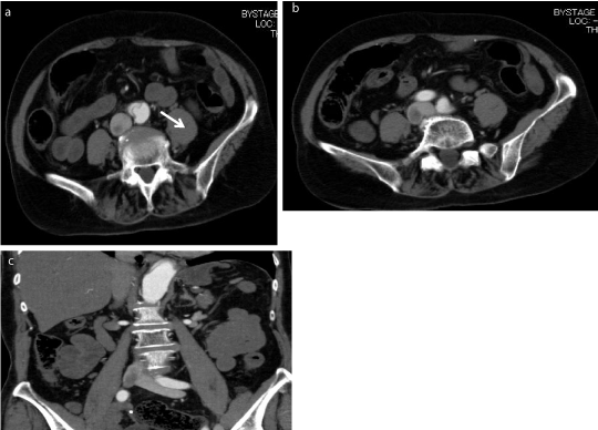
Figure 1a-1c:
he axial and coronal view in arterial phase showed low density area in the middle of right common iliac vain.
Figure 2
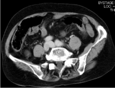
Figure 2
The venous phase showed equally enhanced the right and left common iliac vain.