Case Report
Acute Kidney Injury Following Retro-Cecal Appendectomy in a Male Child
Saleh Al-Ajmi1 and Amal A. Al-Eisa2*
1Department of Pediatrics, Adan Hospital-Ministry of Health, Kuwait
2Department of Pediatrics, Kuwait University, Kuwait
*Corresponding author: Amal A. Al-Eisa, Department of Pediatrics, Kuwait University, P.O. Box: 23924, Safat 13110, Kuwait City, Kuwait
Published: 03 Mar, 2017
Cite this article as: Al-Ajmi S, Al-Eisa AA. Acute Kidney
Injury Following Retro-Cecal
Appendectomy in a Male Child. Clin
Surg. 2017; 2: 1321.
Abstract
Background: Acute kidney injury due to bilateral ureteral obstruction is a rare complication
following appendectomy in children. In this report, we present a 9- year old boy who developed
gross hematuria and anuria 6 days post- appendectomy.
Case: The child presented with acute abdominal pain, fever and vomiting. Acute appendicitis was
suspected and laparoscopic appendectomy was performed. The Appendix was found to be retro-
Cecal in position and perforated with gangrenous tip. On the 6th postoperative day, he developed
AKI with anuria, Serum creatinine peaked to 239 μmol/l after a base- line reading of 30 μmol/l /l on
day of admission. Ultrasound and CT scan of the abdomen revealed bilateral hydronephrosis and
hydroureters with curvilinear hyper dense shadows in course of distal ureters. Cystoscopy revealed
congestions and edema at bladder base, trigon and around both ureteric orifices. AKI resolved
completely within 2 days after cystoscopic bilateral stenting of both ureters.
Conclusion: AKI should be anticipated in male pediatric patients undergoing appendectomy with
a retro-Cecal position. Preemptive ureteric stent insertion might be a good prophylactic option.
Keywords: Acute Kidney Injury (AKI); Appendectomy; Retro-Cecal; Hypercalciuria
Introduction
Appendicitis is a common cause of acute abdomen in children and adolescents [1]. Appendectomy is a common surgical procedure with a well- known complications including abscess formation, peritonitis and wound infection [2]. Other post-operative complications following appendectomy is considered relatively rare such as unilateral ureteric obstruction secondary to appendicular abscess and bilateral ureteral obstruction secondary to bladder wall edema leading to acute kidney injury [3]. In this report, we present a case of gross hematuria associated with anuria and acute kidney injury due to post- appendectomy bilateral ureteral obstruction not related to abscess formation. We also elaborate on the possible causes of mechanical bilateral obstruction other than trigonal edema of the urinary bladder as has been previously postulated.
Case Presentation
A previously healthy 9 -year- old male presented with acute abdominal pain, vomiting and low grade fever of 1 day duration prior to admission. The pain was classical of appendicitis as it started around the umbilicus then shifted to the right iliac fossa. The clinical diagnosis of acute appendicitis was made based on the findings of rebound tenderness at the right iliac fossa with a positive Mc- Burney's sign. Initial laboratory tests pre-operatively showed: Hemoglobin: 121 g/l (137-145), WBC: 11.3 X 10⁹ (4.23-9.07), Platelets: 405 X 109 (150-400), Blood urea 4.4 mmol/l (2.4-6.2) and serum creatinine: 32 μmol/l (normal range: 62-106), serum Sodium: 136 mmol/l (135-145) and serum Potassium: 4.3 mmol/l (3.5-5.2). Urine analysis showed pyuria of 50 WBC/HPF with no protein, RBC, or casts and negative nitrite. Laparoscopic appendectomy was performed. The Appendix was found to be retro-Cecal in position and was perforated with gangrenous tip. The patient received cefuroxime and metronidazole peri-and postoperatively. He was kept in hospital to complete a full course of antibiotic to cover for peritonitis due to perforation. On the 6th postoperative day, the child developed painless gross hematuria and noticed to be anuric. Physical examination at that time showed no sign of fluid overload or dehydration. All vital signs including blood pressure were within normal range. Abdominal examination revealed no palpable masses or signs of peritonitis or surgical wound infection. Blood lab results showed; Hemoglobin: 117 g/l, WBC [14.6 X 10⁹], platelets: 455 X 109, Blood urea 11.40 mmol/l and serum creatinine of 239 μmol/l after a base- line reading of 30 μmol/l /l on first day of admission. No electrolytes derangements were reported. Urine analysis showed RBC >250 /ul, without significant pyuriaor proteinuria. Urine culture was sterile. Ultrasound (Figures 1-3) and CT scan of the abdomen revealed bilateral grade 1 hydronephrosis and hydroureters with curvilinear hyper- dense shadows in the course of distal ureters (Figures 4-7). Accordingly, cystoscopy was performed which revealed the presence significant congestion andedema at bladder base, trigon area and around both ureteric orifices. Double J stents were inserted at both vesico-ureteric junctions to relieve obstruction. This procedure was followed by a prompt increase in urine output within 24 hours followed by marked drop in serum creatinine level with complete normalization within 2 days post stent insertion. The patient was discharged home on day 7. Follow- up ultrasound done 3 weeks after appendectomy was completely normal. As the patient has hyper-echoic shadows bilaterally on CT scan of abdomen during the obstructive phase suggestive of urolithiasis, early morning spot Urine Calcium/Creatinine was performed on 3 occasions over 3 weeks. The sequential results were: 0.73, 0.14 umol/L and 0.23 mmol/umol Cr (Normal range: <0.6). DJ stents were removed 6 weeks after insertion.
Figure 1
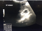
Figure 1
Ultrasound of the right kidney showing hyper echogenicity with
pelvi-calyceal dilation.
Figure 2
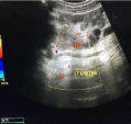
Figure 2
Ultrasound of the pelvis showing dilated left ureter.
Figure 3
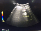
Figure 3
Ultrasound of the pelvis showing dilated right ureter.
Figure 4
Figure 4
CT scan of abdomen showing longitudinal images with hyper
echoic shadows occupying the lower ends of both ureters highly suggestive
of stones.
Figure 5
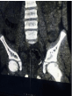
Figure 5
CT scan of abdomen showing longitudinal images with hyperechoic
shadows occupying the lower end of left ureter highly suggestive of
stones.
Figure 6
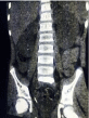
Figure 6
CT scan of abdomen showing longitudinal images with hyperechoic
shadows occupying the lower end of right ureter highly suggestive
of stones.
Discussion
Appendicitis is one the most common medical emergencies requiring urgent surgical intervention. The vermiform appendix can be placed in different positions related to the Cecum part of the bowel. Retro-Cecal positioning of the appendix varies from 26%-65% in normal people [4,5]. This position is known to be affected by many factors including gender, geographic and racial factors. The symptoms of appendicitis due to spread of inflammation depends on the location of the appendix. More than 50% of patients with ascending retro-Cecal appendicitis may have an atypical clinical presentation [6]. Some of these complications include abscess in the para-renal space and spread to bare area of the liver [7]. Post appendectomy complications are not uncommon in children such as acute urinary retention [8], abscess formation and unilateral right ureteric obstruction secondary to appendicular abscess. Few reports in the literature reported unusual cases of post appendectomy bilateral ureteral obstruction [2,8-17]. In all these reports, the appendix was retro-cecal with or without perforation and all patients were males. Our patient showed similar features. Mechanical obstruction at vesico-ureteral junction in such cases has been thought and explained solely on the basis of trigon edema obstructing both ureteral orifices. It is expected that due to its position, retro-cecal appendix can induce pelvic irritation especially if diagnosis is delayed. Missed diagnosis of appendicitis in retro-cecally placed inflamed appendix is a commonly encountered clinical problem as signs of appendicitis are hindered by the overlying Cecum. The interesting finding of hyper-echoic shadows at both low ureteric ends documented on CT in our patient is highly suggestive of urolithiasis. Although other studies had reported similar finding [2,8], but unfortunately none had elaborated on the role of stones in aggravating the obstruction or to the possible pathophysiology of this acute stone formation. Dehydration and stasis are definitely considered as predisposing factors for stone formation especially in the presence of concomitant uretero-vesical junction obstruction due to trigonal edema. Nevertheless we believe these are not the only factors. We did document hypercalciuria in our patient, although only in one urine sample, but we believe it is a significant finding. Transient hypercalciuria is most likely a complication of the bilateral ureteric obstruction and not simply due to supersaturation of concentrated urine of a dehydrated patient. Calcium reabsorption in the distal tubules and the collecting tubules of the kidney might be impaired by back pressure effect mechanical obstruction leading to increased Calcium loss in urine. The need of prophylactic measures to prevent such obstruction in cases diagnosed with retrocecal appendicitis pre or peri-operatively should be assessed. As a pre-operative measure, adequate rehydration of patients is of utmost importance as it will dilute the urine and prevent supersaturation of it with solutes that might precipitate to form gravol and /or stones. Peri-operatively, the surgeon should assess the degree of inflammation induced by the inflamed or the perforated appendix and he should anticipate any further complications that might take place post-operatively. The need of prophylactic bilateral ureteric stent insertion can be a wise option based on the surgeon’s assessment of the degree of pelvic inflammation which can save the patient a second surgical procedure within few days after appendectomy.
Figure 7
Figure 7
CT scan of the abdomen showing transverse image with hyperechoic
shadows at the lower ends of both ureters.
Conclusion
AKI should be anticipated in male pediatric patients undergoing appendectomy with a retro-Cecal position. Close and prolonged postoperative monitoring with proper rehydration of such patients is mandatory to reduce the chances of complicated obstruction with urolithiasis.
References
- Peltokallio P, Tykka H. Evolution of the age distribution and mortality of acute appendicitis. Arch Surg. 1981; 116: 153-156.
- Seeberg LT, Edenberg J, Saetren H. Bilateral ureteral obstruction after appendicectomy. Surgeon. 2005; 3: 45-47.
- Dever DP, Hulbert WC Jr, Emmens RW, Rabinowitz R. Appendiceal abscess masquerading as acute urinary retention in children. Urology. 1985; 25: 289-292.
- Donald c collins. Acute retrocecal appendicitis: based on seven hundred and fifty-one instances. Arch Surg. 1938; 36: 729–743.
- Wakeley CP. The Position of the Vermiform Appendix as Ascertained by an Analysis of 10,000 Cases. J Anat. 1933; 67: 277–283.
- Lee SL, Ho HS. Acute appendicitis: is there a difference between children and adults? Am Surg. 2006; 72: 409-413.
- Morton A Meyers, Michael Oliphant. Ascending retrocecal appendicitis. Radiology. 1974; 110: 295–299.
- Grande M, Lisi G, Bianchi D, Bove P, Miano R, Esser A, et al. Bilateral Ureteral obstruction in children after appendectomy. Case Rep Surg. 2015; 2015: 740795.
- Aronson DC, Moorman-Voesrmans CG, Tiel-van Buul MM, Vos A. A rare complication of acute appendicitis: Complete bilateral distal ureteral obstruction. Lancet. 1994; 344: 99-100.
- Dueholm S, Bagi P, Nordsten M. Ureteric obstruction as a complication to the appendicular abscess. Case report. Acta Chir Scand. 1987; 153: 557-559.
- Tripp BM, Homsy YL, Kiruluta G. Bilateral ureteral obstruction secondary to a perforated appendiceal abscess. J Urol. 1995; 154: 1158.
- Buckley K, Buonomo C. Bilateral ureteral obstruction and renal failure due to a perforated appendix. Pediatr Radiol. 1994; 24: 308-309.
- Aronson DC, Moorman-Voestermans CG, Tiel-van Buul MM, Vos A. A rare complication of acute appendicitis: complete bilateral distal ureteral obstruction. Lancet. 1994; 344: 99-100.
- Hugen CA, Mulders PF, Monnens LA, Dijkman-Neerinex RH, de Vries JD. Bilateral ureteral obstruction after appendectomy in children. J Pediatr Surg. 1995; 30: 1666-1667.
- Timm K, Illi OE, Leumann E, Stauffer UG. Postrenal anuria after appendectomy in childhood. Eur J Pediatr Surg. 1997; 7: 237-238.
- Sandermann J, Hansen LS. Bilateral ureteral obstruction as a complication to a perforated appendix. Report of a case. Acta Chir Scand. 1983; 149: 535-536.
- Kaplan GW, Keiller DL. Ureteral obstruction after appendectomy. J Pediatr Surg. 1974; 9: 559-560.