Research Article
Single Port Access Laparoscopic Right Hemicolectomy in Children: Treatment of Chronic Intussusception Caused by Ileocecal Burkitt Lymphoma
Weili XU, Jintao LI and Suolin Li*
Department of Pediatric Surgery, Hebei Medical University, China
*Corresponding author: Suolin Li, Department of Pediatric Surgery, the Second Hospital of Hebei Medical University, 215 Heping West Road, Shijiazhuang City, 050000, China
Published: 10 Feb, 2017
Cite this article as: Weili XU, Jintao LI, Suolin Li. Single
Port Access Laparoscopic Right
Hemicolectomy in Children: Treatment
of Chronic Intussusception Caused by
Ileocecal Burkitt Lymphoma. Clin Surg.
2017; 2: 1304.
Abstract
Background: The umbilical laparo-endoscopic single-site surgery (LESS) is to minimize the
unnecessary trauma and achieve the combination of minimally invasion with cosmetic advantage
for scarless surgery. We report in this paper a new technique of umbilical single port access (SPA)
laparoscopic right hemicolectomy complying with conventional surgical oncologic principle and
technique of minimally invasive colectomy in children.
Methods: Preliminary experience with umbilical SPA right hemicolectomy in a 5-year-old child
with chronic intussusception caused by ileocecal Burkitt lymphoma.
Results: Umbilical SPA laparoscopic right hemicolectomy was performed successfully with
conventional laparoscopic instruments. Operative time was 90 min and the volume of hemorrhage
was 20 ml. No intraoperative and postoperative complications were recorded. Diet recovered at the
fourth day after operation.
Conclusion: With the plastic cannulas and semi-rigid flexible instruments, umbilical SPA
laparoscopic right hemicolectomy can be performed successfully through the paraumbilical hidden
incision for children colorectal disease, not only follow the traditional surgical principle but also
achieve minimally invasive cosmetic results.
Keywords: Single port access (SPA); Colectomy; Laparoscopy; Minimally invasive surgery; Technique; Scarless surgery; Children
Introduction
The aim of minimally invasive surgical techniques is to minimize the unnecessary trauma and
pain of the operation and achieve the combination of minimally invasion with cosmetic advantage
for scarless surgery. Under the guidance of this concept in recent years, the umbilical laproendoscopic
single-site surgery (LESS) came into being [1].
In this paper, we reported the first cases umbilical SPA laparoscopic right hemicolectomy in the
treatment of children chronic intussusception caused by ileocecal Burkitt lymphoma complying
with conventional surgical oncological principle and technique of minimally invasive colectomy.
Materials and Methods
Case report
A five-year-old male child hospitalized because of an intermittent abdominal pain accompany
with hematochezia for more than one month. During physical examination, there were no abnormal
symptoms on heart and lung, but a mass with ill-defined boundary was touched in the right belly
with pressing pain, but without obvious rebound tenderness and muscle tension. Abdominal CT
examination showed a ileum-ascending colon intussusception, and a mass in anteromedial aspect
of ascending colon. Abdominal ultrasound examination revealed right upper abdominal mass,
multiple solid hypoechoic nodules in the right abdomen and a small amount of effusion in intestinal
interval. The diagnosis of chronic intussusception was definitely established.
Surgical technique
The patient was offered this approach after having given informed consent of his parents. With
a thorough preoperative preparation, laparoscopic exploratory operation was performed under general anesthesia. Firstly, a 5 mm trocar was placed in the middle
of umbilicus after a two-centimeter-incision was cut along the right
edge of umbilical ring and artificial CO2 pneumoperitoneum was
established with a 9 mmHg. Thereafter, two plastic 5 mm trocars were
placed up and down side of the incision respectively (Figure 1A and
1B). Through laparoscopy, one mass of 6 cm × 5 cm × 4 cm size was
confirmed ileum-colon-colonic type of intussusception, with nested
appendix and lots of swelling mesenteric lymph nodes. After restoring
intussusception, we found a tumor with 3 cm × 2 cm × 2 cm size beside
the ileocecus, then resected lateral peritoneum of ascending colon
and gastrocolic ligament in liver area by ultrasonic knife (Figure 2A
and 2B), freed and extracted ileocecus and right hemicolon outside
the abdominal cavity through expanding umbilical incision up to 3
cm (Figure 3A and 3B). Surgical dressings were placed around the
umbilical incision, then resection of terminal ileal and right colon
and ileum-transverse colon anastomosis were performed in vitro
according to the results of frozen biopsy, malignant lymphoma
and reactive proliferative lymph nodes. Moreover, the anastomotic
intestine was return into peritoneal cavity and umbilical incision was
sutured layer by layer (Figure 4A and 4B).
Figure 1
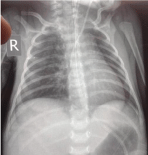
Figure 1
(A) A 5 mm trocar was placed in the middle of umbilicus and
artificial CO2 pneumoperitoneum was established, thereafter, two plastic 5
mm trocars were placed up and down side of the incision. (B) Therefore, the
semi-rigid flexible instruments were put into abdominal cavity through plastic
cannulas and "chopstick effect" was overcame inspite of collisions between
the instruments and small space in children abdominal cavity.
Figure 2
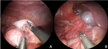
Figure 2
(A) Ileum-colon-colonic type of intussusception, with nested
appendix and lots of swelling mesenteric lymph nodes, was confirmed and
restored. (B) The right half colon was dissociated by resecting the lateral
peritoneum of ascending colon and gastrocolic ligament in liver area with
ultrasonic knife.
Figure 3
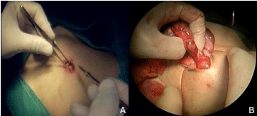
Figure 3
The ileocecus and right hemicolon were freed and extracted
outside the abdominal cavity through expanding umbilical incision up to 3 cm.
Figure 4
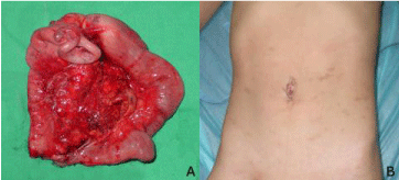
Figure 4
(A) Specimen of the resected right half colon was shown. (B)
Moreover, the anastomotic intestine was returned into peritoneal cavity and
umbilical incision was sutured layer by layer, achieving minimally invasive
cosmetic results.
Results
The operation had been lasted for ninety minutes with a twenty milliliters hemorrhage. The duration of hospital stay was six days. Diet recovered at the fourth day after operation. No intraoperative and postoperative complications occurred. The immune histopathologic diagnosis of the resected specimen was ileocecal Burkitt lymphoma. This children was then transferred to the department of pediatrics for continue chemotherapy. With a 9 month postoperative follow-up, no tumor recurred by CT review.
Discussion
As a special type of non-Hodgkin's disease, Burkitt's lymphoma
was scattered reported in China, with high grade malignancy, rapid
progress and poor prognosis [2]. Clinical manifestations of Burkitt’s
lymphoma varied very much due to a different position of its lesion;
however, ileocecal tumor combined with intussusception was rarely
reported in previous literature. In present study, the diagnosis of
chronic ileocecal intussusception was delayed because the ileocecal
tumor only results in the symptoms of incomplete intestinal
obstruction.
With the development of minimally invasive surgical techniques,
laparoscopic right hemicolectomy has become the first choice for
treatment of ileocecal tumor disease, but single-incision access
laparoscopic right hemicolectomy can only be seen in few case reports
[3,4], particularly has not been reported in children. To Ileocecal
malignant lymphoma, pediatric surgeons preferred to early perform
laparoscopy exploratory operation because of its earlier metastasis
and poorer life quality for children. In adults, the glove-port technique
was often regarded as a simple, low-cost, reproducible, and sure
method to perform single-incision laparoscopic surgery (SILS) in
some high-experienced laparoscopic surgical centres [5-7]. But in this
LESS, the plastic cannulas, with a more simple, safe and inexpensive
superiority, were successfully placed in the paraumbilical hidden
incision. The semi-rigid flexible instruments were put into abdominal
cavity through plastic cannulas placed on both sides of umbilicus
and "chopstick effect" was overcome in spite of collisions between
the instruments and small space in children abdominal cavity. After
dissociation of the right peritoneum and gastrocolic ligament in liver
area by 5 mm ultrasonic knife, the right colon can be dragged out
and resected easily through the slightly expanded umbilical incision.
The following operation of right colon outside abdominal cavity
and dressings setting around the umbilical incision not only make
operation more easily but also prevent the implantation metastasis of
intra-abdominal tumor cells and possible intestinal contents leaking.
Thus, umbilical SPA laparoscopic right hemicolectomy can
not only follow the traditional surgical principle but also achieve
minimally invasive cosmetic results may have the advantage over
NOTES approach to offer the safety of laparoscopic colectomy
especially for children colorectal disease.
Acknowledgement
The authors like to thank the members of the Operation Centre at the Second Hospital of Hebei Medical University for help with recording a video of SPA operation. We also thank the members of the Pathological Center at the Second Hospital of Hebei Medical University for a Quick Intraoperative Diagnosis of frozen biopsy.
References
- Hansen EN, Muensterer OJ, Georgeson KE, Harmon CM. Single-incision pediatric endosurgery: lessons learned from our first 224 laparoendoscopic single-site procedures in children. Pediatr Surg Int. 2011; 27: 643-648.
- Wang X, Zhai Y, Mao X. Clinical pathological features and differential diagnosis of Burkitt's lymphoma. Chinese Clinical Oncology. 2007; 12: 869-873.
- Bucher P, Pugin F, Morel P. Single port access laparoscopic right hemicolectomy. Int J Colorectal Dis. 2008; 23: 1013-1016.
- Remzi FH, Kirat HT, Kaouk JH, Geisler DP. Single-port laparoscopy in colorectal surgery. Colorectal Dis. 2008; 10: 823-826.
- Livraghi L, Berselli M, Bianchi V, Latham L, Farassino L, Cocozza E. Glove technique in single-port access laparoscopic surgery: results of an initial experience. Minim Invasive Surg. 2012; 2012: 415430.
- Day W, Lau P. Novel "glove" access port for single port surgery in right hemicolectomy: a pilot study. Surg Laparosc Endosc Percutan Tech. 2011; 21: 145-147.
- Hompes R, Lindsey I, Jones OM, Guy R, Cunningham C, Mortensen NJ, et al. Step-wise integration of single-port laparoscopic surgery into routine colorectal surgical practice by use of a surgical glove port. Tech Coloproctol. 2011; 15: 165-171.