Case Report
Dieulafoy Lesion of Rectum: an Uncommon Pathology not to be Missed
Pallavi Huma Arya1, Joseph Sebastian2, Sudhakar Mangam2 and Narasimhaiah Srinivasaiah3*
1University Hospital London, UK
2QEQM Hospital, UK
3St. Mark’s Hospital, UK
*Corresponding author: Narasimhaiah Srinivasaiah, St. Mark’s Hospital, No.21, Kenton court, Kenton, Harrow, HA3 8AQ, UK
Published: 02 Nov, 2016
Cite this article as: Arya PH, Sebastian J, Mangam S,
Srinivasaiah N. Dieulafoy Lesion of
Rectum: an Uncommon Pathology not
to be Missed. Clin Surg. 2016; 1: 1170.
Abstract
Introduction: Dieulafoy lesions of the upper gastrointestinal (UGI) tract are well known. Rectal
lesions are less common. We present a case of a rectal Dieulafoy lesion causing significant rectal
bleeding, managed successfully.
Methods and Results: A case note and literature review was done. An 82-year-old female was
referred to the surgeons for management of rectal bleeding. She was treated by medical team for
pneumonia; was intubated, ventilated, central venous access gained and on inotropes. She developed
fresh per rectal bleeding with clots, leading to haemo-dynamic instability. She underwent an UGI
endoscopy, followed by on-table colonoscopy with caecal intubation. Distal blood streaking of the
mucosa till 30-35 cms was noted but no blood in the lumen beyond this. A 2-3 mm raised mucosal
lesion- Dieulafoy lesion was noted in the rectal mucosa above the dentate line at 5 o’clock with an
arterial spurter (Figure 1 and 2). This was over sewn with three 2.0 PDS sutures. This stopped the
bleeding completely.
Discussion: Dieulafoy lesion of rectum is an uncommon entity that needs to be considered as a
cause of lower GI bleeding. It is very vital to visualise the distal rectum and anal canal for the cause
of the bleed could be ano-rectal.
Keywords: Rectal bleeding; Colonoscopy; Dieulafoy lesion; Diagnosis; Gastrointestinal surgery; Vascular surgery
Case Presentation
An 82-year-old female was referred to the surgeons for management of per rectal bleeding. She
was critically unwell, having been admitted under the general physicians with a one-week history
of lower abdominal pain and diarrhoea. Her past medically history included hypertension and
recurrent urinary tract infections. Socially she lived on her own, was a non-smoker and a social
drinker. She was being treated for urosepsis and acute kidney injury. Markers of inflammation were
raised with a leukocytosis of 16.5 10^9/l, CRP of 435 mg/L, Urea of 11.6 umol/L and creatinine
of 176 umol/L. The clinical picture was complicated by a possible CAP. She was catheterised and
commenced on intravenous fluids and broad-spectrum antibiotics.
Following her initial management, she deteriorated with high-grade pyrexia of 40 degrees, and
hypoxia with a saturation of 85% on air, Oliguria of 10ml/hour and hypotension of 73/56 mm/Hg
despite aggressive resuscitation. Care was escalated to Intensive care unit, where she was intubated,
ventilated, central venous access gained and supported on Inotropes. Based on blood cultures from
admission, which grew Staphylococcus Aureus, Flucloxacillin and Rifampicin were added.
Day 14 of admission, she developed fresh per rectal bleeding with clots. At this point she was
haemodynamically stable maintaining a blood pressure of 110/70 mm/Hg and a pulse of 120
beats per minute. Her abdomen was soft, non-tender with normal bowel sounds. On digital rectal
examination fresh blood was noted with a large haemorrhoid at the 7’o clock position. Bloods
showed mild anaemia with an Hb of 94 g/L, platelets of 319x10^9/l, PT of 17.2 secs and APTT of
35.4 secs. With such a large, sudden and fresh PR bleed, initial inclination was towards an upper
gastrointestinal bleed. Patient was resuscitated; anticoagulation stopped and was commenced on
IV PPIs. An urgent oesophagogastroduodenoscopy (OGD) followed a further deterioration in the
patient’s condition. Patient was haemodynamically unstable with a blood pressure of 50/38mm/Hg
and heart rate of 130 beats per minute. Haemoglobin had dropped
to 64 g/L that was brought back with 4 units of blood. Following a
normal OGD, a CT angiography was planned. Dictated by the clinical
state of the patient, she had to be rushed to Theatres.
Patient underwent an on table colonoscopy reaching the caecum.
Some blood streaking of the mucosa till 30-35 cms was noted but no
blood in the lumen beyond this. A 2-3 mm raised mucosal lesion-
Dieulafoy lesion was noted in the rectal mucosa just above the dentate
line at 5’o clock with an arterial spurter (Figure 1 and 2). This was
transfixed with three 2.0 PDS sutures followed by the application of
two spongostan and surgicel. This stopped the bleeding completely.
Post procedure patient was haemodynamically stable and no further
immediate bleeds were noted. Five days post the first bleed however
patient had a further fresh per rectal bleed where she underwent a
flexible sigmoidoscopy and oversowing of the previous anal suture
was performed. She had no further bleeds following this.
Figure 1
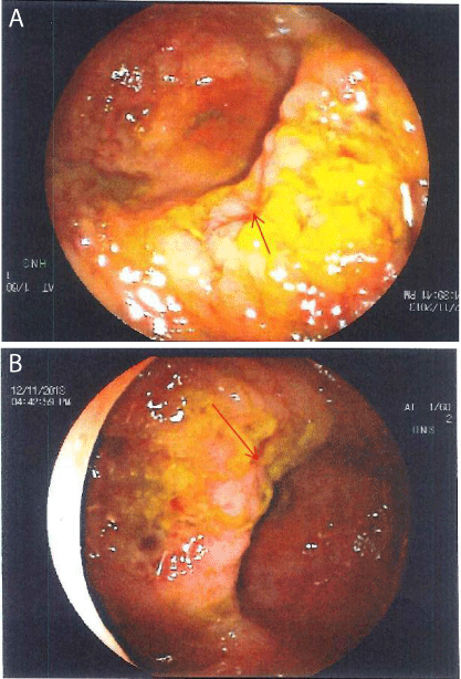
Figure 1
(a) and (b) Rectal Dieulafoy lesion, pointed by the arrow.
Figure 2
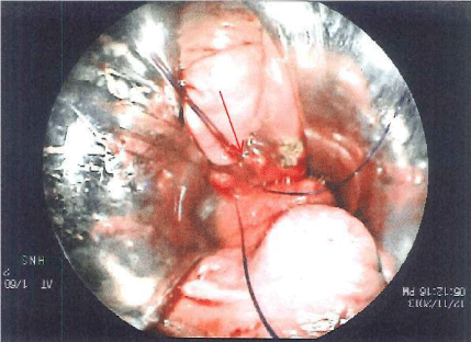
Figure 2
Rectal Dieulafoy lesion over sewn with three 2.0 PDS sutures..
Discussion
Acute massive gastrointestinal bleed is often seen in the form
of haematemesis, melaena or hematochezia eventually leading to
haemodynamic instability [1]. Massive lower gastrointestinal bleeds
can be a challenge to manage. It can often be impossible to identify
the source of the bleeding despite a wide spectrum of diagnostic tests
and it is not unusual for a subtotal colectomy to be undertaken with
an end Ileostomy. Attention to detail and suspicion of a possible
pathology is vital. An unwary decision would have landed with a
major surgery without recognition and management of local rectal
pathology.
Dieulafoy lesion is a rare but well-recognized cause of
gastrointestinal bleeding. The lesion is a submucosal artery that
protrudes through a mucosal defect of the lumen [2]. It most
commonly affects the mucosa within close proximity to the gastrooesophageal
junction on the lesser curve of the stomach but can rarely
be the cause of massive lower GI bleeds due to lesions at the anorectal
junctions.
Over the years many causes of Dieulafoy lesions have been
suggested. It was initially thought to be due to aneurysms in a vessel
of the mucosal wall in combination with atherosclerosis [3,4]. It
has also been thought to be due to acquired or congenital vascular
malformations however over time the general consensus appears
to be that it is due to an abnormally large/torturous submucosal
artery at a very close proximity to the mucosal surface which is
often visualised on endoscopy [5-7]. The artery can be found to be
protruding through a small mucosal defect (2-5 mm) and when
ruptured can lead to massive unexpected haemorrhage. Interestingly
rectal Dieulafoy lesions have been associated with age, renal disease,
burns, liver transplantation and GI stromal tumours [1]. The
diagnosis of a Dieulafoy lesion is most commonly made by endoscopy
or laparotomy [8]. Endoscopic criteria that defines these lesions
are as follows [9] 1) active arterial bleeding from a small (< 3 mm)
mucosal defect with normal surrounding mucosa; 2) visualization
of the protruding artery with or without bleeding within the defect;
3) the presence of fresh, adherent clot attached to a minute mucosal
defect or mucosa of normal appearance. Occasionally diagnosis can
be made by angiography when initial endoscopy has failed [10-12].
Primarily a digital rectal examination is used to detect anal
pathology [11,12]. Urgent oesophagogastroduodenoscopy is vital to
rule out upper GI pathology including peptic ulcer disease or varices.
Colonoscopy or flexible sigmoidoscopies are vital to determine
whether the source of the bleed is proximal to the rectum [11,12].
Submucosal injection of epinephrine is the most commonly described
endoscopic technique for achieving haemostasis although it may
provide only temporary control of haemorrhage [1]. Haemostatic
clips have longer lasting effect than epinephrine and can also be
applied endoscopically. Endoscopic band ligation is the treatment of
choice for oesophageal varices [13] and has also been used for the
treatment of rectal abnormalities including varices and Dieulafoy
lesions [13,14]. be excellent. There have been no
reports of recurrence to date.
Conclusion
Dieulafoy lesion of Rectum is an uncommon entity that needs to be considered as a cause of lower GI bleeding. It is very vital to visualise the distal rectum and anal canal for the cause of the bleed could be ano-rectal.
References
- Kim M, Song HJ, Kim S, Cho YK, Kim HU, Song BC, et al. Massive lifethreatening lower gastrointestinal hemorrhage caused by an internal hemorrhoid in a patient receiving antiplatelet therapy: a case report. Korean J Gastroenterol. 2012; 60: 253-257.
- Kim HH, Kim JH, Kim SE, Park SJ, Park MI, Moon W. Rectal dieulafoy lesion managed by hemostatic clips. J Clin Med Res. 2012; 4: 439-441.
- Donaldson GA, Hamlin E Jr. Massive hematemesis resulting from rupture of a gastric-artery aneurysm; report of 3 cases. N Engl J Med. 1950; 243: 369-373.
- al-Mishlab T, Amin AM, Ellul JP. Dieulafoy's lesion: an obscure cause of GI bleeding. J R Coll Surg Edinb. 1999; 44: 222-225.
- Baettig B, Haecki W, Lammer F, Jost R. Dieulafoy's disease: endoscopic treatment and follow up. Gut. 1993; 34: 1418-1421.
- Katz PO, Salas L. Less frequent causes of upper gastrointestinal bleeding. Gastroenterol Clin North Am. 1993; 22: 875-889.
- Miko TL, Thomazy VA. The caliber persistent artery of the stomach: a unifying approach to gastric aneurysm, Dieulafoy's lesion, and submucosal arterial malformation. Hum Pathol. 1988; 19: 914-921.
- Bielicki D, Wasilewicz MP. Acute lower gastrointestinal tract bleeding caused by Dieulafoy lesion in rectum--case report. Pol Merkur Lekarski. 2006; 20: 708-709.
- Ruiz-Tovar J, Die-Trill J, Lopez-Quindos P, Rey A, Lopez-Hervas P, Devesa JM. Massive low gastrointestinal bleeding due to a Dieulafoy rectal lesion. Colorectal Dis. 2008; 10: 624-625.
- Strong RW. Dieulafoy's disease--a distinct clinical entity. Aust N Z J Surg. 1984; 54: 337-339.
- Veldhuyzen van Zanten SJ, Bartelsman JF, Schipper ME, Tytgat GN. Recurrent massive haematemesis from Dieulafoy vascular malformations- -a review of 101 cases. Gut. 1986; 27: 213-222.
- Juler GL, Labitzke HG, Lamb R, Allen R. The pathogenesis of Dieulafoy's gastric erosion. Am J Gastroenterol. 1984; 79: 195-200.
- Fixa B, Komárková O, Dvorácková I. Submucosal arterial malformation of the stomach as a cause of gastrointestinal bleeding. Gastroenterologia. 1966; 105: 357-365.
- Saur K. The solitary exulceratio simplex (Dieulafoy) causing a severe acute gastric bleeding. 1973; 44: 293-299.