Case Report
Adult Onset Asthma and Periocular Xanthogranuloma: A Difficult Diagnosis
Daniela Reyes-Capό, Kimberly D. Tran, Juan Ayala-Haedo, Oliver Fischer, Sander A. Dubovy and Sara Wester*
Department of Ophthalmology, University of Miami Miller School of Medicine, USA
*Corresponding author: Sara Wester, Department of Ophthalmology, Bascom Palmer Eye Institute, University of Miami Miller School of Medicine, 900 NW 17th St Miami, FL 33136, USA
Published: 27 Oct, 2016
Cite this article as: Reyes-Capό D, Tran KD, Ayala-Haedo
J, Fischer O, Dubovy SA, Wester S.
Adult Onset Asthma and Periocular
Xanthogranuloma: A Difficult Diagnosis.
Clin Surg. 2016; 1: 1168.
Abstract
Purpose: This case report describes the presentation, diagnosis, and treatment of adult onset asthma
and periocular xanthogranuloma (AAPOX) in a 41 year-old male and highlights the diagnostic and
therapeutic challenges of this disease.
Observations: The authors report a case of a 41 year-old male with a 12 year history of episodic
bilateral upper eyelid edema, and mechanical ptosis of uncertain etiology. He had previously
undergone biopsy and failed trials of surgical excision, systemic corticosteroids, methotrexate, and
radiation therapy. He was initially diagnosed in 2004 with bilateral idiopathic orbital inflammation
by biopsy. Due to concomitant sinus congestion, shortness of breath, and hilar adenopathy on
chest x-ray, the patient underwent endoscopic sinus surgery and transbronchial biopsy, which
showed vague granulomatous chronic inflammation suggestive of sarcoidosis. Serologic workup
was notable for peripheral blood eosinophilia (17%); however, infectious and rheumatologic
serologies, including angiotensin converting enzyme (ACE) and antinuclear antibody (ANA), were
unremarkable. The patient presented to the Bascom Palmer Eye Institute (BPEI) for evaluation due
to recurrence of bilateral lid swelling. He underwent repeat biopsy which disclosed fibro vascular
and adipose tissue containing xanthomatous infiltrate of foamy histiocytes without necrosis
consistent with xanthogranuloma. The overall presentation was suggestive of AAPOX. The patient
was started on methotrexate and long-acting bronchodilators, and at 8-month follow-up the patient
reports improved left-sided peripheral vision, and continues to be followed by the Pulmonology,
Rheumatology, and Oculoplastics services.
Conclusion and Importance: AAPOX is a rare, non-Langerhans histiocytic disorder with
characteristic orbital and systemic findings. This patient was followed for 12 years by multiple
providers with innumerable work-up, biopsies, and imaging without a definitive diagnosis. This
case illustrates the diagnostic challenges and important histopathologic clues in the diagnosis of this
rare disease.
Keywords: Orbital mass; Eyelid swelling; Asthma; Xanthogranulomatous disease
Introduction
Adult onset asthma and periocular xanthogranuloma (AAPOX) is a rare, non-Langerhans histiocytic disorder with characteristic orbital and systemic findings. This case report explores the diagnostic and therapeutic challenges of this disorder, and the importance of a multidisciplinary approach in recognizing and treating this disease.
Case Presentation
A 41 year-old male with a history of diabetes and keratoconus presented to the Bascom Palmer
Eye Institute (BPEI) Oculoplastics service due to chronic, intermittent upper eyelid swelling and
diplopia in all fields of gaze of uncertain etiology. His symptoms began 12 years prior to presentation
with bilateral upper lid swelling. He underwent bilateral anterior orbitotomies with biopsies and
was diagnosed with bilateral idiopathic orbital inflammatory disease (IOIS). The patient was only
partially responsive to high dose oral steroids and methotrexate, and reported clinical worsening
with radiation therapy of 10 fractions for a total of 2000 cGy. Two years later, the patient underwent
repeat bilateral upper lid blepharoplasties for surgical debulking and biopsy, and histopathologic
examination showed a xanthomatous and chronic inflammatory infiltrate. The diagnosis was
unclear at this point, and the patient was continued on oral corticosteroids for presumed IOIS.
Concomitant with his ocular symptoms, the patient complained
of sinus congestion and shortness of breath, with x-rays showing
sinusitis and hilar adenopathy. The patient underwent endoscopic
sinus surgery, and transbronchial biopsy revealing vague
granulomatous chronic inflammation suggestive of sarcoidosis, thus
the patient was diagnosed with sinusitis and pulmonary sarcoidosis.
After 12 years of symptoms, he developed worsening of his
eyelid swelling and presented to the BPEI Oculoplastics service. He
reported bilateral eyelid enlargement and diplopia that obstructed
his visual axis and impacted his activities of daily living. On review
of symptoms, he endorsed sinusitis and pulmonary disease, but was
otherwise pain free and without evidence of systemic, arthritic, renal,
or dermatologic disease. Best corrected visual acuity was 20/30 OD,
20/25 OS, with normal pupils and intraocular pressure. No proptosis
was noted, and the patient was orthophoric. External exam revealed
fullness of both superotemporal upper lids, with a palpable soft
preseptal mass in the left upper lid extending to the superotemporal
quadrant into the lacrimal gland (Figure 1). The patient demonstrated
limited supraduction OS, and left superior hemifield defect. Parotid
gland enlargement was notable on the left greater than right. Anterior
segment examination was notable only for bilateral papillary
conjunctivitis and the rest of the ocular exam was unremarkable in
both eyes.
Serologic workup was notable for peripheral blood eosinophilia
(17%); however, infectious and rheumatologic serologies, including
ACE and ANA, were unremarkable. Contact B-scan and diagnostic
A-scan were not consistent with IOIS nor with orbital sarcoidosis,
demonstrating a well-circumscribed, irregular, variably reflective,
soft tissue lesion in the superior/anterior left orbit involving the
levator muscle and extending superotemporally over the orbital rim.
MRI showed diffuse sinus disease, extraocular muscle enlargement,
and contrast-enhancing superotemporal eyelids and lacrimal gland/
orbital masses, left greater than right without fat stranding (Figure 2).
Given the patient’s chronic disease and unclear clinical picture,
the decision was made for a third incision biopsy of the left orbital
lesion to evaluate for IgG4 and xanthogranulomatous-related disease.
Flow cytometry revealed no immunophenotypically abnormal B or
T-cell populations. Histopathologic exam revealed xanthogranuloma
with fibro vascular and adipose tissue with foci of skeletal muscle
containing xanthomatous infiltrate of foamy histiocytes without
necrosis. The lesion stained positive for CD68, CD3, and CD20; and
stained variably positive for CD8 and CD4. Due to the lack
of plasma cell infiltration, IgG4 was not stained for on histopathologic
exam, and was not tested in the serum.
The differential diagnoses included adult onset
xanthogranulomatous disease, IgG4-related disease, eosinophilic
granuloma, Kimura’s disease, and Mikulicz’s syndrome. However,
based on the patient’s clinical presentation, histopathologic analysis,
and associated systemic disease, the overall presentation was
suggestive of AAPOX. The patient was started on methotrexate and
long-acting bronchodilators, and at 8-month follow-up, the patient
reported significant resolution of the left orbital lesion, improved
left-sided peripheral vision, and continues to be followed by the
Pulmonology, Rheumatology, and Oculoplastics services (Figure 4).
Figure 1
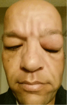
Figure 1
Clinical photograph at presentation demonstrating fullness of bilateral upper lids, with a left upper lid palpable soft preseptal mass in the super temporal quadrant. Parotid gland enlargement was notable on the left greater than right..
Figure 2
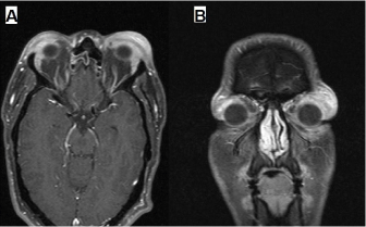
Figure 2
Axial (A) and Coronal (B) T1 magnetic resonance imaging with contrast and fat suppression showing diffuse sinus disease, extraocular muscle enlargement, and contrast-enhancing super temporal eyelids and lacrimal gland / orbital masses, left greater than right without fat stranding.
Discussion
Adult onset xanthogranulomatous (AOX) disease is an
uncommon spectrum of non-Langherhans histiocytic disorders
characterized by the presence of foamy non-Langerhans histiocytes,
Touton giant cells, and varying degrees of fibrosis [1,2]. Non-
Langerhans histiocytic disorders are often associated with other
systemic manifestations, which allow sub classification into four
syndromes: Adult onset xanthogranuloma (AOX), necrobiotic
xanthogranuloma (NBX), Erdheim-Chester disease (ECD), and
adult onset asthma and periocular xanthogranuloma (AAPOX). First
reported in 1993 [2], AAPOX remains an uncommon entity with
fewer than 50 cases reported in the English literature to date [1,3-12].
The clinical and histologic features in this case are consistent with
AAPOX.
AAPOX is characterized by periocular xanthogranuloma with
asthma. Patients are often observed with systemic lymphadenopathy,
salivary gland enlargement, and cases of elevated IgG serum levels are
reported [1,8]. In the largest review of published reports to date, pooled
data analysis of 21 AAPOX cases revealed mean age at diagnosis of
46 years (SD 13, range 22-74) and no gender preference [1]. Patients
typically had periocular skin manifestations, preseptal, and anterior
orbital involvement, and evidence of immune dysfunction including
asthma, lymphadenopathy, paraproteinemia, and B cell lymphoid
aggregates on histology [1]. These systemic characteristics of AAPOX
overlap with those of IgG4-related ophthalmic disease, and it has
been suggested that AAPOX may be associated with IgG4-related
disease [8]. AAPOX has not been a reported cause of mortality [1].
Empiric treatment modalities include surgical excision,
corticosteroids, chemotherapeutic agents, and radiation with varying
effectiveness. In Sivak’s case series of 8 AAPOX patients [1], surgery
alone was effective in 75% (6 of 8), corticosteroids were useful in 100%
(2 of 2), and cyclosporine (T cell suppression) and cyclophosphamide
(B cell suppression) were also found to be beneficial. In a review of
the literature, cases reporting management with cyclophosphamide,
melphalan, chlorambucil, vincristine, vinblastine, interferon,
etoposide, methotrexate [3], etanercept, gammaglobulin, clofazimine,
and doxorubicine have also been reported. Given the apparent B cell
dysfunction demonstrated in patients with AAPOX, there may be
rationale for systemic B cell suppression [4].
This patient demonstrated the typical signs of AAPOX
with submandibular and hilar lymphadenopathy, parotid gland
enlargement, asthma, and preseptal and anterior orbital involvement.
However, his diagnosis was delayed for 12 years from presentation,
due in part to biopsy results thought to be consistent with IOIS.
IOIS and AAPOX share histologically similar features, such as
an inflammatory cell infiltrate with predominance of mature
lymphocytes admixed with plasma cells, eosinophils and macrophages
[13]. Thus, upon receiving biopsy results consistent with IOIS, it is
still important to consider AAPOX within the differential diagnosis,
most notably when systemic symptoms are present, as was the case
in this patient. Interestingly, B cell lymphoid aggregates were not
noted on this patient’s histology, which may be related with his
long-term treatment with systemic corticosteroids. Additionally, our
patient further demonstrates the uncertainty of radiation therapy and
reinforces the use of methotrexate in treating AAPOX.
Conclusion
This patient was followed for 12 years by multiple providers with innumerable work-up, biopsies, and imaging without a definitive diagnosis. The rarity of AAPOX and other AOX clinical presentations precludes prospective investigation and meta-analysis for therapeutic studies. This case presentation of AAPOX adds incremental knowledge to our collective understanding of this uncommon entity, and demonstrates the diagnostic challenges, important histopathologic tools, and potential treatment modalities used particularly in the case of rare diseases with difficult diagnoses.
References
- Sivak-Callcott JA, Rootman J, Rasmussen SL, Nugent RA, White VA, Paridaens D, et al. Adult xanthogranulomatous disease of the orbit and ocular adnexa: new immunohistochemical findings and clinical review. Br J Ophthalmol. 2006; 90: 602-608.
- Jakobiec FA, Mills MD, Hidayat AA, Dallow RL, Townsend DJ, Brinker EA, et al. Periocular xanthogranulomas associated with severe adult-onset asthma. Trans Am Ophthalmol Soc. 1993; 91: 99-125.
- Cavallazzi R, Hirani A, Vasu TS, Sergott RC, Bilyk JR, Eagle RC, et al. Clinical manifestations and treatment of adult-onset asthma and periocular xanthogranuloma. Can Respir J. 2009; 16: 159-162.
- Papagoras C, Kitsos G, Voulgari PV, Zikou AK, Argyropoulou MI, Zioga A, et al. Periocular xanthogranuloma: A forgotten entity? Clin Ophthalmol. 2010; 4: 105-110.
- Tokuhara KG, Agarwal MR, Rao NA. Adult-onset asthma and severe periocular xanthogranuloma: a case report. Ophthal Plast Reconstr Surg. 2011; 27: e63-64.
- Minami-Hori M, Takahashi I, Honma M, Ito Y, Takahashi H, Ishida- Yamamoto A, et al. Adult orbital xanthogranulomatous disease: adultonset xanthogranuloma of periorbital location. Clin Exp Dermatol. 2011; 36: 628-631.
- Agi CU, Gober MD, Ferenczi K, Takach P, Eagle RC, Rosenbach M. A case of adult-onset asthma with periocular xanthogranulomas. Arch Dermatol. 011; 147: 1230-1231.
- London J, Martin A, Soussan M, Badelon I, Gille T, Uzunhan Y, et al. Adult Onset Asthma and Periocular Xanthogranuloma (AAPOX), a Rare Entity With a Strong Link to IgG4-Related Disease: An Observational Case Report Study. Medicine (Baltimore). 2015; 94: e1916.
- London J, Soussan M, Gille T, Badelon I, Warzocha U, Galatoire O, et al. Adult-onset asthma associated with periocular xanthogranuloma: new diagnostic and therapeutic approaches in a very rare systemic disease. Ophthal Plast Reconstr Surg. 2013; 29: 104-108.
- Kubota T, Moritani S, Ichihara S, Terasaki H. Association of systemic characteristics and histological variations in a case study of adult-onset asthma and periocular xanthogranuloma. J Clin Pathol. 2014; 67: 92-94.
- Shams PN, Rasmussen SL, Dolman PJ. Adult-Onset Asthma Associated With Simultaneous Conjunctival, Eyelid, and Orbital Xanthogranulomatosis Responsive to Systemic Immunosuppression. Ophthal Plast Reconstr Surg. 2015; 31: e162-163.
- Kiratli H, Kiliç M, Tarlan B, Söylemezoglu F. Adult orbital xanthogranulomas: clinical features and management. Eur J Ophthalmol. 2015; 25: 288-292.
- Swamy BN, McCluskey P, Nemet A, Crouch R, Martin P, Benger R, et al. Idiopathic orbital inflammatory syndrome: clinical features and treatment outcomes. Br J Ophthalmol. 2007; 91: 1667-1670.