Case Report
Primary Extracranial Meningioma of Middle Ear
Chien WW1,2*, Saleh J1 and Kumar A3
1Neurotology Program, National Institutes of Health, USA
2Department of Otolaryngology-Head and Neck Surgery, Johns Hopkins School of Medicine, USA
3Department of Pathology, Suburban Hospital, USA
*Corresponding author: Wade W Chien, Department of Otolaryngology-Head and Neck, John Hopkins School of Medicine, Baltimore, MD 21287, USA
Published: 29 Sep, 2016
Cite this article as: Chien WW, Saleh J, Kumar A. Primary
Extracranial Meningioma of Middle Ear.
Clin Surg. 2016; 1: 1142.
Abstract
The differential diagnosis of middle ear tumor is numerous and variable. In this care report, we describe a patient who presented with conductive hearing loss and was found to have a middle ear meningioma. This is one of the few reports of this rare middle ear tumor, and it serves to remind otolaryngologists that meningioma can sometimes occur in the middle ear.
Introduction
Meningiomas are one of most common benign tumors of the central nervous system [1]. They account for approximately 13% to 26% of all intracranial neoplasms; however, less than 2% of meningiomas have extracranial extension or temporal bone involvement [1]. Due to the rare occurrence of middle ear meningiomas, the diagnosis of these tumors can be challenging [1]. We present a rare case of primary extracranial meningioma located in the middle ear to highlight its clinical, imaging, immunohistochemical, and histological features. This case report was exempt from IRB review per institutional policy.
Case Presentation
A 65-year-old female was referred for evaluation of an enlarging, painful mass in her left middle
ear. She had a five-year history of left-sided hearing loss and pulsatile tinnitus that occurred during
strenuous physical activity. She denied otorrhea, vertigo, or any neurological or constitutional
symptoms. She did not have any previous history of ear surgery, recurrent ear infections, or
exposure to loud noise.
Binocular microscopy showed normal right external auditory canal and right tympanic
membrane. Left external auditory canal was also found to be clear; however, are d mass behind an
intact left tympanic membrane was revealed. Cranial nerve examination was normal bilaterally.
Audiometry demonstrated a left conductive hearing loss. Speech Reception Threshold (SRT)
was 5dB on the right and 25dB on the left. Speech discrimination score remained 100% on the right
and 100% on the left.
A contrast-enhanced temporal bone computed tomographic revealed a 9mm lobulated mass
in the left middle ear with extension into the hypotympanum. There was no bony erosion. The
ossicular chain was intact but was being pressed by the middle ear mass.
The patient underwent complete surgical resection of the mass. Histological examination
showed whorls and lobules of cells with indistinct cellular borders forming syncytium (Figure 1). No
nuclear pleomorphism and mitotic activity was identified. Immunoperoxidase stains were positive
for epithelial membrane antigen (EMA) and CD56 (Figure 2). S-100 protein, chromogranin, and
synaptophysin were negative. These findings were consistent with meningioma.
Postoperatively, the patient reported improvement in her hearing, which was confirmed by her
3-month postoperative audiogram. The pulsatile tinnitus had resolved. The patient had shown no
clinical evidence of recurrence during a follow-up period of 12 months.
Figure 1
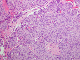
Figure 1
Histological examination demonstrating whorls and lobules of cells with indistinct cellular borders forming syncytium.
Figure 2
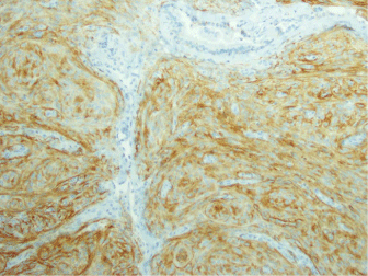
Figure 2
Positive immunohistochemical staining for EMA.
Discussion
Extracranial meningiomas are classified into primary or secondary types based on their origins
[1]. Primary meningiomas arise from ectopic arachnoid cells and therefore are not associated with
an intracranial mass, whereas secondary tumors result from direct extensions of intracranial masses
[1]. In this case, the patient experienced a left conductive hearing loss and pulsatile tinnitus without
any symptoms of cranial nerve involvement, which is consistent
with the diagnosis of primary middle ear meningioma. This suggests
that meningiomas should be considered in the differential diagnosis
of middle ear mass. Other differential diagnosis may include
schwannomas, paragangliomas, middle ear adenomas, metastatic
carcinomas, cholesteatomas, glomus tumors, retrotympanic vascular
tumors, and lipoma [2].
Imaging modalities may aid the diagnosis of primary extracranial
meningioma. While CT may reveal the presence of calcification and
bony hyperostosis, MRI has a higher sensitivity than CT and may
show an enhancing dural-based soft-tissue mass with a characteristic
dural tail [3]. In this case, the CT did not reveal calcification or bony
hyperostosis.
Immunohistochemistry and histology are crucial to differentiate
extracranial meningiomas from other differential diagnosis. In this
case, the tumor was characterized by positive immunostaining for
EMA, which is a characteristic finding of meningioma [1]. While
meningiomas exhibit similar immunohistochemical profiles, they
may express a variety of different histological patterns. The most
common histological pattern is meningothelial [4]. Moreover, cells
are arranged in syncytial sheets with indistinct cytoplasmic margins,
whorl formation, and clear to faintly eosinophilic cytoplasm [4].
Occasional psammoma bodies may be present, but mitotic activity
and necrosis are absent [4]. These findings in this case confirms the
diagnosis.
As demonstrated in this case, patients with symptomatic
meningiomas or consistently growing tumors should be managed
with complete surgical excision. Because meningiomas are slowgrowing
tumors, long-term follow-up is necessary to exclude the
recurrence [4]. The recurrence rate for meningiomas is reported to
vary widely from 7% to 84% [4]. Nevertheless, the prognosis remains
excellent, and to date, there are no published cases indicting that
recurrence can reduce life expectancy [5].
Conclusion
Meningioma can rarely present in the middle ear. This should be kept in the differential diagnosis when evaluating patients with middle ear mass. Patients with growing and symptomatic middle ear meningioma should undergo surgical excision and postoperative surveillance to monitor tumor recurrence.
References
- Maeng JW, Kim YH, SeoJinwon, Si Whan Kim. Primary Extracranial Meningioma Presenting as a Cheek Mass. Clin Exp Otorhinolaryngol. 2013; 6: 266-268.
- Zan E, Limb CJ, Koehler JF, Yousem Dm. Middle ear adenoma: a challenging diagnosis. AJNR Am J Neuroradiol. 2009; 20: 1602-1603.
- Alzahrani M, Gaboury L, Saliba I. A rare case of large skull base meningioma mimicking otitis media with effusion. Case Rep Med. 2013; 2013: 396905.
- Thompson LDR, Bouffard JP, Sandberg GD. Primary Ear and Temporal Bone Meningiomas: A Clinicopathologic Study of 36 Cases with a Review of the Literature. Mod Pathol. 2003; 16: 236-245.
- Radley MG, di Sant’Agnese PA, Eskin TA, Wilbur DC. Epithelial differentiation in meningiomas. An immunohistochemical, histochemical, and ultrastructural study—with review of the literature. Am J ClinPathol. 1989; 92: 266-272.